Abstract
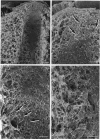
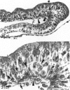
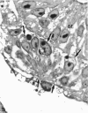
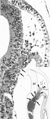
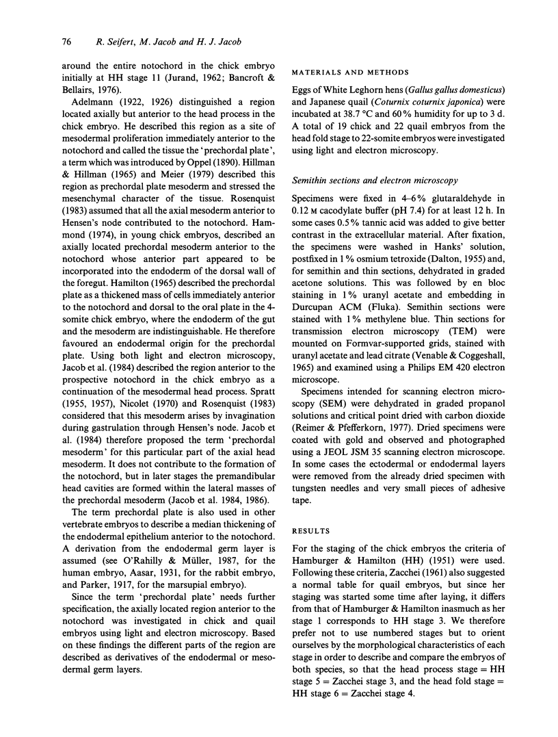
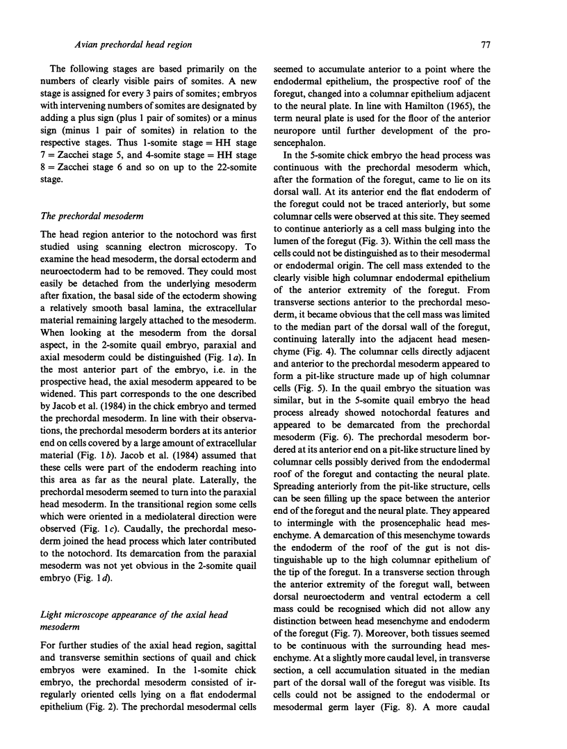
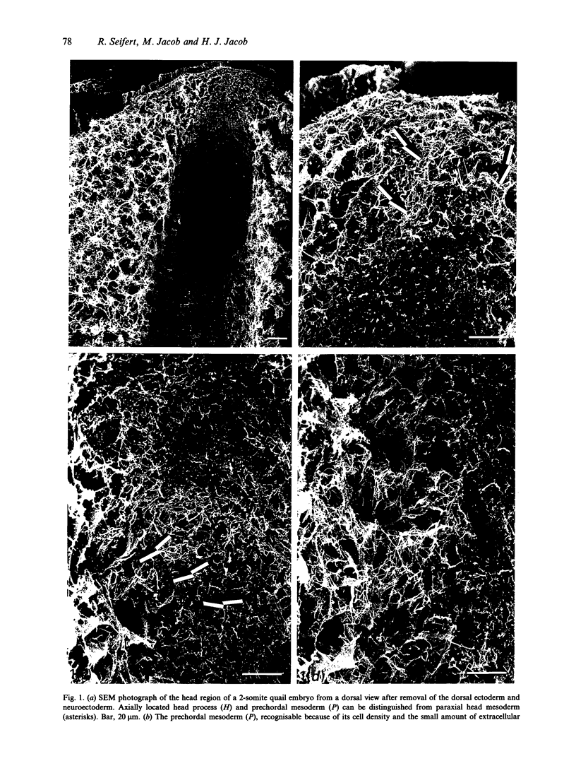
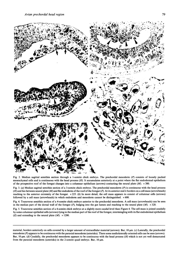
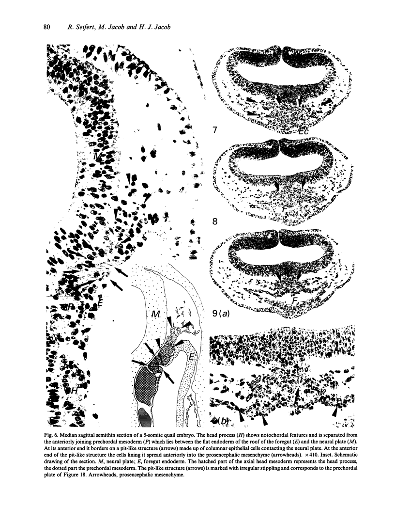
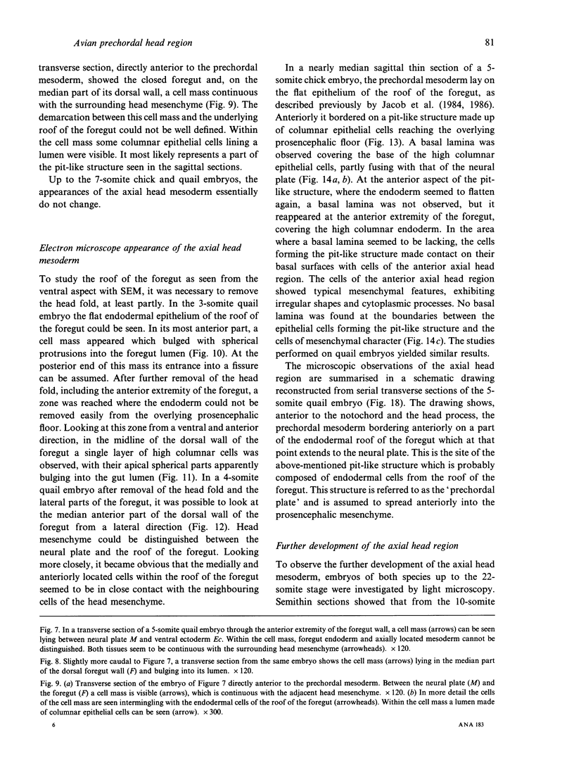
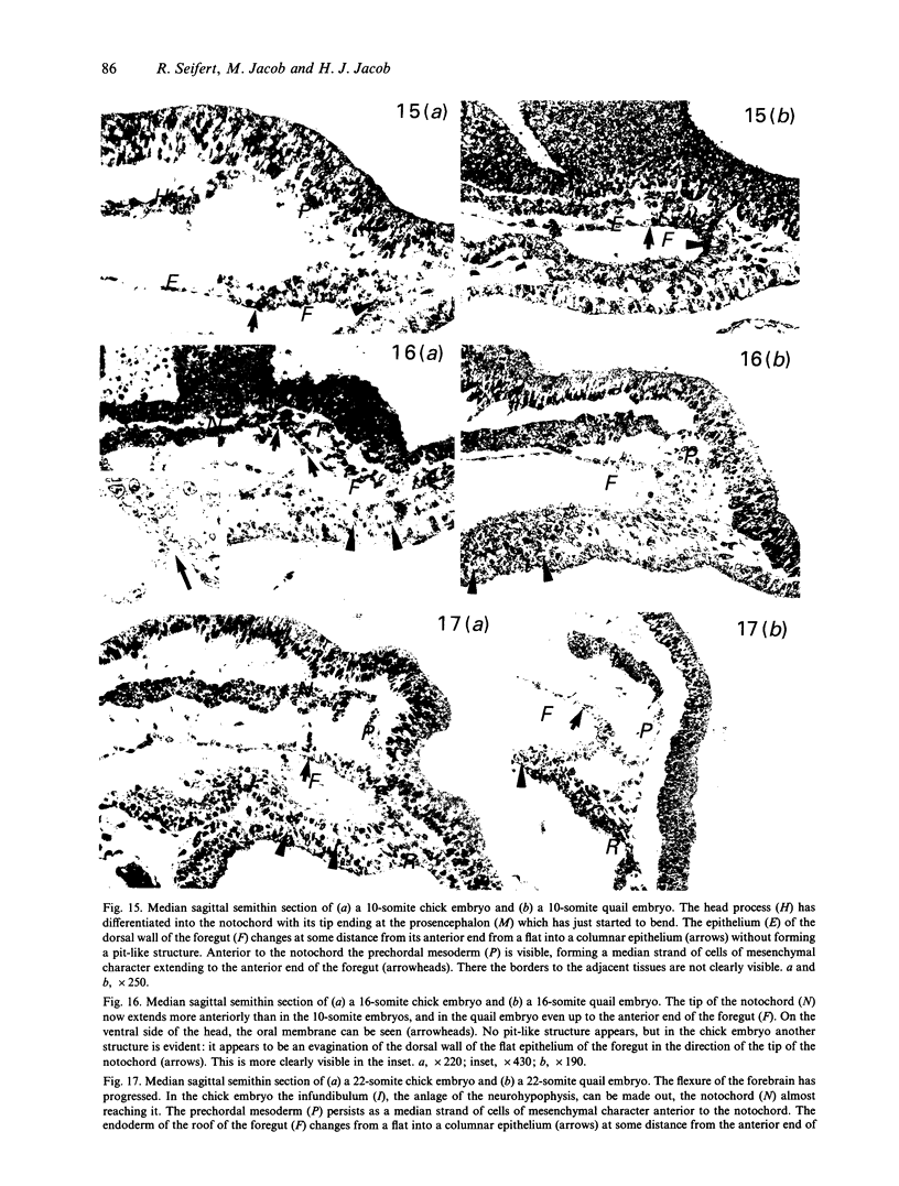
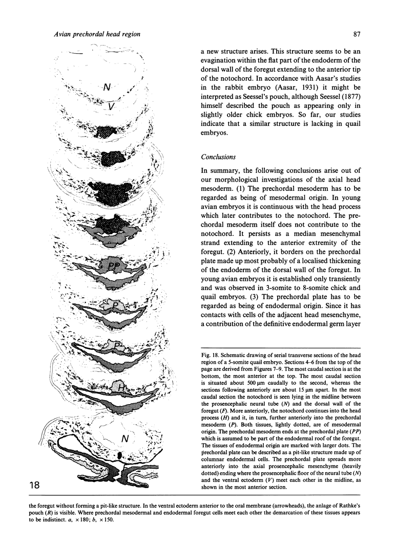
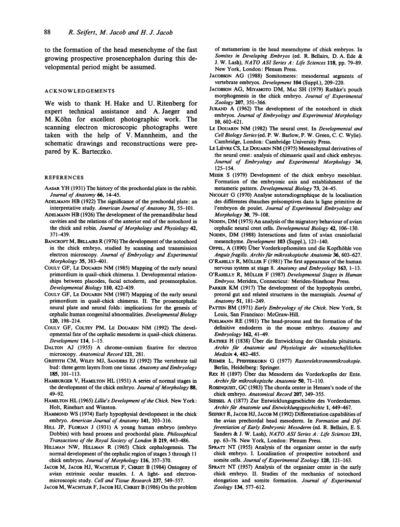
The axial mesoderm of the anterior head region was investigated in young chick and quail embryos by light and electron microscopy. Semithin sections showed that the axial head mesoderm consists of the head process and prechordal mesoderm. At the anterior end of the prechordal mesoderm, a group of columnar epithelial cells formed a pit-like structure. The bases of these columnar cells extended to the neural plate, thus limiting the prechordal mesoderm anteriorly. The cells lining the pit-like structure at its anterior end joined a cell accumulation made up of cells of mesenchymal character. Electron microscopy revealed that the columnar cells forming the pit-like structure were covered by a basal lamina which was discontinuous on its anterior aspect. No basal lamina was recognisable between the columnar epithelial cells and mesenchymal cells joining them anteriorly. The columnar epithelial cells bordering the prechordal mesoderm anteriorly were therefore assumed to be part of the endodermal germ layer. In agreement with the findings of other authors, it is proposed to term these axially located columnar cells of the endoderm the prechordal plate and to distinguish them from the prechordal mesoderm arising during gastrulation. For the mesenchymal cell accumulation anterior to the prechordal plate, participation in the formation of the prosencephalic mesenchyme is assumed. This implies that the definitive endodermal germ layer, like the ectodermal one represented by the neural crest, may also be able to contribute to mesenchyme formation in the head.
Full text
PDF
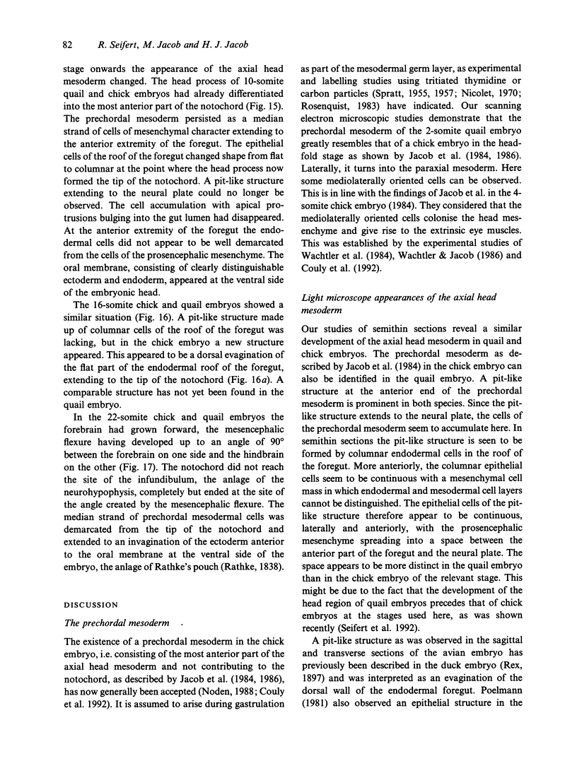
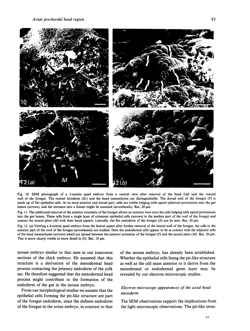
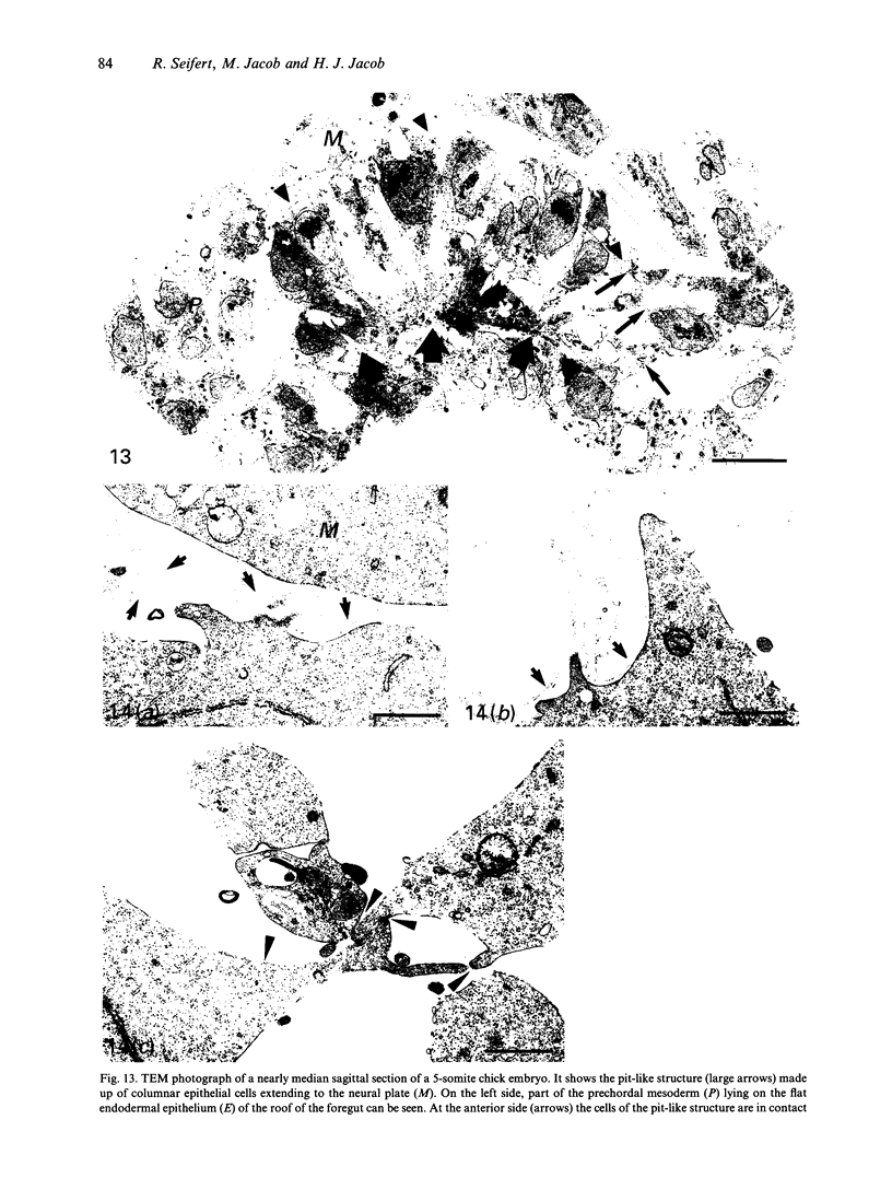
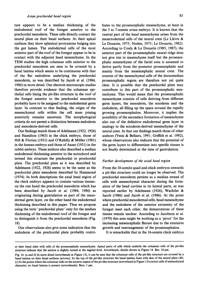

Images in this article
Selected References
These references are in PubMed. This may not be the complete list of references from this article.
- Aasar Y. H. The History of the Prochordal Plate in the Rabbit. J Anat. 1931 Oct;66(Pt 1):14–i3. [PMC free article] [PubMed] [Google Scholar]
- Bancroft M., Bellairs R. The development of the notochord in the chick embryo, studied by scanning and transmission electron microscopy. J Embryol Exp Morphol. 1976 Apr;35(2):383–401. [PubMed] [Google Scholar]
- Couly G. F., Coltey P. M., Le Douarin N. M. The developmental fate of the cephalic mesoderm in quail-chick chimeras. Development. 1992 Jan;114(1):1–15. doi: 10.1242/dev.114.1.1. [DOI] [PubMed] [Google Scholar]
- Couly G. F., Le Douarin N. M. Mapping of the early neural primordium in quail-chick chimeras. I. Developmental relationships between placodes, facial ectoderm, and prosencephalon. Dev Biol. 1985 Aug;110(2):422–439. doi: 10.1016/0012-1606(85)90101-0. [DOI] [PubMed] [Google Scholar]
- Couly G. F., Le Douarin N. M. Mapping of the early neural primordium in quail-chick chimeras. II. The prosencephalic neural plate and neural folds: implications for the genesis of cephalic human congenital abnormalities. Dev Biol. 1987 Mar;120(1):198–214. doi: 10.1016/0012-1606(87)90118-7. [DOI] [PubMed] [Google Scholar]
- Griffith C. M., Wiley M. J., Sanders E. J. The vertebrate tail bud: three germ layers from one tissue. Anat Embryol (Berl) 1992;185(2):101–113. doi: 10.1007/BF00185911. [DOI] [PubMed] [Google Scholar]
- HILLMAN N. W., HILLMAN R. CHICK CEPHALOGENESIS. THE NORMAL DEVELOPMENT OF THE CEPHALIC REGION OF STAGES 3 THROUGH 11 CHICK EMBRYOS. J Morphol. 1965 May;116:357–369. doi: 10.1002/jmor.1051160304. [DOI] [PubMed] [Google Scholar]
- Hammond W. S. Early hypophysial development in the chick embryo. Am J Anat. 1974 Nov;141(3):303–315. doi: 10.1002/aja.1001410303. [DOI] [PubMed] [Google Scholar]
- JURAND A. The development of the notochord in chick embryos. J Embryol Exp Morphol. 1962 Dec;10:602–621. [PubMed] [Google Scholar]
- Jacob M., Jacob H. J., Wachtler F., Christ B. Ontogeny of avian extrinsic ocular muscles. I. A light- and electron-microscopic study. Cell Tissue Res. 1984;237(3):549–557. doi: 10.1007/BF00228439. [DOI] [PubMed] [Google Scholar]
- Jacobson A. G. Somitomeres: mesodermal segments of vertebrate embryos. Development. 1988;104 (Suppl):209–220. doi: 10.1242/dev.104.Supplement.209. [DOI] [PubMed] [Google Scholar]
- Le Lièvre C. S., Le Douarin N. M. Mesenchymal derivatives of the neural crest: analysis of chimaeric quail and chick embryos. J Embryol Exp Morphol. 1975 Aug;34(1):125–154. [PubMed] [Google Scholar]
- Noden D. M. An analysis of migratory behavior of avian cephalic neural crest cells. Dev Biol. 1975 Jan;42(1):106–130. doi: 10.1016/0012-1606(75)90318-8. [DOI] [PubMed] [Google Scholar]
- Noden D. M. Interactions and fates of avian craniofacial mesenchyme. Development. 1988;103 (Suppl):121–140. doi: 10.1242/dev.103.Supplement.121. [DOI] [PubMed] [Google Scholar]
- O'Rahilly R., Müller F. The first appearance of the human nervous system at stage 8. Anat Embryol (Berl) 1981;163(1):1–13. doi: 10.1007/BF00315766. [DOI] [PubMed] [Google Scholar]
- Parker K. M. The Development of the Hypophysis Cerebri, Pre-Oral Gut, and Related Structures in the Marsupialia. J Anat. 1917 Apr;51(Pt 3):181–249. [PMC free article] [PubMed] [Google Scholar]
- Poelmann R. E. The head-process and the formation of the definitive endoderm in the mouse embryo. Anat Embryol (Berl) 1981;162(1):41–49. doi: 10.1007/BF00318093. [DOI] [PubMed] [Google Scholar]
- Rosenquist G. C. The chorda center in Hensen's node of the chick embryo. Anat Rec. 1983 Oct;207(2):349–355. doi: 10.1002/ar.1092070214. [DOI] [PubMed] [Google Scholar]
- SPRATT N. T., Jr Analysis of the organizer center in the early chick embryo. II. Studies of the mechanics of notochord elongation and somite formation. J Exp Zool. 1957 Apr;134(3):577–612. doi: 10.1002/jez.1401340309. [DOI] [PubMed] [Google Scholar]
- VENABLE J. H., COGGESHALL R. A SIMPLIFIED LEAD CITRATE STAIN FOR USE IN ELECTRON MICROSCOPY. J Cell Biol. 1965 May;25:407–408. doi: 10.1083/jcb.25.2.407. [DOI] [PMC free article] [PubMed] [Google Scholar]
- Veini M., Bellairs R. Early mesoderm differentiation in the chick embryo. Anat Embryol (Berl) 1991;183(2):143–149. doi: 10.1007/BF00174395. [DOI] [PubMed] [Google Scholar]
- Wachtler F., Jacob H. J., Jacob M., Christ B. The extrinsic ocular muscles in birds are derived from the prechordal plate. Naturwissenschaften. 1984 Jul;71(7):379–380. doi: 10.1007/BF00410750. [DOI] [PubMed] [Google Scholar]
- Wachtler F., Jacob M. Origin and development of the cranial skeletal muscles. Bibl Anat. 1986;(29):24–46. [PubMed] [Google Scholar]
- ZACCHEI A. M. [The embryonal development of the Japanese quail (Coturnix coturnix japonica T. and S.)]. Arch Ital Anat Embriol. 1961;66:36–62. [PubMed] [Google Scholar]


