

Abstract
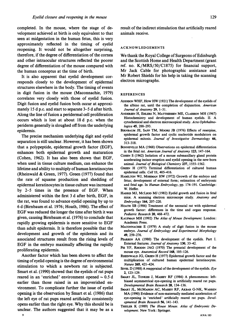
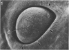
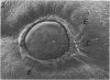
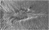
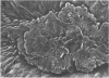
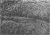
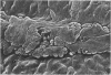
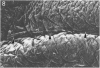
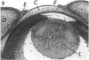
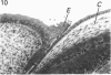
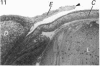
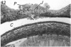
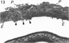
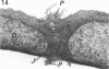
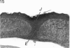
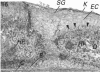
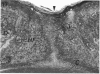
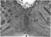
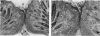
The process of eyelid development was studied in the mouse. The critical events occur between about 15.5 d postcoitum (p.c.) and 12 d after birth, and were studied by conventional histology and by scanning electron microscopy. At about 15.5 d p.c. the cornea of the eye is clearly visible with the primitive eyelids being represented by protruding ridges of epithelium at its periphery. Over the next 24 h, eyelid development proceeds to the stage when the cornea is completely covered by the fused eyelids. Periderm cells stream in to fill the gap between the developing eyelids. Their proliferative activity is such that they produce a cellular excrescence on the outer surface of the line of fusion of the eyelids. This excrescence had almost disappeared by about 17.5 d p.c. Keratinisation is first evident at this stage on the surface of the eyelids and passes continuously from one eyelid to the other. Evidence of epidermal differentiation is more clearly seen in the newborn, where a distinctive stratum granulosum now occupies about one third of its entire thickness. Within the subjacent dermis, hair follicles are differentiating. By about 5 d after birth, a thick layer of keratin extends without interruption across the junctional region. While a noticeable surface indentation overlies the latter, a similar depression is only seen on the conjunctival surface by about 10 d after birth. Keratinisation is also observed to extend in from the epidermal surface to involve the entire region between the 2 eyelids at about this time.(ABSTRACT TRUNCATED AT 250 WORDS)
Full text
PDF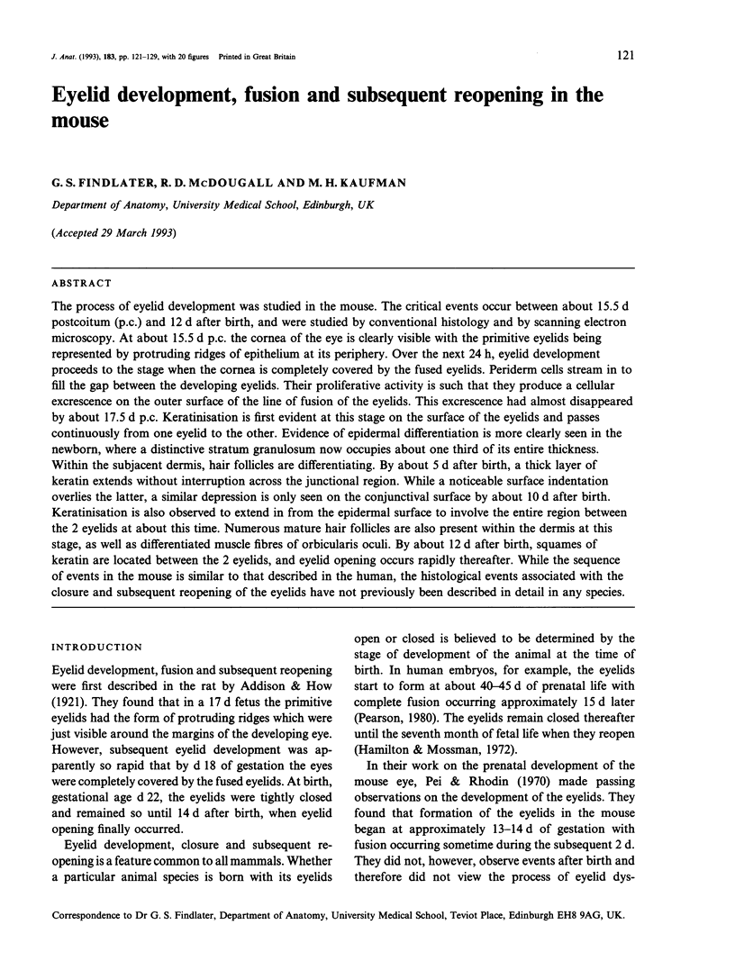
Images in this article
Selected References
These references are in PubMed. This may not be the complete list of references from this article.
- Andersen H., Ehlers N., Matthiessen M. E., Claesson M. H. Histochemistry and development of the human eyelids. II. A cytochemical and electron microscopical study. Acta Ophthalmol (Copenh) 1967;45(3):288–293. doi: 10.1111/j.1755-3768.1967.tb06492.x. [DOI] [PubMed] [Google Scholar]
- Birnbaum J. E., Sapp T. M., Moore J. B., Jr Effects of reserpine, epidermal growth factor, and cyclic nucleotide modulators on epidermal mitosis. J Invest Dermatol. 1976 May;66(5):313–318. doi: 10.1111/1523-1747.ep12482297. [DOI] [PubMed] [Google Scholar]
- Bonneville M. A. Observations on epidermal differentiation in the fetal rat. Am J Anat. 1968 Jul;123(1):147–164. doi: 10.1002/aja.1001230107. [DOI] [PubMed] [Google Scholar]
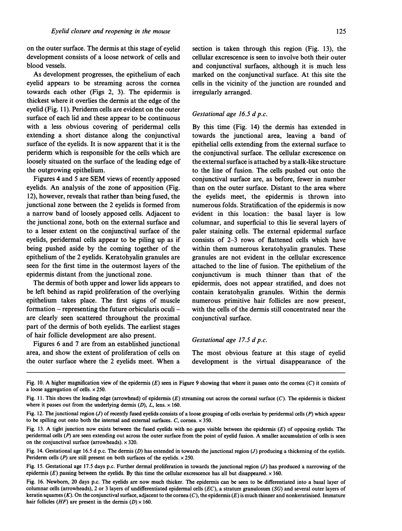
- COHEN S. Isolation of a mouse submaxillary gland protein accelerating incisor eruption and eyelid opening in the new-born animal. J Biol Chem. 1962 May;237:1555–1562. [PubMed] [Google Scholar]
- Green H. Terminal differentiation of cultured human epidermal cells. Cell. 1977 Jun;11(2):405–416. doi: 10.1016/0092-8674(77)90058-7. [DOI] [PubMed] [Google Scholar]
- Harris M. J., McLeod M. J. Eyelid growth and fusion in fetal mice. A scanning electron microscope study. Anat Embryol (Berl) 1982;164(2):207–220. doi: 10.1007/BF00318505. [DOI] [PubMed] [Google Scholar]
- Hoath S. B. Treatment of the neonatal rat with epidermal growth factor: differences in time and organ response. Pediatr Res. 1986 May;20(5):468–472. doi: 10.1203/00006450-198605000-00017. [DOI] [PubMed] [Google Scholar]
- Maconnachie E. A study of digit fusion in the mouse embryo. J Embryol Exp Morphol. 1979 Jan;49:259–276. [PubMed] [Google Scholar]
- Pearson A. A. The development of the eyelids. Part I. External features. J Anat. 1980 Jan;130(Pt 1):33–42. [PMC free article] [PubMed] [Google Scholar]
- Pei Y. F., Rhodin J. A. The prenatal development of the mouse eye. Anat Rec. 1970 Sep;168(1):105–125. doi: 10.1002/ar.1091680109. [DOI] [PubMed] [Google Scholar]
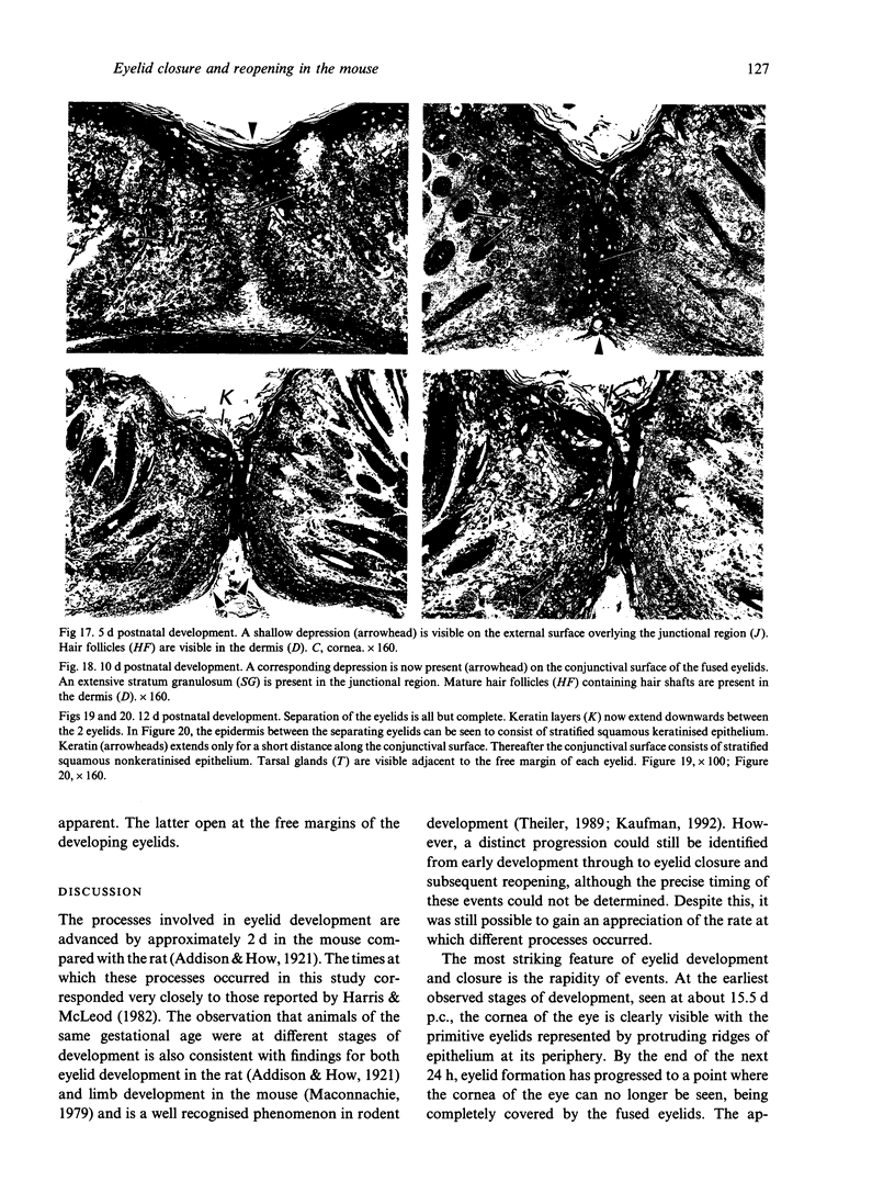
- Rheinwald J. G., Green H. Epidermal growth factor and the multiplication of cultured human epidermal keratinocytes. Nature. 1977 Feb 3;265(5593):421–424. doi: 10.1038/265421a0. [DOI] [PubMed] [Google Scholar]
- Sevel D. A reappraisal of the development of the eyelids. Eye (Lond) 1988;2(Pt 2):123–129. doi: 10.1038/eye.1988.25. [DOI] [PubMed] [Google Scholar]
- Smart J. L., McMahon A. C., Massey R. F., Akbar G. N., Warren M. A. Evidence of non-maternally mediated acceleration of eye-opening in 'enriched' artificially reared rat pups. Brain Res Dev Brain Res. 1990 Oct 1;56(1):141–143. doi: 10.1016/0165-3806(90)90174-w. [DOI] [PubMed] [Google Scholar]
- Smart J. L., Tonkiss J., Massey R. F. A phenomenon: left-biassed asymmetrical eye-opening in artificially reared rat pups. Brain Res. 1986 Jul;393(1):134–136. doi: 10.1016/0165-3806(86)90073-8. [DOI] [PubMed] [Google Scholar]