Abstract
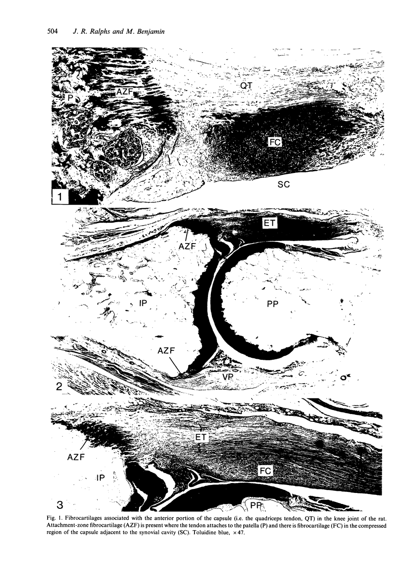
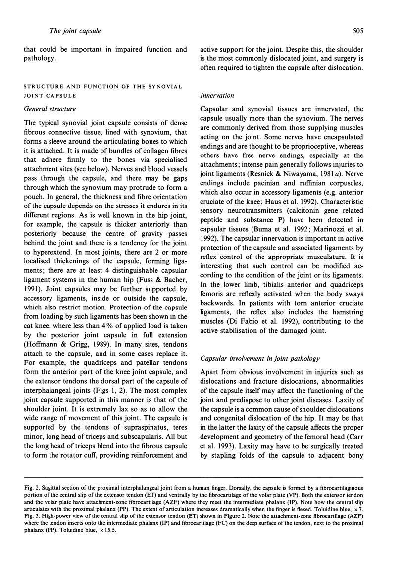
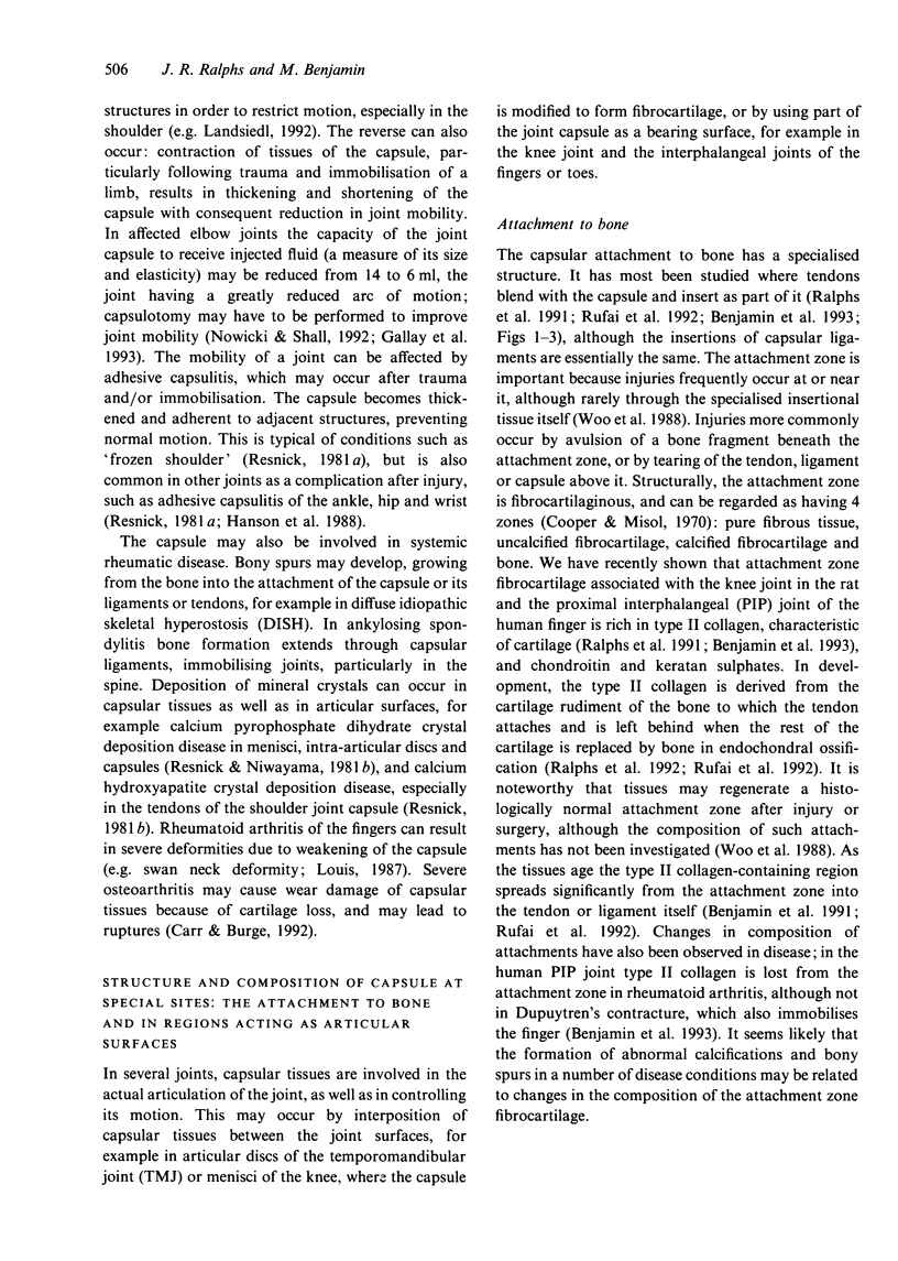
The joint capsule is vital to the function of synovial joints. It seals the joint space, provides passive stability by limiting movements, provides active stability via its proprioceptive nerve endings and may form articular surfaces for the joint. It is a dense fibrous connective tissue that is attached to the bones via specialised attachment zones and forms a sleeve around the joint. It varies in thickness according to the stresses to which it is subject, is locally thickened to form capsular ligaments, and may also incorporate tendons. The capsule is often injured, leading to laxity, constriction and/or adhesion to surrounding structures. It is also important in rheumatic disease, including rheumatoid arthritis and osteoarthritis, crystal deposition disorders, bony spur formation and ankylosing spondylitis. This article concentrates on the specialised structures of the capsule--where capsular tissues attach to bone or form part of the articulation of the joint. It focuses on 2 joints: the rat knee and the proximal interphalangeal (PIP) joint of the human finger. The attachments to bone contain fibrocartilage, derived from the cartilage of the embryonic bone rudiment and rich in type II collagen and glycosaminoglycans. The attachment changes with age, when type II collagen spreads into the capsular ligament or tendon, or pathology--type II collagen is lost from PIP capsular attachments in rheumatoid arthritis. Parts of the capsule that are compressed during movement adapt by becoming fibrocartilaginous. Such regions accumulate cartilage-like glycosaminoglycans and may contain type II collagen, especially in aged material.(ABSTRACT TRUNCATED AT 250 WORDS)
Full text
PDF
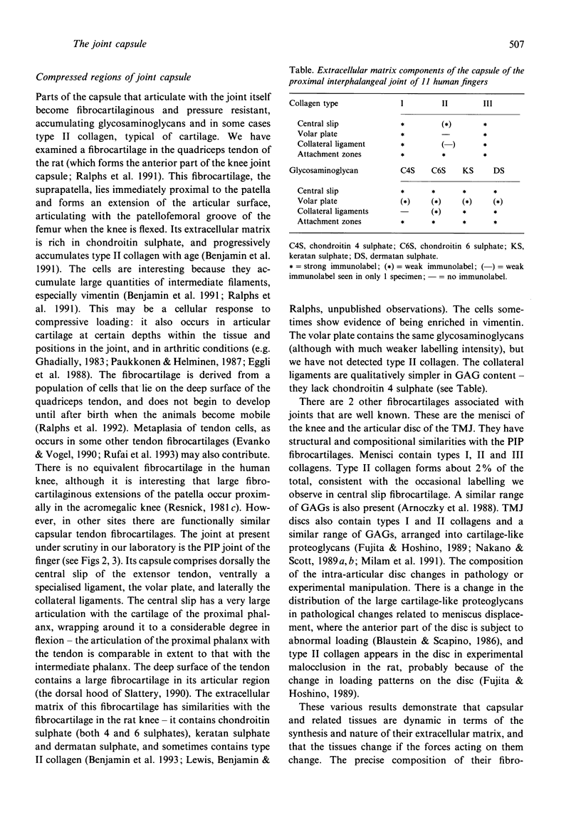
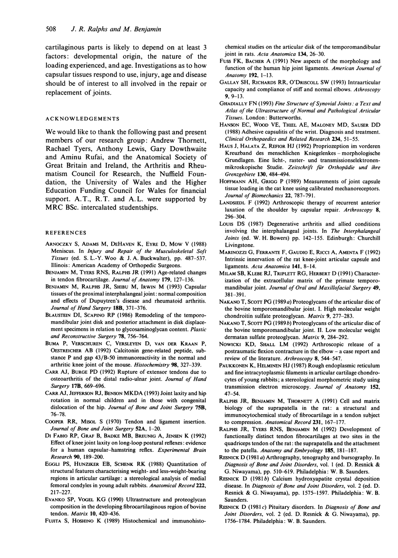
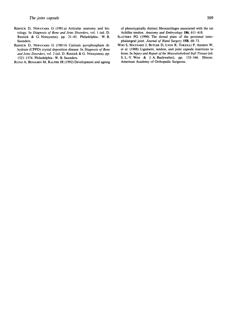
Images in this article
Selected References
These references are in PubMed. This may not be the complete list of references from this article.
- Benjamin M., Ralphs J. R., Shibu M., Irwin M. Capsular tissues of the proximal interphalangeal joint: normal composition and effects of Dupuytren's disease and rheumatoid arthritis. J Hand Surg Br. 1993 Jun;18(3):371–376. doi: 10.1016/0266-7681(93)90067-p. [DOI] [PubMed] [Google Scholar]
- Benjamin M., Tyers R. N., Ralphs J. R. Age-related changes in tendon fibrocartilage. J Anat. 1991 Dec;179:127–136. [PMC free article] [PubMed] [Google Scholar]
- Blaustein D. I., Scapino R. P. Remodeling of the temporomandibular joint disk and posterior attachment in disk displacement specimens in relation to glycosaminoglycan content. Plast Reconstr Surg. 1986 Dec;78(6):756–764. doi: 10.1097/00006534-198678060-00008. [DOI] [PubMed] [Google Scholar]
- Buma P., Verschuren C., Versleyen D., Van der Kraan P., Oestreicher A. B. Calcitonin gene-related peptide, substance P and GAP-43/B-50 immunoreactivity in the normal and arthrotic knee joint of the mouse. Histochemistry. 1992 Dec;98(5):327–339. doi: 10.1007/BF00270017. [DOI] [PubMed] [Google Scholar]
- Carr A. J., Burge P. D. Rupture of extensor tendons due to osteoarthritis of the distal radio-ulnar joint. J Hand Surg Br. 1992 Dec;17(6):694–696. doi: 10.1016/0266-7681(92)90203-e. [DOI] [PubMed] [Google Scholar]
- Carr A. J., Jefferson R. J., Benson M. K. Joint laxity and hip rotation in normal children and in those with congenital dislocation of the hip. J Bone Joint Surg Br. 1993 Jan;75(1):76–78. doi: 10.1302/0301-620X.75B1.8421041. [DOI] [PubMed] [Google Scholar]
- Cooper R. R., Misol S. Tendon and ligament insertion. A light and electron microscopic study. J Bone Joint Surg Am. 1970 Jan;52(1):1–20. [PubMed] [Google Scholar]
- Di Fabio R. P., Graf B., Badke M. B., Breunig A., Jensen K. Effect of knee joint laxity on long-loop postural reflexes: evidence for a human capsular-hamstring reflex. Exp Brain Res. 1992;90(1):189–200. doi: 10.1007/BF00229271. [DOI] [PubMed] [Google Scholar]
- Eggli P. S., Hunziker E. B., Schenk R. K. Quantitation of structural features characterizing weight- and less-weight-bearing regions in articular cartilage: a stereological analysis of medial femoral condyles in young adult rabbits. Anat Rec. 1988 Nov;222(3):217–227. doi: 10.1002/ar.1092220302. [DOI] [PubMed] [Google Scholar]
- Evanko S. P., Vogel K. G. Ultrastructure and proteoglycan composition in the developing fibrocartilaginous region of bovine tendon. Matrix. 1990 Dec;10(6):420–436. doi: 10.1016/s0934-8832(11)80150-2. [DOI] [PubMed] [Google Scholar]
- Fujita S., Hoshino K. Histochemical and immunohistochemical studies on the articular disk of the temporomandibular joint in rats. Acta Anat (Basel) 1989;134(1):26–30. doi: 10.1159/000146729. [DOI] [PubMed] [Google Scholar]
- Fuss F. K., Bacher A. New aspects of the morphology and function of the human hip joint ligaments. Am J Anat. 1991 Sep;192(1):1–13. doi: 10.1002/aja.1001920102. [DOI] [PubMed] [Google Scholar]
- Gallay S. H., Richards R. R., O'Driscoll S. W. Intraarticular capacity and compliance of stiff and normal elbows. Arthroscopy. 1993;9(1):9–13. doi: 10.1016/s0749-8063(05)80336-6. [DOI] [PubMed] [Google Scholar]
- Hanson E. C., Wood V. E., Thiel A. E., Maloney M. D., Sauser D. D. Adhesive capsulitis of the wrist. Diagnosis and treatment. Clin Orthop Relat Res. 1988 Sep;(234):51–55. [PubMed] [Google Scholar]
- Haus J., Halata Z., Refior H. J. Propriozeption im vorderen Kreuzband des menschlichen Kniegelenkes--morphologische Grundlagen. Eine licht-, raster- und transmissionselektronenmikroskopische Studie. Z Orthop Ihre Grenzgeb. 1992 Nov-Dec;130(6):484–494. doi: 10.1055/s-2008-1039657. [DOI] [PubMed] [Google Scholar]
- Hoffman A. H., Grigg P. Measurement of joint capsule tissue loading in the cat knee using calibrated mechanoreceptors. J Biomech. 1989;22(8-9):787–791. doi: 10.1016/0021-9290(89)90062-6. [DOI] [PubMed] [Google Scholar]
- Landsiedl F. Arthroscopic therapy of recurrent anterior luxation of the shoulder by capsular repair. Arthroscopy. 1992;8(3):296–304. doi: 10.1016/0749-8063(92)90059-k. [DOI] [PubMed] [Google Scholar]
- Marinozzi G., Ferrante F., Gaudio E., Ricci A., Amenta F. Intrinsic innervation of the rat knee joint articular capsule and ligaments. Acta Anat (Basel) 1991;141(1):8–14. doi: 10.1159/000147091. [DOI] [PubMed] [Google Scholar]
- Milam S. B., Klebe R. J., Triplett R. G., Herbert D. Characterization of the extracellular matrix of the primate temporomandibular joint. J Oral Maxillofac Surg. 1991 Apr;49(4):381–391. doi: 10.1016/0278-2391(91)90376-w. [DOI] [PubMed] [Google Scholar]
- Nakano T., Scott P. G. Proteoglycans of the articular disc of the bovine temporomandibular joint. I. High molecular weight chondroitin sulphate proteoglycan. Matrix. 1989 Aug;9(4):277–283. doi: 10.1016/s0934-8832(89)80023-x. [DOI] [PubMed] [Google Scholar]
- Nowicki K. D., Shall L. M. Arthroscopic release of a posttraumatic flexion contracture in the elbow: a case report and review of the literature. Arthroscopy. 1992;8(4):544–547. doi: 10.1016/0749-8063(92)90024-6. [DOI] [PubMed] [Google Scholar]
- Paukkonen K., Helminen H. J. Rough endoplasmic reticulum and fine intracytoplasmic filaments in articular cartilage chondrocytes of young rabbits; a stereological morphometric study using transmission electron microscopy. J Anat. 1987 Jun;152:47–54. [PMC free article] [PubMed] [Google Scholar]
- Ralphs J. R., Benjamin M., Thornett A. Cell and matrix biology of the suprapatella in the rat: a structural and immunocytochemical study of fibrocartilage in a tendon subject to compression. Anat Rec. 1991 Oct;231(2):167–177. doi: 10.1002/ar.1092310204. [DOI] [PubMed] [Google Scholar]
- Ralphs J. R., Tyers R. N., Benjamin M. Development of functionally distinct fibrocartilages at two sites in the quadriceps tendon of the rat: the suprapatella and the attachment to the patella. Anat Embryol (Berl) 1992;185(2):181–187. doi: 10.1007/BF00185920. [DOI] [PubMed] [Google Scholar]
- Rufai A., Benjamin M., Ralphs J. R. Development and ageing of phenotypically distinct fibrocartilages associated with the rat Achilles tendon. Anat Embryol (Berl) 1992 Dec;186(6):611–618. doi: 10.1007/BF00186984. [DOI] [PubMed] [Google Scholar]
- Slattery P. G. The dorsal plate of the proximal interphalangeal joint. J Hand Surg Br. 1990 Feb;15(1):68–73. doi: 10.1016/0266-7681_90_90052-6. [DOI] [PubMed] [Google Scholar]


