Abstract
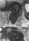
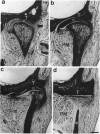
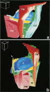
The anatomy of the temporomandibular joint capsule and its possible relationships to other structures near the joint are not fully understood. A 3-dimensional analysis based on sagittal, frontal and horizontal serial sections through the human temporomandibular joint region was therefore undertaken. Capsular elements which directly connect the temporal bone with the mandible were seen only on the lateral side of the joint. In the posterior, anterior and medial regions of the joint the upper and lower laminae of the articular disc are attached separately either to the temporal bone or to the mandibular condyle. The shaping of the articular cavities and the texture of the joint capsule permit movements of the articular disc predominantly in the anteromedial direction. On the entire medial side of the joint the articular disc and its capsular attachments are in close contact with the fascia of the lateral pterygoid muscle whereby a small portion of the upper head of this muscle inserts directly into the anteromedial part of the articular disc. Thus both the upper and the lower heads of the lateral pterygoid muscle are likely to influence the position of the articular disc directly during temporomandibular joint movements. Laterally, the articular disc is attached to the fascia of the masseter muscle, and part of the lateral ligament inserts into the temporalis fascia. Since these attachments are relatively weak, neither the temporalis nor the masseter muscles are considered to act directly on the articular disc; instead, via afferents from muscle spindles, they may take part in signalling the position of the temporomandibular joint components, including that of the articular disc.
Full text
PDF
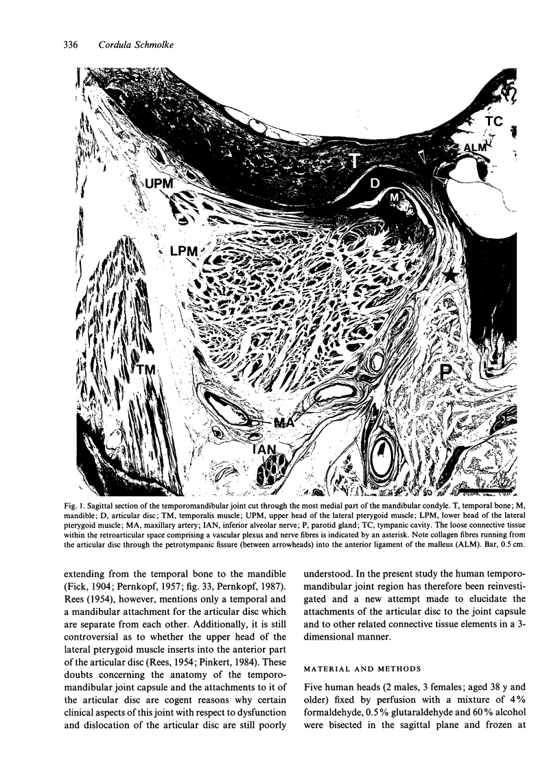
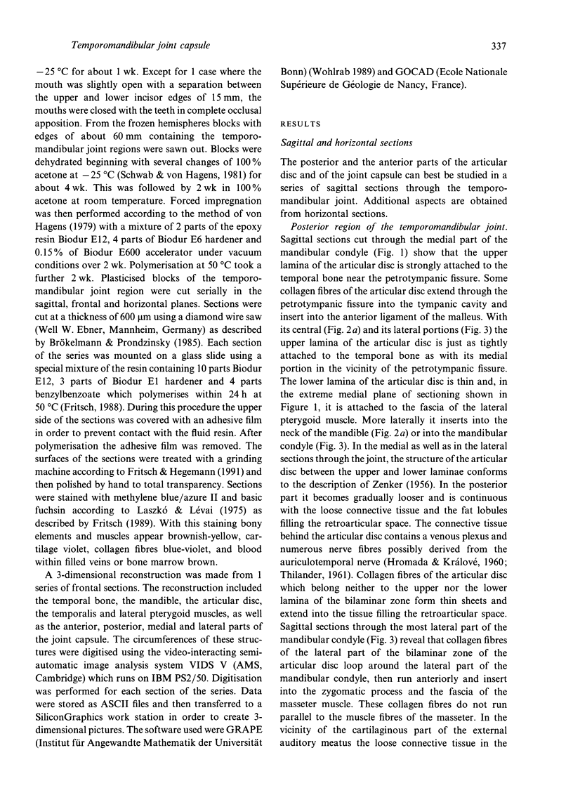
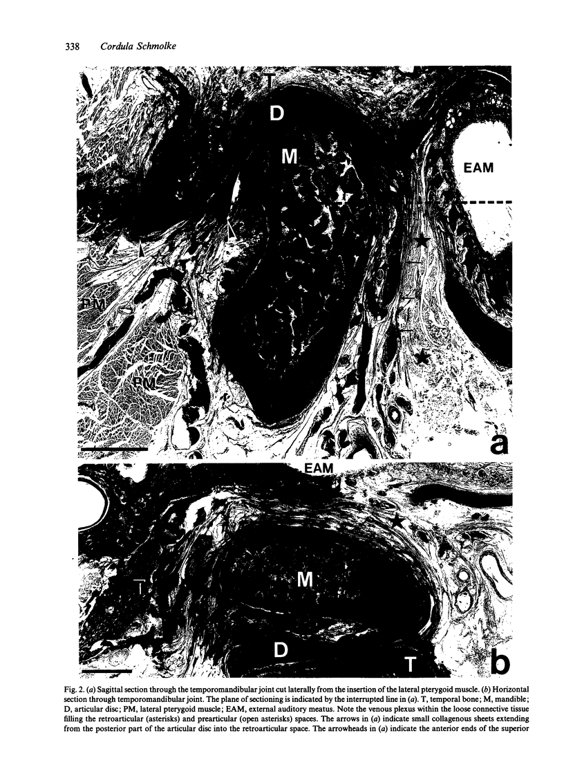
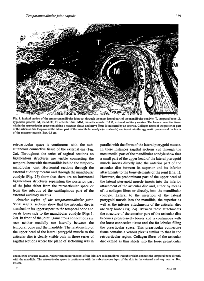
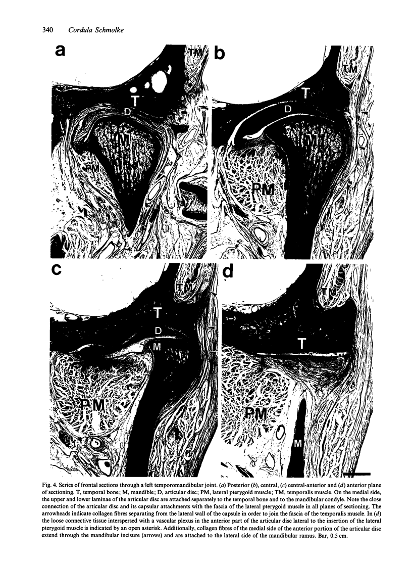
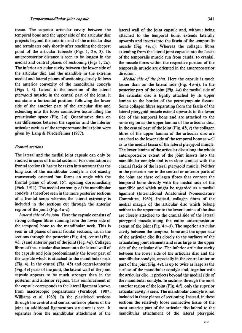
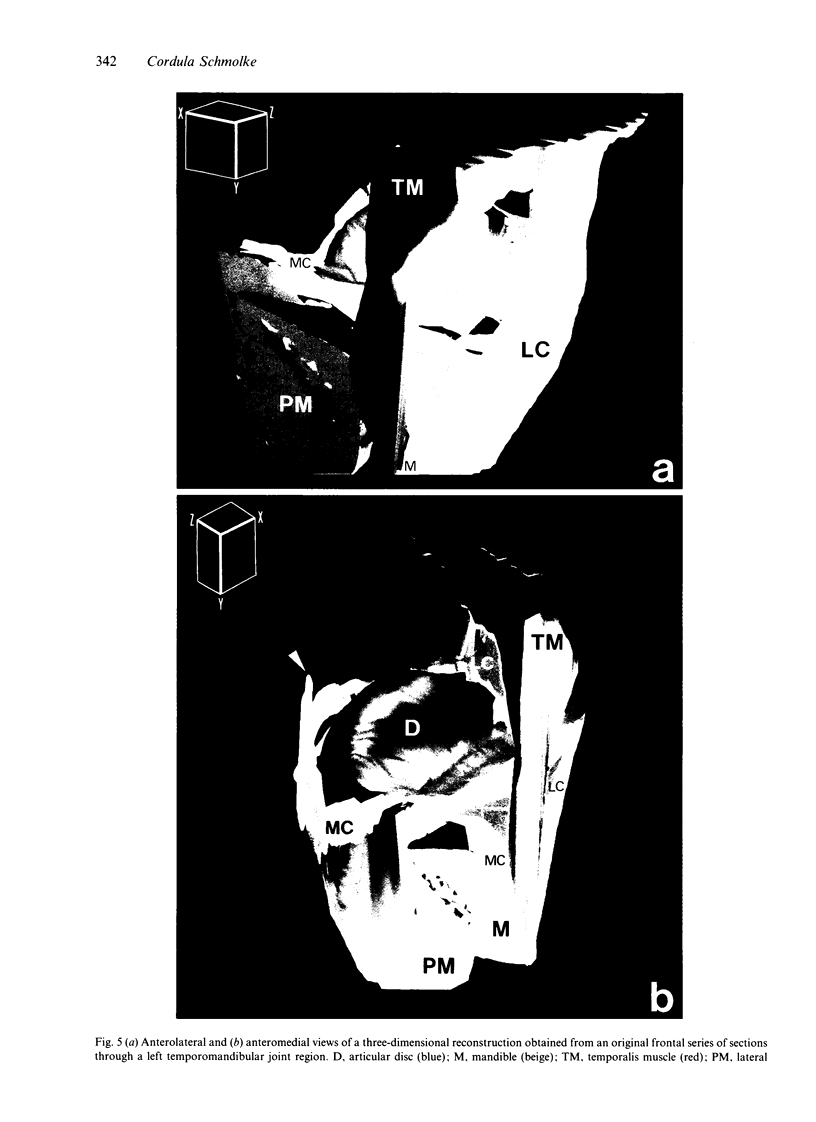
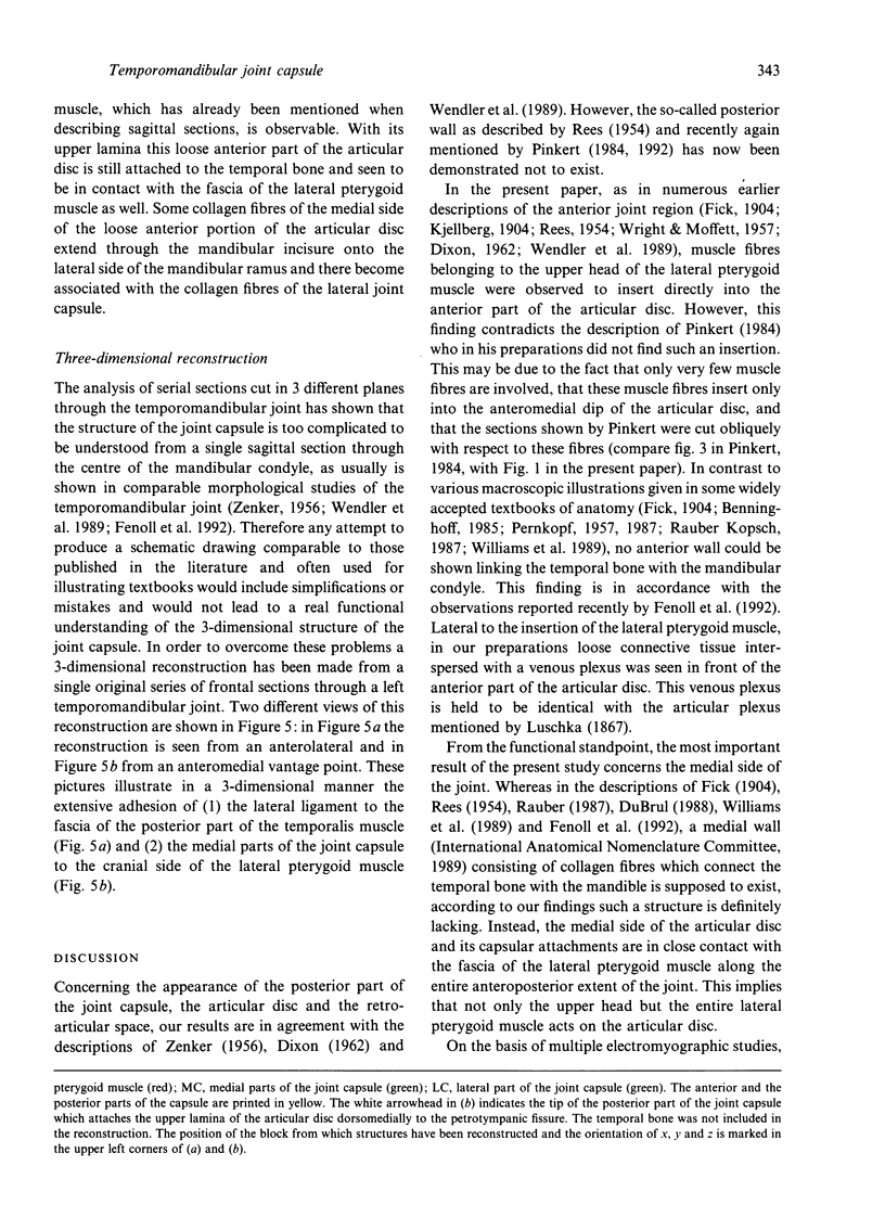
Images in this article
Selected References
These references are in PubMed. This may not be the complete list of references from this article.
- Bermejo Fenoll A., González Sequeros O., González González J. M. Histological study of the temporomandibular joint capsule: theory of the articular complex. Acta Anat (Basel) 1992;145(1):24–28. doi: 10.1159/000147337. [DOI] [PubMed] [Google Scholar]
- Coleman R. D. Temporomandibular joint: relation of the retrodiskal zone to Meckel's cartilage and lateral pterygoid muscle. J Dent Res. 1970 May-Jun;49(3):626–630. doi: 10.1177/00220345700490032701. [DOI] [PubMed] [Google Scholar]
- DIXON A. D. Structure and functional significance of the intraarticular disc of the human temporomandibular joint. Oral Surg Oral Med Oral Pathol. 1962 Jan;15:48–61. doi: 10.1016/0030-4220(62)90063-4. [DOI] [PubMed] [Google Scholar]
- Fritsch H. Developmental changes in the retrorectal region of the human fetus. Anat Embryol (Berl) 1988;177(6):513–522. doi: 10.1007/BF00305138. [DOI] [PubMed] [Google Scholar]
- Fritsch H., Hegemann L. Vereinfachung der Herstellung plastinationshistologischer Präparate durch Einsatz einer Schleifmaschine. Anat Anz. 1991;173(3):161–165. [PubMed] [Google Scholar]
- Fritsch H. Staining of different tissues in thick epoxy resin-impregnated sections of human fetuses. Stain Technol. 1989 Mar;64(2):75–79. doi: 10.3109/10520298909108049. [DOI] [PubMed] [Google Scholar]
- Jenö L., Géza L. A simple differential staining method for semi-thin sections of ossifying cartilage and bone tissues embedded in epoxy resin. Mikroskopie. 1975 Apr;31(1-2):1–4. [PubMed] [Google Scholar]
- Kubota K., Masegi T. Muscle spindle supply to the human jaw muscle. J Dent Res. 1977 Aug;56(8):901–909. doi: 10.1177/00220345770560081201. [DOI] [PubMed] [Google Scholar]
- Körber E., Freesmeyer W. B., Hüls A. Betrachtungen zu funktionsanalytischen Massnahmen. Zahnarztl Prax. 1987 Jan 9;38(1):6, 8-13. [PubMed] [Google Scholar]
- Lang J., Niederfeilner J. Uber Flächenwerte der Kiefergelenkspalte. Anat Anz. 1977;141(4):398–400. [PubMed] [Google Scholar]
- Mahan P. E., Wilkinson T. M., Gibbs C. H., Mauderli A., Brannon L. S. Superior and inferior bellies of the lateral pterygoid muscle EMG activity at basic jaw positions. J Prosthet Dent. 1983 Nov;50(5):710–718. doi: 10.1016/0022-3913(83)90214-7. [DOI] [PubMed] [Google Scholar]
- McNamara J. A., Jr The independent functions of the two heads of the lateral pterygoid muscle. Am J Anat. 1973 Oct;138(2):197–205. doi: 10.1002/aja.1001380206. [DOI] [PubMed] [Google Scholar]
- Scheuner G., Schuster T. Zur polarisationsoptischen Untersuchung von Semidünnschnitten. Anat Anz. 1985;158(2):199–200. [PubMed] [Google Scholar]
- Schwaighofer B. W., Tanaka T. T., Klein M. V., Sartoris D. J., Resnick D. MR imaging of the temporomandibular joint: a cadaver study of the value of coronal images. AJR Am J Roentgenol. 1990 Jun;154(6):1245–1249. doi: 10.2214/ajr.154.6.2110737. [DOI] [PubMed] [Google Scholar]
- Smeele L. E. Ontogeny of relationship of middle ear and temporomandibular (squamomandibular) joint in mammals [corrected]. I. Morphology and ontogeny in man. Acta Anat (Basel) 1988;131(4):338–341. [PubMed] [Google Scholar]
- Wright D. M., Moffett B. C., Jr The postnatal development of the human temporomandibular joint. Am J Anat. 1974 Oct;141(2):235–249. doi: 10.1002/aja.1001410206. [DOI] [PubMed] [Google Scholar]
- von Hagens G. Impregnation of soft biological specimens with thermosetting resins and elastomers. Anat Rec. 1979 Jun;194(2):247–255. doi: 10.1002/ar.1091940206. [DOI] [PubMed] [Google Scholar]


