Abstract
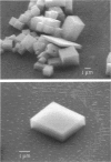
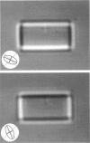
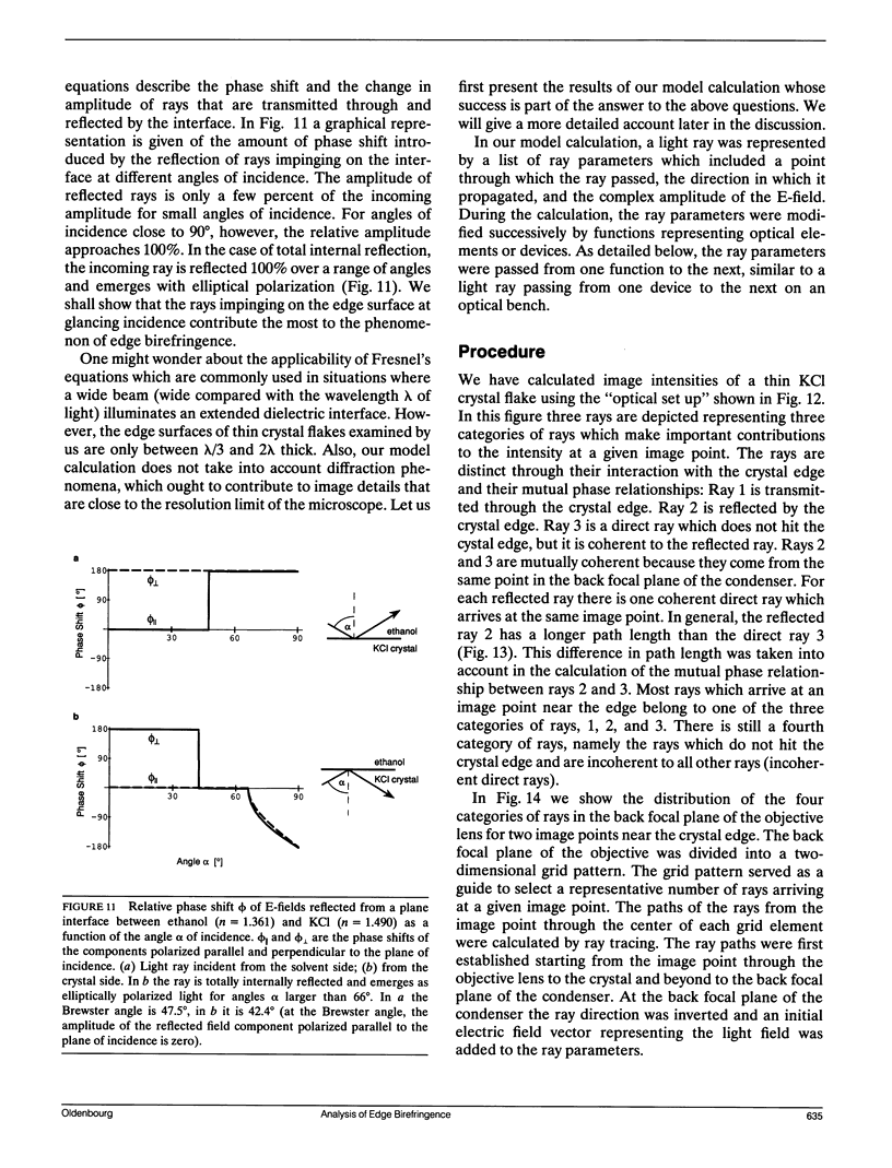
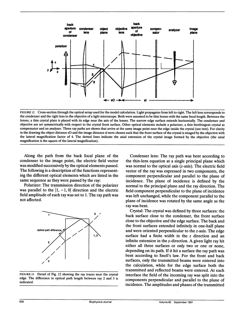
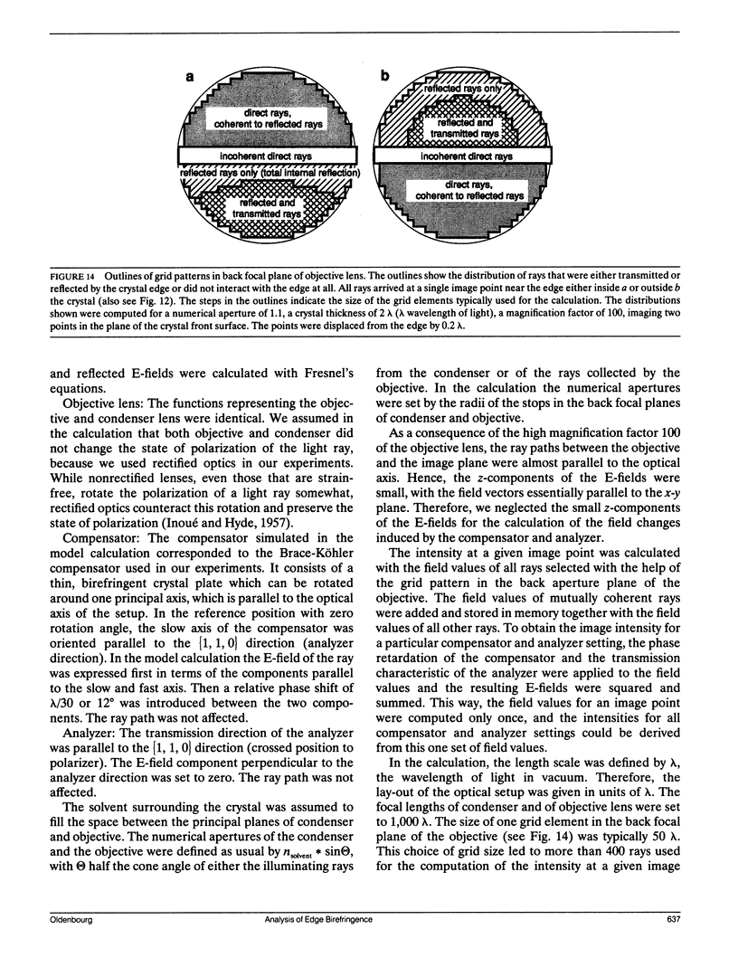
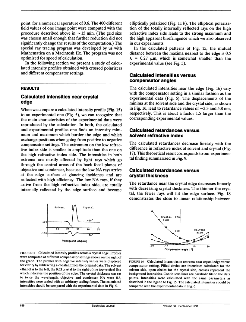
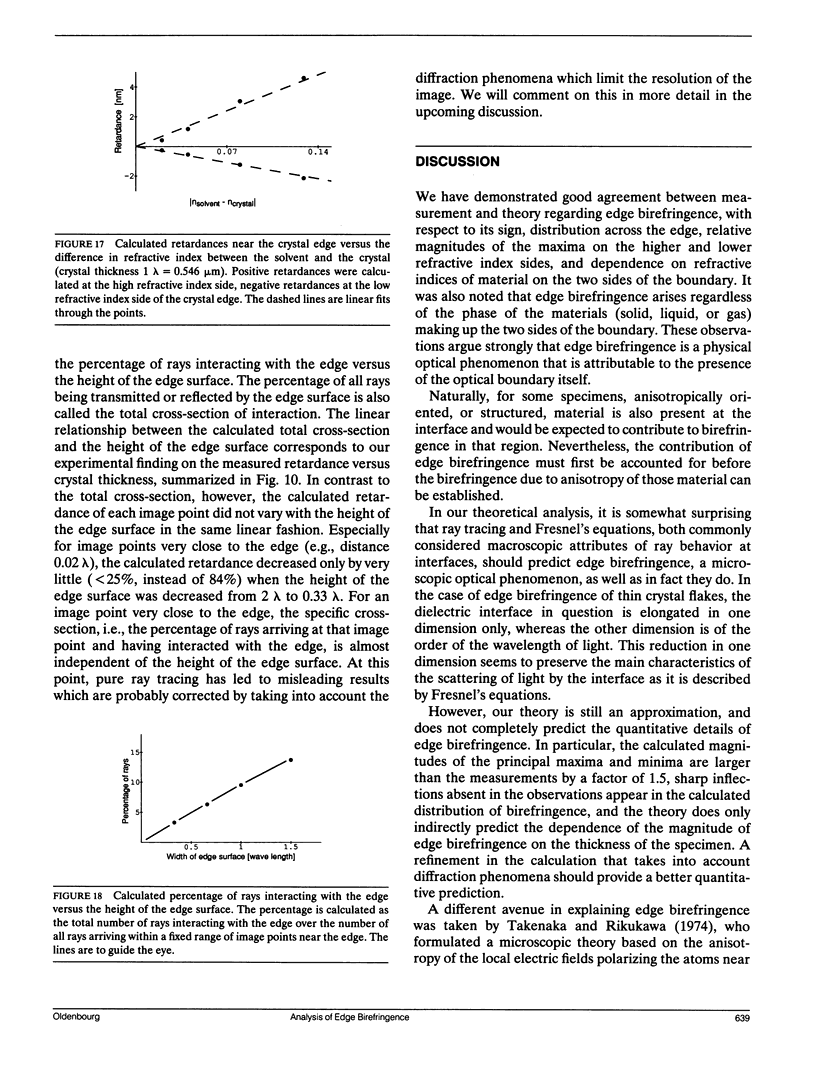
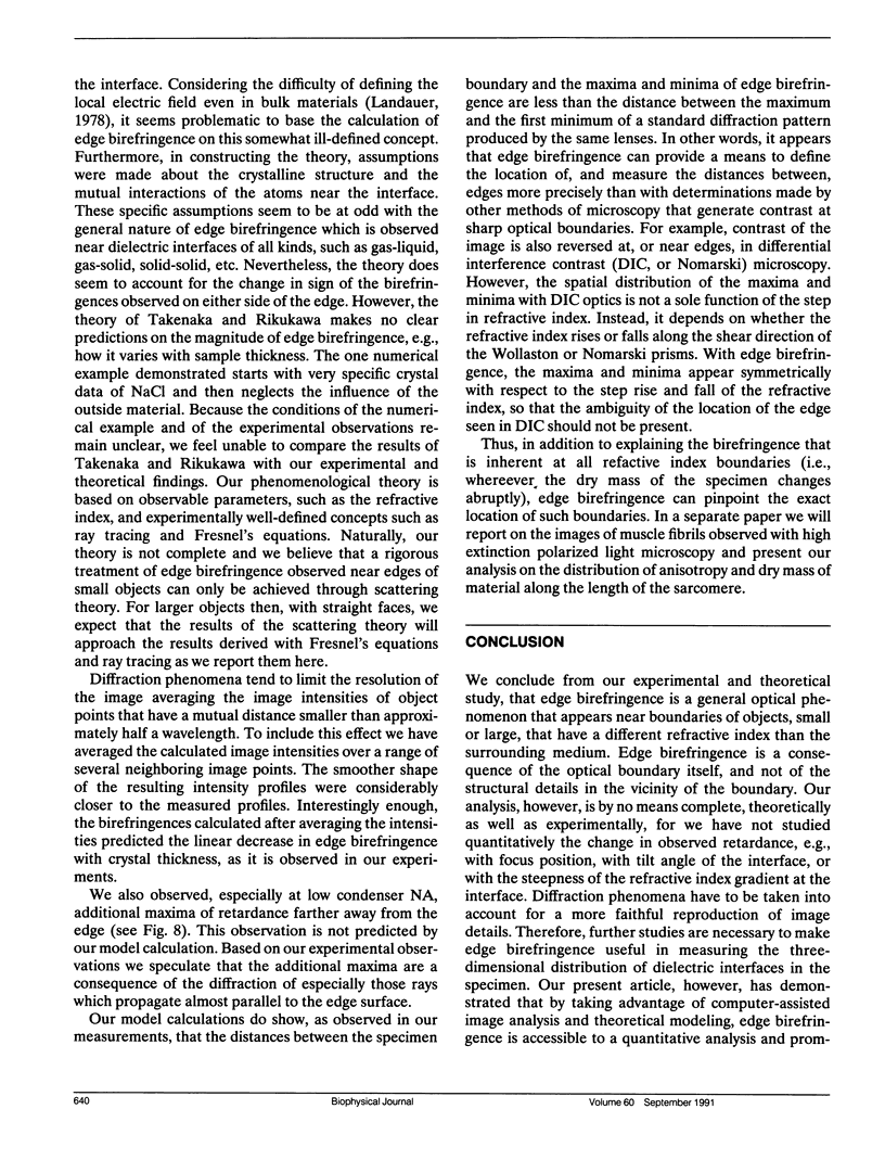
We present an experimental and theoretical study of the phenomenon of edge birefringence that appears near boundaries of transparent objects which are observed with high extinction and high resolution polarized light microscopy. As test objects, thin flakes of isotropic KCl crystals were immersed in media of various refractive indices. The measured retardation near crystal edges increased linearly with both the crystal thickness (tested between 0.3 and 1 micron), and the difference in refractive indices n between crystal (n = 1.49) and immersion liquids (n between 1.36 and 1.62). The specific edge birefringence, i.e., the retardation per thickness and per refractive index difference, is 0.029 on the high refractive index side of the boundary and -0.015 on the low refractive index side. The transition through zero birefringence specifies the position of a boundary at a much higher precision than predicted by the diffraction limit of the optical setup. The theoretical study employs a ray tracing procedure modeling the change in phase and polarization of rays passing through the specimen. We find good agreement between the model calculations and the experimental results indicating that edge birefringence can be attributed to the change in polarization of light that is refracted and reflected by dielectric interfaces.
Full text
PDF
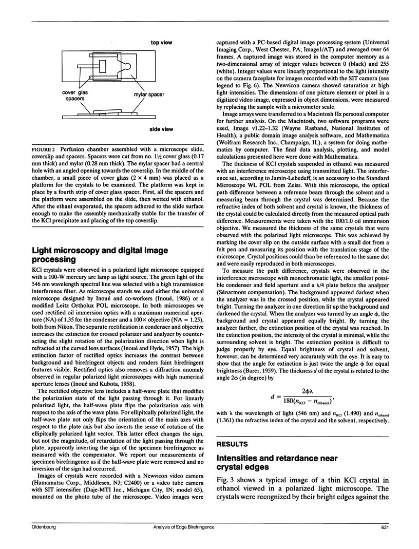
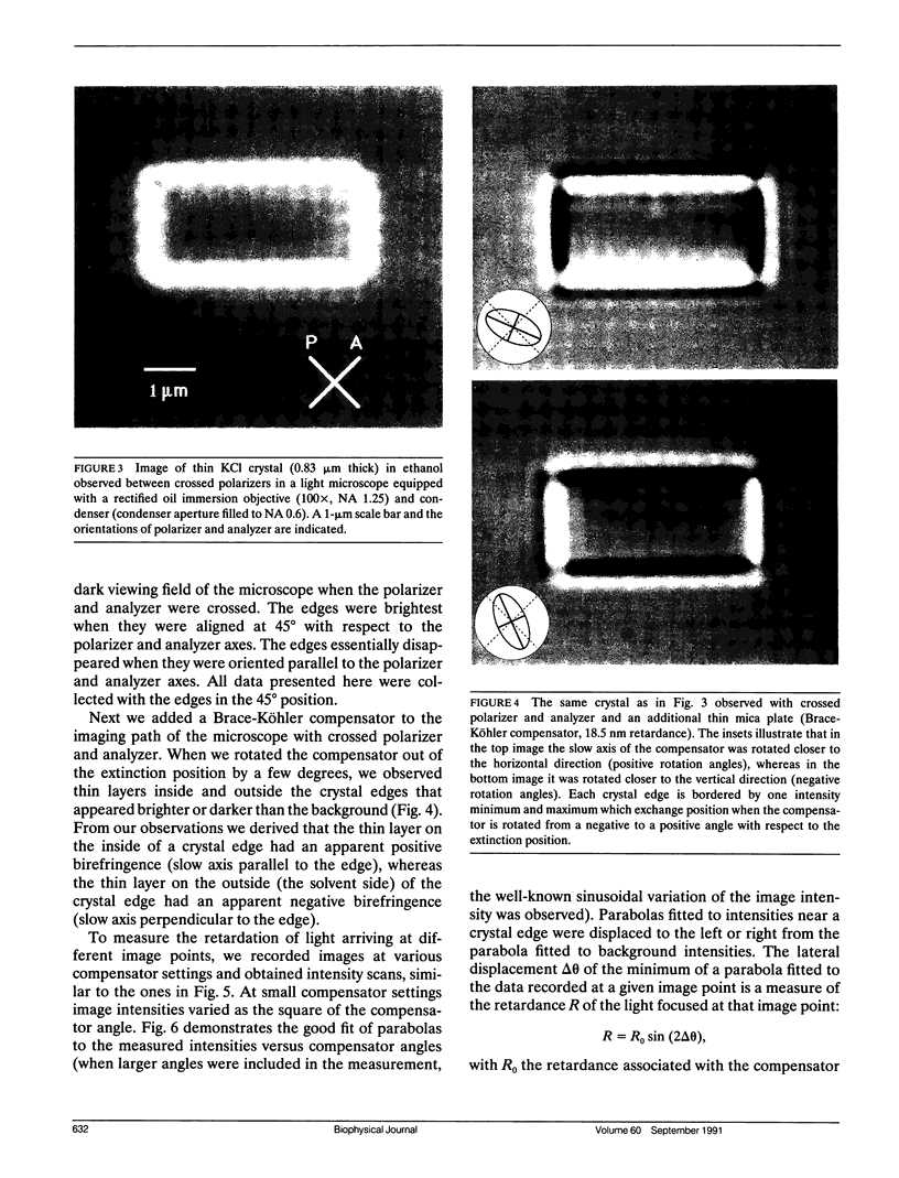
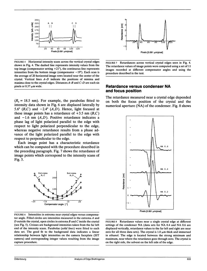
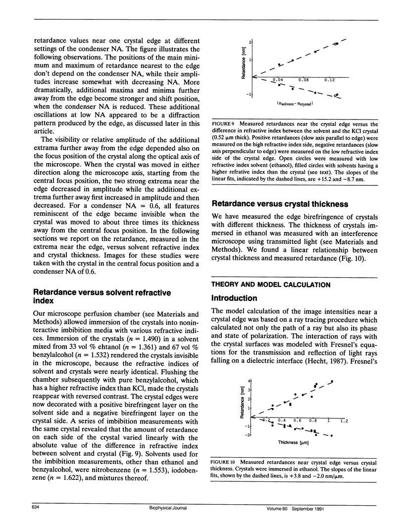
Images in this article
Selected References
These references are in PubMed. This may not be the complete list of references from this article.
- Block S. M., Fahrner K. A., Berg H. C. Visualization of bacterial flagella by video-enhanced light microscopy. J Bacteriol. 1991 Jan;173(2):933–936. doi: 10.1128/jb.173.2.933-936.1991. [DOI] [PMC free article] [PubMed] [Google Scholar]
- INOUE S., HYDE W. L. Studies on depolarization of light at microscope lens surfaces. II. The simultaneous realization of high resolution and high sensitivity with the polarizing microscope. J Biophys Biochem Cytol. 1957 Nov 25;3(6):831–838. doi: 10.1083/jcb.3.6.831. [DOI] [PMC free article] [PubMed] [Google Scholar]
- INOUE S., KUBOTA H. Diffraction anomaly in polarizing microscopes. Nature. 1958 Dec 20;182(4651):1725–1726. doi: 10.1038/1821725a0. [DOI] [PubMed] [Google Scholar]
- INOUE S. [Polarization optical studies of the mitotic spindle. I. The demonstration of spindle fibers in living cells]. Chromosoma. 1953;5(5):487–500. doi: 10.1007/BF01271498. [DOI] [PubMed] [Google Scholar]
- Walker R. A., Inoué S., Salmon E. D. Asymmetric behavior of severed microtubule ends after ultraviolet-microbeam irradiation of individual microtubules in vitro. J Cell Biol. 1989 Mar;108(3):931–937. doi: 10.1083/jcb.108.3.931. [DOI] [PMC free article] [PubMed] [Google Scholar]


