

Abstract
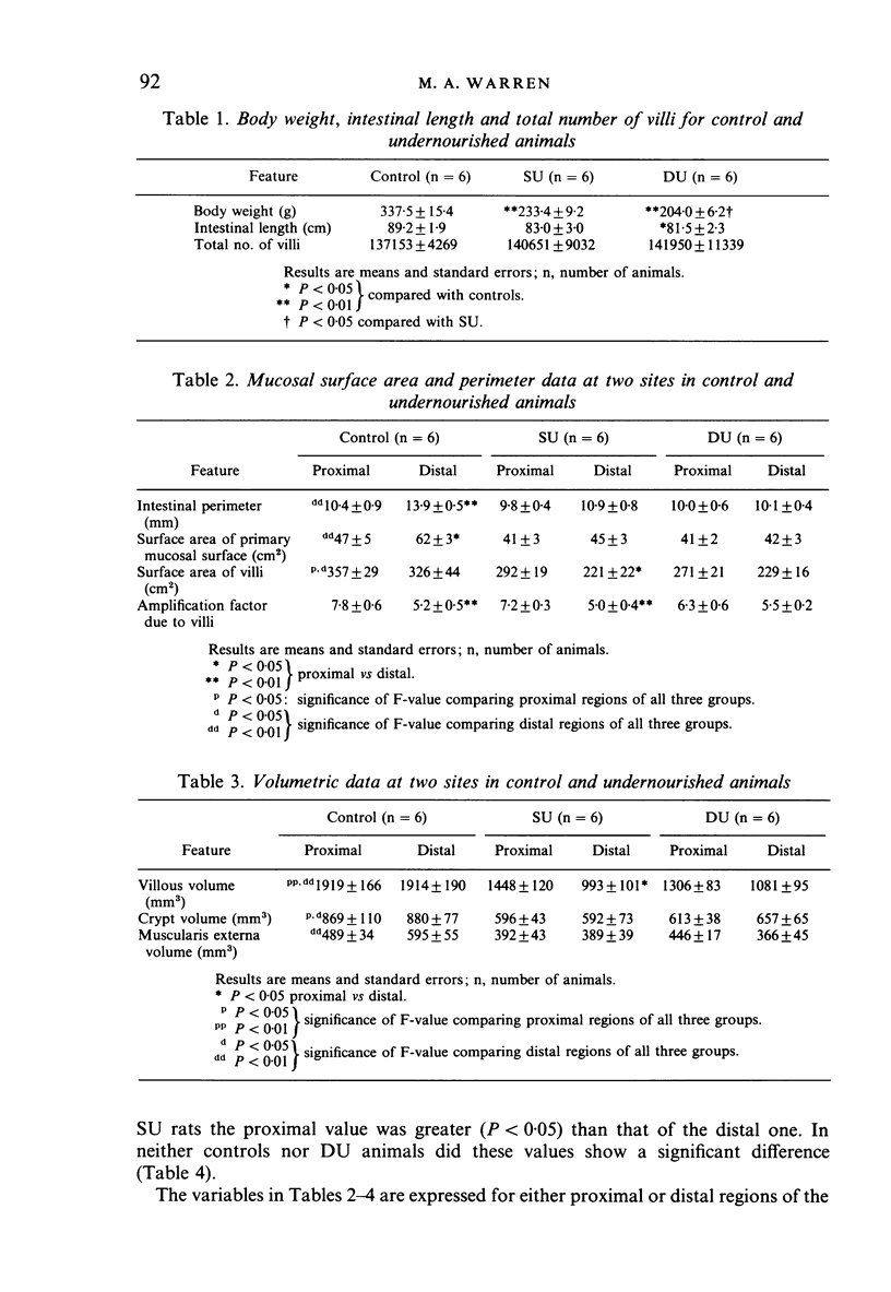
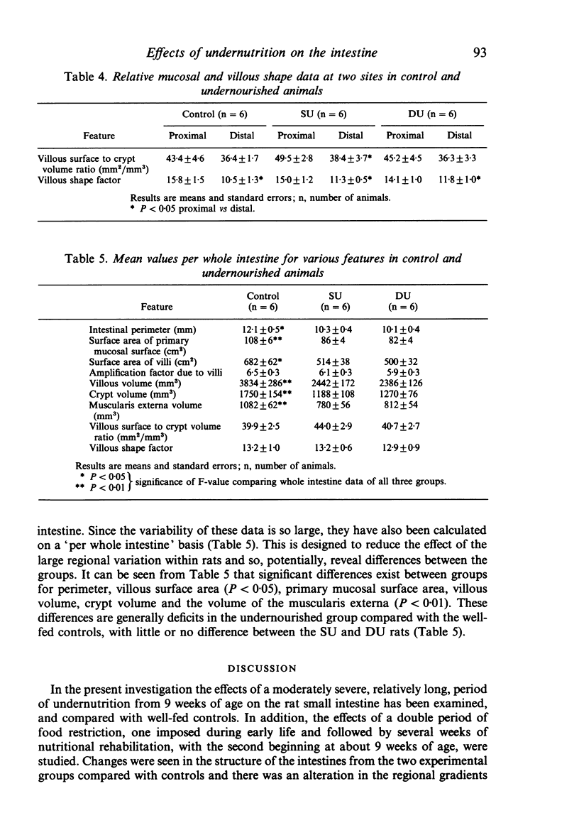
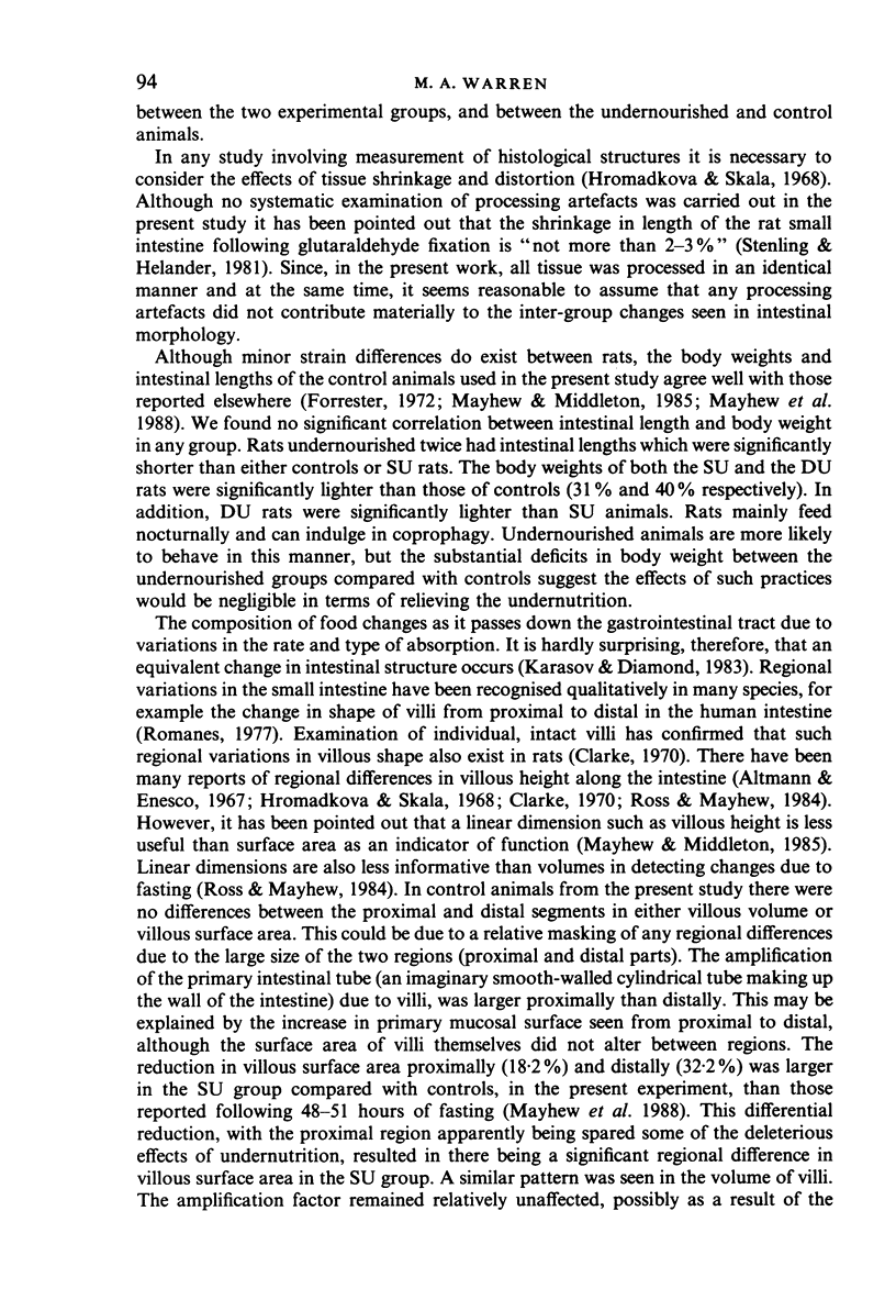
Male rats were undernourished from birth to 3 weeks of age and then allowed a period of unrestricted access to food. These animals were subjected to a second period of undernutrition from 9 to 12 weeks. A group of rats undernourished from 9 to 12 weeks only, and a group well-fed throughout life were used for comparison. Six rats from each group were anaesthetised by intraperitoneal injection with sodium pentobarbitone and killed by perfusion with glutaraldehyde at 12 weeks of age and the small intestine removed and its length recorded. Resin-embedded sections 1 micron thick were cut in the transverse plane from proximal and distal regions of each intestine. Point and intersection counting was performed to estimate villous surface area, volume and shape as well as crypt and muscle volume. The body weight of the undernourished rats was significantly (P less than 0.01) reduced compared to controls, with doubly undernourished rats weighing significantly (P less than 0.05) less than singly deprived animals. Rats undernourished once showed larger deficits in villous surface area and volume in distal regions than in proximal ones. However, in rats undernourished during the weaning period and again in later life the proximal part also became affected, while the distal region was not affected further. Therefore, it appears that the distal region of the rat small intestine is more vulnerable to a single period of food restriction during adult life than is the proximal part.
Full text
PDF
Selected References
These references are in PubMed. This may not be the complete list of references from this article.
- Aldewachi H. S., Wright N. A., Appleton D. R., Watson A. J. The effect of starvation and refeeding on cell population kinetics in the rat small bowel mucosa. J Anat. 1975 Feb;119(Pt 1):105–121. [PMC free article] [PubMed] [Google Scholar]
- Altmann G. G., Enesco M. Cell number as a measure of distribution and renewal of epithelial cells in the small intestine of growing and adult rats. Am J Anat. 1967 Sep;121(2):319–336. doi: 10.1002/aja.1001210210. [DOI] [PubMed] [Google Scholar]
- Baddeley A. J., Gundersen H. J., Cruz-Orive L. M. Estimation of surface area from vertical sections. J Microsc. 1986 Jun;142(Pt 3):259–276. doi: 10.1111/j.1365-2818.1986.tb04282.x. [DOI] [PubMed] [Google Scholar]
- Clarke R. M. Mucosal architecture and epithelial cell production rate in the small intestine of the albino rat. J Anat. 1970 Nov;107(Pt 3):519–529. [PMC free article] [PubMed] [Google Scholar]
- Clarke R. M. The effect of growth and of fasting on the number of villi and crypts in the small intestine of the albino rat. J Anat. 1972 May;112(Pt 1):27–33. [PMC free article] [PubMed] [Google Scholar]
- Forrester J. M. The number of villi in rat's jejunum and ileum: effect of normal growth, partial enterectomy, and tube feeding. J Anat. 1972 Feb;111(Pt 2):283–291. [PMC free article] [PubMed] [Google Scholar]
- Hromádková V., Skála I. Factors influencing the assessment of size of the mucosal surface and length of the small intestine in rats. Digestion. 1968;1(3):149–158. doi: 10.1159/000196847. [DOI] [PubMed] [Google Scholar]
- Karasov W. H., Diamond J. M. Adaptive regulation of sugar and amino acid transport by vertebrate intestine. Am J Physiol. 1983 Oct;245(4):G443–G462. doi: 10.1152/ajpgi.1983.245.4.G443. [DOI] [PubMed] [Google Scholar]
- Mayhew T. M. A geometric model for estimating villous surface area in rat small bowel is justified by unbiased estimates obtained using vertical sections. J Anat. 1988 Dec;161:187–193. [PMC free article] [PubMed] [Google Scholar]
- Mayhew T. M., Carson F. L. Mechanisms of adaptation in rat small intestine: regional differences in quantitative morphology during normal growth and experimental hypertrophy. J Anat. 1989 Jun;164:189–200. [PMC free article] [PubMed] [Google Scholar]
- Mayhew T. M. Geometric model of the rat intestinal mucosa for stereological evaluation of villus amplification factors. J Microsc. 1984 Sep;135(Pt 3):337–346. doi: 10.1111/j.1365-2818.1984.tb02538.x. [DOI] [PubMed] [Google Scholar]
- Mayhew T. M., Middleton C. Crypts, villi and microvilli in the small intestine of the rat. A stereological study of their variability within and between animals. J Anat. 1985 Aug;141:1–17. [PMC free article] [PubMed] [Google Scholar]
- Ross G. A., Mayhew T. M. Effects of fasting on mucosal dimensions in the duodenum, jejunum and ileum of the rat. J Anat. 1985 Oct;142:191–200. [PMC free article] [PubMed] [Google Scholar]
- Ross G. A., Mayhew T. M. Effects of fasting on villi along the small intestine: a stereological approach to the problem of quantifying villus 'shape'. Experientia. 1984 Aug 15;40(8):856–858. doi: 10.1007/BF01951993. [DOI] [PubMed] [Google Scholar]
- Stenling R., Helander H. F. Stereological studies on the small intestinal epithelium of the rat. 1. The absorptive cells of the normal duodenum and jejunum. Cell Tissue Res. 1981;217(1):11–21. doi: 10.1007/BF00233821. [DOI] [PubMed] [Google Scholar]
- Stewart R. J., Preece R. F., Sheppard H. G. Twelve generations of marginal protein deficiency. Br J Nutr. 1975 Mar;33(2):233–253. doi: 10.1079/bjn19750027. [DOI] [PubMed] [Google Scholar]