

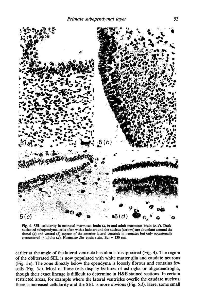
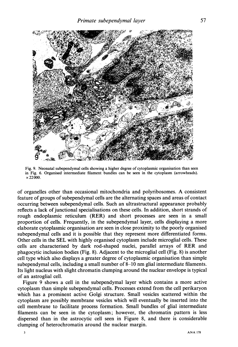
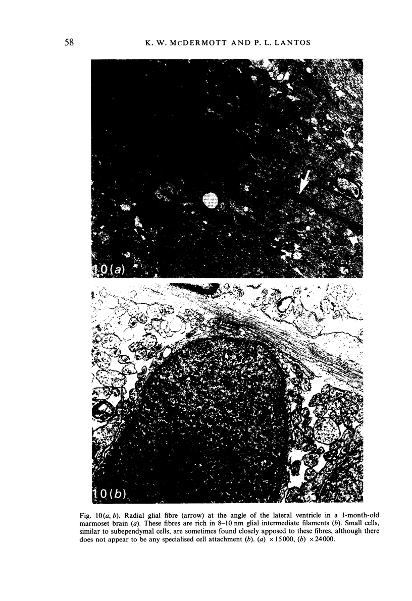
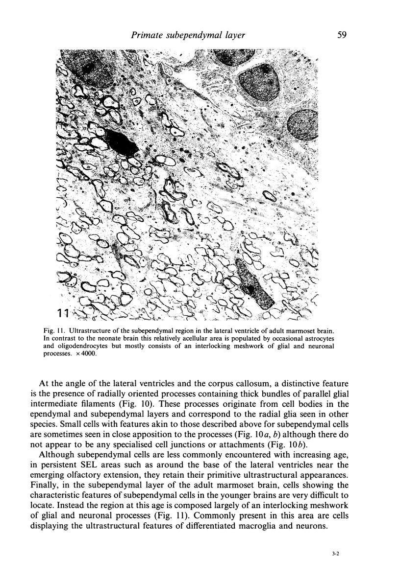
Abstract
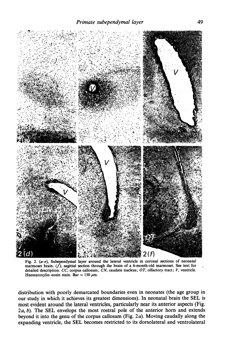
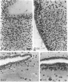
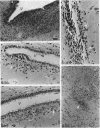
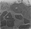
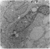
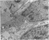
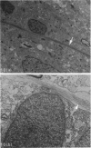
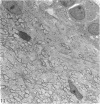
The subependymal layer (SEL) of the postnatal marmoset, a simian primate, has been investigated by histological and electron microscopic techniques. Although well documented in rodents, little is known about this layer in primates. The distribution of the SEL in marmosets is most extensive at birth around the anterior lateral ventricles, where the layer is generally 5-10 cells deep; however, there is considerable regional variation. With age the size of the SEL decreases dramatically, becoming very poorly demarcated in adult animals. Ultrastructurally, many subependymal cells in neonatal and young brains display the features of undifferentiated cells, although neurons and glia are also present. Cells displaying features intermediate between astrocytes and undifferentiated SEL cells are occasionally encountered. In adults undifferentiated cells are seen rarely and the former SEL is composed primarily of glial and neuronal processes. Thus the layer in primates probably represents a site of continued cellular differentiation in the postnatal brain and as such must play an important role in the final stages of cortical development.
Full text
PDF
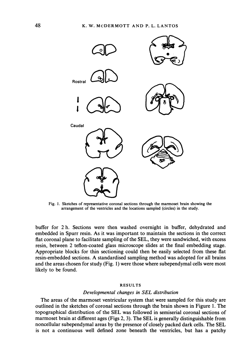
Images in this article


Selected References
These references are in PubMed. This may not be the complete list of references from this article.
- ALTMAN J. Autoradiographic investigation of cell proliferation in the brains of rats and cats. Anat Rec. 1963 Apr;145:573–591. doi: 10.1002/ar.1091450409. [DOI] [PubMed] [Google Scholar]
- Altman J. Autoradiographic and histological studies of postnatal neurogenesis. IV. Cell proliferation and migration in the anterior forebrain, with special reference to persisting neurogenesis in the olfactory bulb. J Comp Neurol. 1969 Dec;137(4):433–457. doi: 10.1002/cne.901370404. [DOI] [PubMed] [Google Scholar]
- Blakemore W. F. The ultrastructure of the subependymal plate in the rat. J Anat. 1969 May;104(Pt 3):423–433. [PMC free article] [PubMed] [Google Scholar]
- Cammermeyer J. The hypependymal microglia cell. Z Anat Entwicklungsgesch. 1965;124(6):543–561. doi: 10.1007/BF00520846. [DOI] [PubMed] [Google Scholar]
- Coppoletta J. M., Wolbach S. B. Body Length and Organ Weights of Infants and Children: A Study of the Body Length and Normal Weights of the More Important Vital Organs of the Body between Birth and Twelve Years of Age. Am J Pathol. 1933 Jan;9(1):55–70. [PMC free article] [PubMed] [Google Scholar]
- Das Gupta V., Cadwallader D. E. Acid dye method for the analysis of thiamine. J Pharm Sci. 1968 Jan;57(1):112–117. doi: 10.1002/jps.2600570122. [DOI] [PubMed] [Google Scholar]
- Demêmes D., Marty R. La plaque subépendymaire chez le chat: topographie et histologie; aptitudes réactionnelles. C R Acad Sci Hebd Seances Acad Sci D. 1971 Apr 26;272(17):2201–2204. [PubMed] [Google Scholar]
- Fischer K. Subependymale Zellproliferationen und Tumordisposition brachycephaler Hunderassen. Acta Neuropathol. 1967 May 5;8(3):242–254. doi: 10.1007/BF00688826. [DOI] [PubMed] [Google Scholar]
- Hajós F., Gallatz K. Immunocytochemical demonstration of radial glia in the developing rat olfactory bulb with antibodies to glial fibrillary acidic protein. Brain Res. 1987 Nov;433(1):131–138. doi: 10.1016/0165-3806(87)90071-x. [DOI] [PubMed] [Google Scholar]
- Ichikawa M., Shiga T., Hirata Y. Spatial and temporal pattern of postnatal proliferation of glial cells in the parietal cortex of the rat. Brain Res. 1983 Aug;285(2):181–187. doi: 10.1016/0165-3806(83)90050-0. [DOI] [PubMed] [Google Scholar]
- Kishi K. Golgi studies on the development of granule cells of the rat olfactory bulb with reference to migration in the subependymal layer. J Comp Neurol. 1987 Apr 1;258(1):112–124. doi: 10.1002/cne.902580109. [DOI] [PubMed] [Google Scholar]
- Lewis P. D. Mitotic activity in the primate subependymal layer and the genesis of gliomas. Nature. 1968 Mar 9;217(5132):974–975. doi: 10.1038/217974a0. [DOI] [PubMed] [Google Scholar]
- Lewis P. D. The fate of the subependymal cell in the adult rat brain, with a note on the origin of microglia. Brain. 1968;91(4):721–736. doi: 10.1093/brain/91.4.721. [DOI] [PubMed] [Google Scholar]
- Ling E. A. The subependyma of the primate, slow loris (Nycticebus coucang coucang). Tissue Cell. 1974;6(2):371–381. doi: 10.1016/0040-8166(74)90059-7. [DOI] [PubMed] [Google Scholar]
- McDermott K. W., Lantos P. L. Cell proliferation in the subependymal layer of the postnatal marmoset, Callithrix jacchus. Brain Res Dev Brain Res. 1990 Dec 15;57(2):269–277. doi: 10.1016/0165-3806(90)90053-2. [DOI] [PubMed] [Google Scholar]
- McDermott K. W., Lantos P. L. The distribution of glial fibrillary acidic protein and vimentin in postnatal marmoset (Callithrix jacchus) brain. Brain Res Dev Brain Res. 1989 Feb 1;45(2):169–177. doi: 10.1016/0165-3806(89)90036-9. [DOI] [PubMed] [Google Scholar]
- NOETZEL H., ROX J. AUTORADIOGRAPHISCHE UNTERSUCHUNGEN UEBER ZELLTEILUNG UND ZELLENTWICKLUNG IM GEHIRN DER ERWACHSENEN MAUS UND DES ERWACHSENEN RHESUS-AFFEN NACH INJEKTION VON RADIOAKTIVEM THYMIDIN. Acta Neuropathol. 1964 Mar 4;3:326–342. doi: 10.1007/BF00691841. [DOI] [PubMed] [Google Scholar]
- Prado Reis F., Abrantes Erhart E. The brain of the marmoset (Callithrix jacchus). Acta Anat (Basel) 1979;103(3):350–357. [PubMed] [Google Scholar]
- Privat A., Leblond C. P. The subependymal layer and neighboring region in the brain of the young rat. J Comp Neurol. 1972 Nov;146(3):277–302. doi: 10.1002/cne.901460302. [DOI] [PubMed] [Google Scholar]
- Stensaas L. J., Gilson B. C. Ependymal and subependymal cells of the caudato-pallial junction in the lateral ventricle of the neonatal rabbit. Z Zellforsch Mikrosk Anat. 1972;132(3):297–322. doi: 10.1007/BF02450711. [DOI] [PubMed] [Google Scholar]
- Sturrock R. R., Smart I. H. A morphological study of the mouse subependymal layer from embryonic life to old age. J Anat. 1980 Mar;130(Pt 2):391–415. [PMC free article] [PubMed] [Google Scholar]