Abstract
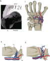
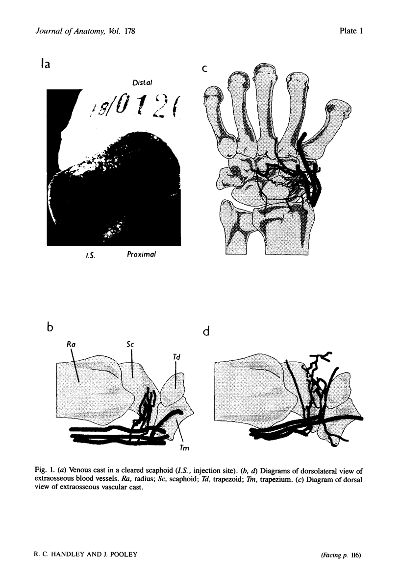
The intraosseous injection of coloured latex allows the venous drainage of a particular area of a bone to be studied. The extraosseous anatomy is visualised by chemical digestion of the soft tissues, the intraosseous anatomy by clearing the bone using the Spalteholz technique. When applied to the proximal pole of the scaphoid, this showed the venous drainage to be via the dorsal ridge into the venae comitantes of the radial artery.
Full text
PDF
Images in this article
Selected References
These references are in PubMed. This may not be the complete list of references from this article.
- Gelberman R. H., Menon J. The vascularity of the scaphoid bone. J Hand Surg Am. 1980 Sep;5(5):508–513. doi: 10.1016/s0363-5023(80)80087-6. [DOI] [PubMed] [Google Scholar]
- HARRISON R. G., GOSSMAN H. H. The fate of radiopaque media injected into the cancellous bone of the extremities. J Bone Joint Surg Br. 1955 Feb;37-B(1):150–156. doi: 10.1302/0301-620X.37B1.150. [DOI] [PubMed] [Google Scholar]
- Panagis J. S., Gelberman R. H., Taleisnik J., Baumgaertner M. The arterial anatomy of the human carpus. Part II: The intraosseous vascularity. J Hand Surg Am. 1983 Jul;8(4):375–382. doi: 10.1016/s0363-5023(83)80195-6. [DOI] [PubMed] [Google Scholar]
- Taleisnik J., Kelly P. J. The extraosseous and intraosseous blood supply of the scaphoid bone. J Bone Joint Surg Am. 1966 Sep;48(6):1125–1137. [PubMed] [Google Scholar]


