

Abstract
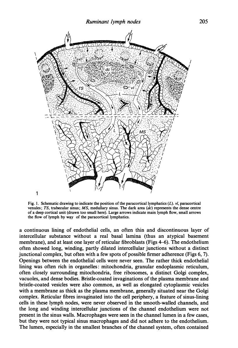
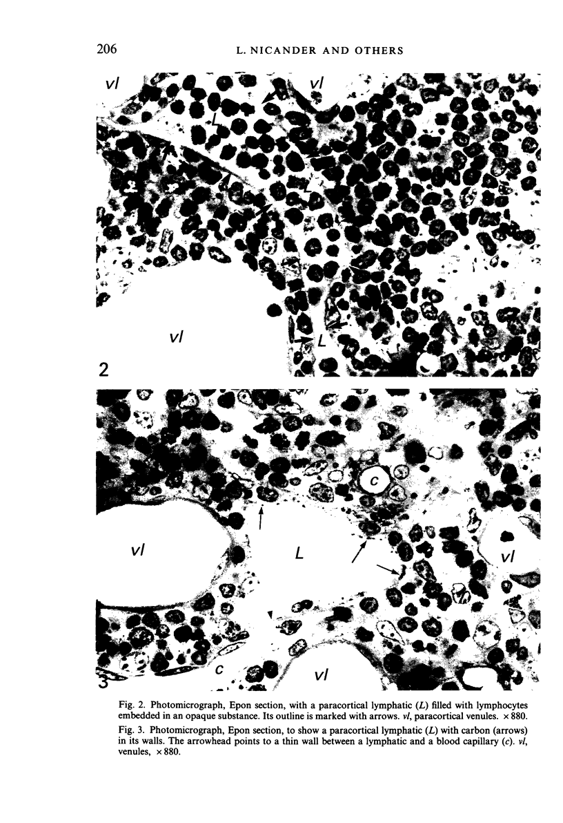
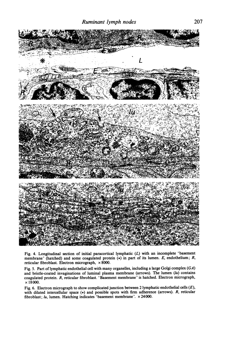
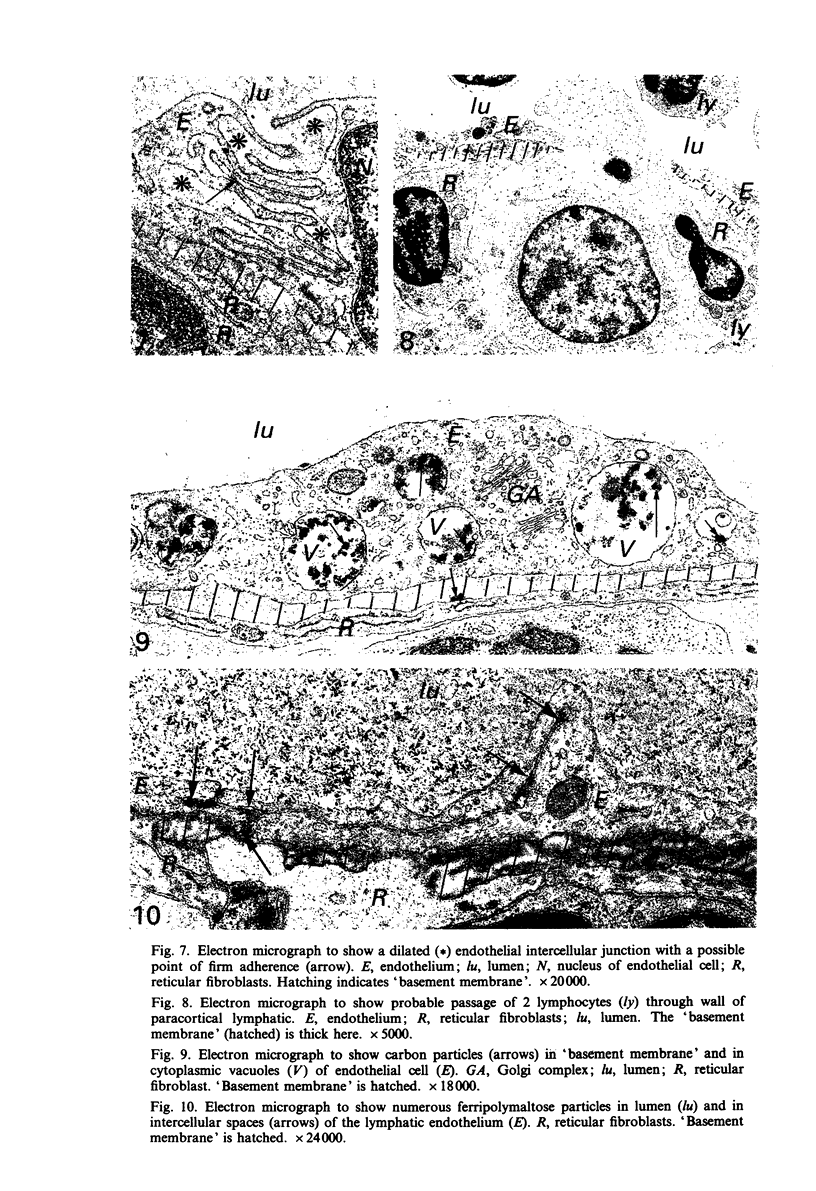
Ruminant lymph nodes, except when very small, were found to have a system of smooth-walled channels in the periphery of the 'deep cortical units' defined by Bélisle & Sainte-Marie (1981 a,b). Each channel originated with many 'blind' branches in the subnodular layer of the cortex and ended by joining a medullary sinus. The wall consisted of a continuous endothelial lining, a sometimes thin or discontinuous basement membrane without a basal lamina, and at least one layer of flattened reticular fibroblasts. The endothelium was higher than in most typical lymphatics, with a cytoplasmic fine structure similar to that of sinus-lining cells in the medullary sinuses. The intercellular junctions were generally long and elaborate. The lumen often contained opaque material, especially in the branches, as for initial lymphatics, as well as a few lymphocytes and an occasional nonlymphoid cell, but sinus macrophages were never seen. In some lymph nodes the lumen was crowded with lymphocytes. When small ferripolymaltose particles arrived in the node with the afferent lymph, many of them rapidly passed through the outer cortex and reached the lumen of the smooth-walled channels by way of the intercellular junctions of the endothelium. When colloidal carbon was introduced the same way, some of it also reached the channels where it accumulated in the basement membrane and in vesicles and vacuoles of the endothelium. These channels are interpreted as initial lymphatics of the same type as in other lymphoid organs rather than lymph node sinuses. They seem to play an important role for the exit of lymphocytes from the nodes and also for the passage of particulate material, including antigens, through those areas where recirculating lymphocytes arrive in the cortex.
Full text
PDF
Images in this article
Selected References
These references are in PubMed. This may not be the complete list of references from this article.
- Boucher Y., Roberge S., Roy P. E. Ultrastructural comparative study on lymphatic capillaries of the subendocardium, myocardium, and subepicardium of the heart left ventricle. Microvasc Res. 1985 May;29(3):305–319. doi: 10.1016/0026-2862(85)90021-4. [DOI] [PubMed] [Google Scholar]
- Bélisle C., Sainte-Marie G. Topography of the deep cortex of the lymph nodes of various mammalian species. Anat Rec. 1981 Nov;201(3):553–561. doi: 10.1002/ar.1092010311. [DOI] [PubMed] [Google Scholar]
- Bélisle C., Sainte-Marie G. Tridimensional study of the deep cortex of the rat lymph node. III. Morphology of the deep cortex units. Anat Rec. 1981 Feb;199(2):213–226. doi: 10.1002/ar.1091990206. [DOI] [PubMed] [Google Scholar]
- Casley-Smith J. R. The fine structure and functioning of tissue channels and lymphatics. Lymphology. 1980 Dec;13(4):177–183. [PubMed] [Google Scholar]
- Compton C. C., Raviola E. Structure of the sinus-lining cells in the popliteal lymph node of the rabbit. Anat Rec. 1985 Aug;212(4):408–423. doi: 10.1002/ar.1092120412. [DOI] [PubMed] [Google Scholar]
- Heath T. J., Kerlin R. L., Spalding H. J. Afferent pathways of lymph flow within the popliteal node in sheep. J Anat. 1986 Dec;149:65–75. [PMC free article] [PubMed] [Google Scholar]
- Heath T. J., Spalding H. J. Pathways of lymph flow to and from the medulla of lymph nodes in sheep. J Anat. 1987 Dec;155:177–188. [PMC free article] [PubMed] [Google Scholar]
- Kelly R. H. Functional anatomy of lymph nodes. I. The paracortical cords. Int Arch Allergy Appl Immunol. 1975;48(6):836–849. doi: 10.1159/000231371. [DOI] [PubMed] [Google Scholar]
- Kelly R. H., Wolstencroft R. A., Dumonde D. C., Balfour B. M. Role of lymphocyte activation products (LAP) in cell-mediated immunity. II. Effects of lymphocyte activation products on lymph node architecture and evidence for peripheral release of LAP following antigenic stimulation. Clin Exp Immunol. 1972 Jan;10(1):49–65. [PMC free article] [PubMed] [Google Scholar]
- Kurokawa T., Ogata T. A scanning electron microscopic study on the lymphatic microcirculation of the rabbit mesenteric lymph node. A corrosion cast study. Acta Anat (Basel) 1980;107(4):439–466. doi: 10.1159/000145272. [DOI] [PubMed] [Google Scholar]
- Soda R., Tavassoli M. Transendothelial transport (transcytosis) of iron-transferrin complex in the bone marrow. J Ultrastruct Res. 1984 Jul;88(1):18–29. doi: 10.1016/s0022-5320(84)90178-3. [DOI] [PubMed] [Google Scholar]
- Söderström N., Stenström A. Outflow paths of cells from the lymph node parenchyma to the efferent lymphatics--observations in thin section histology. Scand J Haematol. 1969;6(3):186–196. doi: 10.1111/j.1600-0609.1969.tb01825.x. [DOI] [PubMed] [Google Scholar]