Abstract
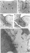
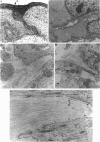
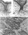
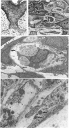
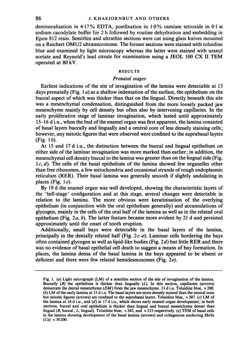
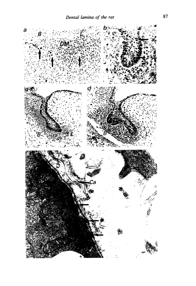
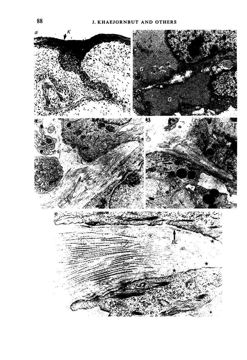
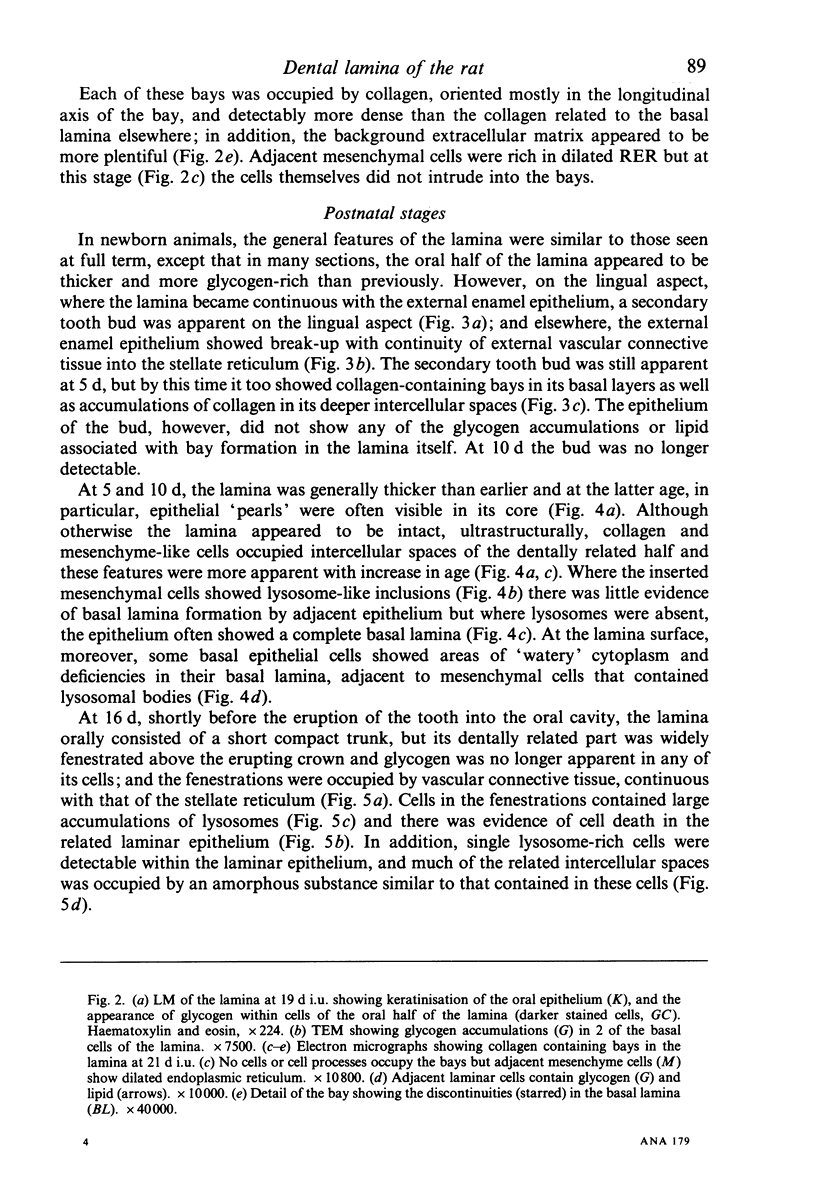
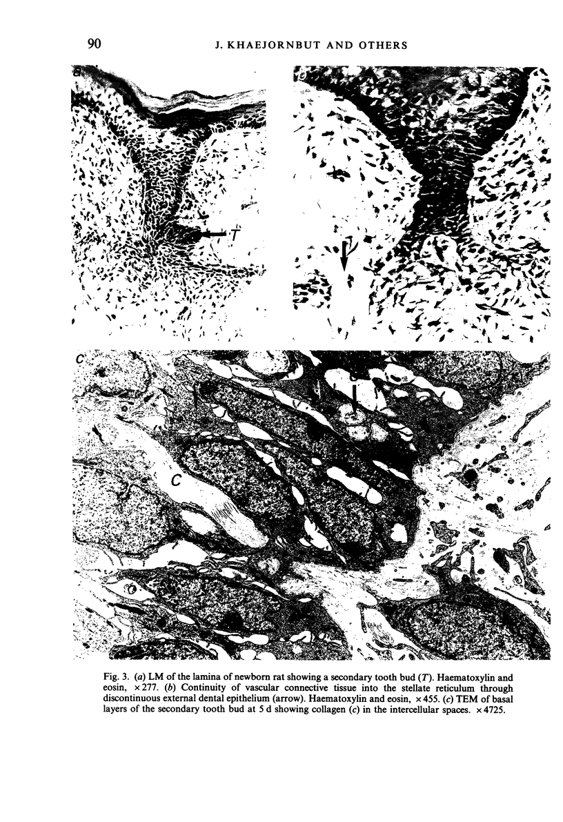
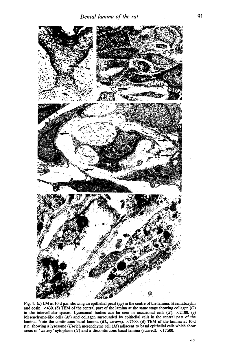
The lamina of the first mandibular molar teeth of rats, age range 13 d intrauterine (i.u.) to 16 d postnatal (p.n.), was examined by light and transmission electron microscopy to establish histological baselines of its development and fate. All material was obtained from animals anaesthetised with ether, killed by cervical dislocation and prepared by routine methods for both types of examination. Contrary to earlier reports that the lamina remains intact throughout development, mesenchymal elements disrupt the lamina. These were seen first at 19 d i.u., as collagen-filled bays in the basal epithelial layers, associated with partial loss of related basal lamina. In the early stages, collagen deposition was limited and it was not obviously preceded by epithelial cell death or transformation, even though many bay-related cells showed lipid and glycogen accumulations. Later disruption of the lamina showed more mesenchymal cells as well as collagen in deeper spaces. After the onset of tooth eruption, mesenchymal cells external to and within the lamina contained lysosomal bodies and these plus evidence of related epithelial cell death and capillaries in the laminar spaces became more and more apparent. Similar collagen deposits were observed in a successional tooth primordium, which appeared at term but eventually aborted between days 5 and 10 p.n. Thus disruption of the lamina by connective tissue began earlier than has been reported previously and progressed as the tooth erupted towards the oral cavity. The evidence suggests that this disruption is initiated and sustained by mesenchymal cell activity rather than by programmed cell death or transformation of the epithelium.
Full text
PDF
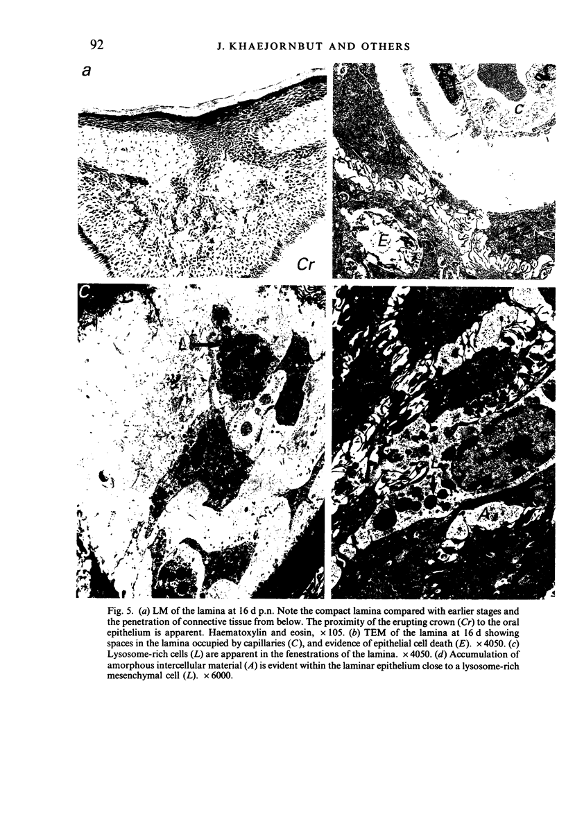
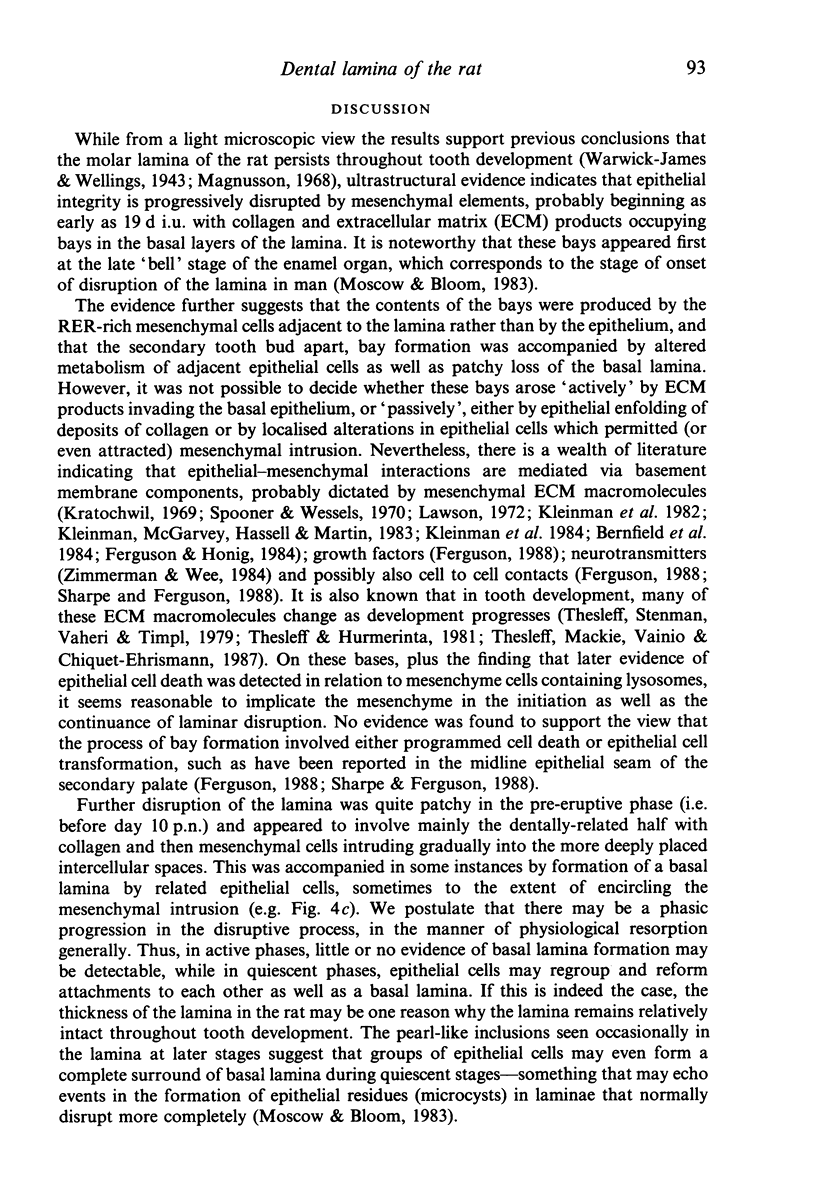
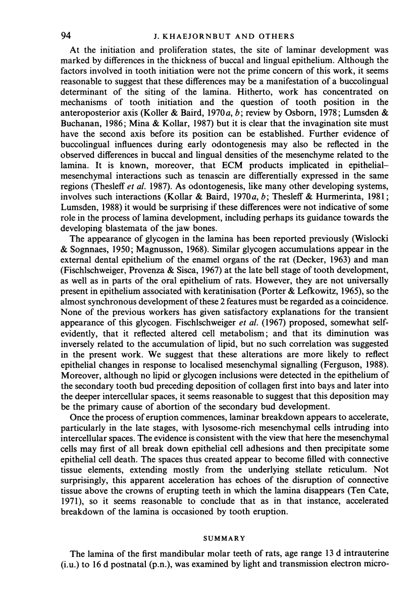
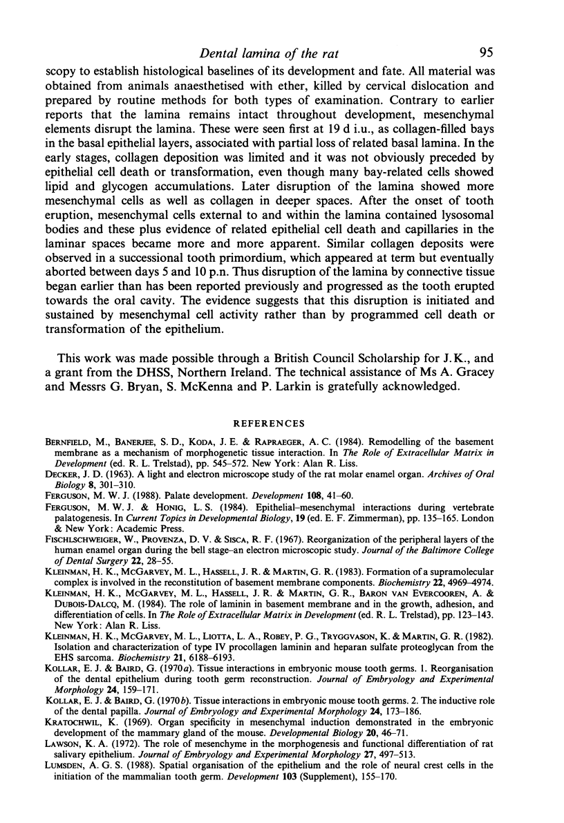
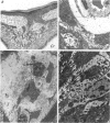
Images in this article
Selected References
These references are in PubMed. This may not be the complete list of references from this article.
- DECKER J. D. A light and electron microscope study of the rat molar enamel organ. Arch Oral Biol. 1963 May-Jun;8:301–310. doi: 10.1016/0003-9969(63)90022-0. [DOI] [PubMed] [Google Scholar]
- Ferguson M. W. Palate development. Development. 1988;103 (Suppl):41–60. doi: 10.1242/dev.103.Supplement.41. [DOI] [PubMed] [Google Scholar]
- Fischlschweiger W., Provenza D. V., Sisca R. F. Reorganization of the peripheral layers of the human enamel organ during the bell stage--an electron microscopic study. J Baltimore Coll Dent Surg. 1967 Jul;22(1):28–55. [PubMed] [Google Scholar]
- Kleinman H. K., McGarvey M. L., Hassell J. R., Martin G. R. Formation of a supramolecular complex is involved in the reconstitution of basement membrane components. Biochemistry. 1983 Oct 11;22(21):4969–4974. doi: 10.1021/bi00290a014. [DOI] [PubMed] [Google Scholar]
- Kleinman H. K., McGarvey M. L., Liotta L. A., Robey P. G., Tryggvason K., Martin G. R. Isolation and characterization of type IV procollagen, laminin, and heparan sulfate proteoglycan from the EHS sarcoma. Biochemistry. 1982 Nov 23;21(24):6188–6193. doi: 10.1021/bi00267a025. [DOI] [PubMed] [Google Scholar]
- Kollar E. J., Baird G. R. Tissue interactions in embryonic mouse tooth germs. I. Reorganization of the dental epithelium during tooth-germ reconstruction. J Embryol Exp Morphol. 1970 Aug;24(1):159–171. [PubMed] [Google Scholar]
- Kollar E. J., Baird G. R. Tissue interactions in embryonic mouse tooth germs. II. The inductive role of the dental papilla. J Embryol Exp Morphol. 1970 Aug;24(1):173–186. [PubMed] [Google Scholar]
- Kratochwil K. Organ specificity in mesenchymal induction demonstrated in the embryonic development of the mammary gland of the mouse. Dev Biol. 1969 Jul;20(1):46–71. doi: 10.1016/0012-1606(69)90004-9. [DOI] [PubMed] [Google Scholar]
- Lawson K. A. The role of mesenchyme in the morphogenesis and functional differentiation of rat salivary epithelium. J Embryol Exp Morphol. 1972 Jun;27(3):497–513. [PubMed] [Google Scholar]
- Lumsden A. G., Buchanan J. A. An experimental study of timing and topography of early tooth development in the mouse embryo with an analysis of the role of innervation. Arch Oral Biol. 1986;31(5):301–311. doi: 10.1016/0003-9969(86)90044-0. [DOI] [PubMed] [Google Scholar]
- Lumsden A. G. Spatial organization of the epithelium and the role of neural crest cells in the initiation of the mammalian tooth germ. Development. 1988;103 (Suppl):155–169. doi: 10.1242/dev.103.Supplement.155. [DOI] [PubMed] [Google Scholar]
- Mina M., Kollar E. J. The induction of odontogenesis in non-dental mesenchyme combined with early murine mandibular arch epithelium. Arch Oral Biol. 1987;32(2):123–127. doi: 10.1016/0003-9969(87)90055-0. [DOI] [PubMed] [Google Scholar]
- Moskow B. S., Bloom A. Embryogenesis of the gingival cyst. J Clin Periodontol. 1983 Mar;10(2):119–130. doi: 10.1111/j.1600-051x.1983.tb02200.x. [DOI] [PubMed] [Google Scholar]
- Porter K., Lefkowitz W. Glycogen in developing and young rat oral epithelium. J Dent Res. 1965 Sep-Oct;44(5):954–958. doi: 10.1177/00220345650440053401. [DOI] [PubMed] [Google Scholar]
- Sharpe P. M., Ferguson M. W. Mesenchymal influences on epithelial differentiation in developing systems. J Cell Sci Suppl. 1988;10:195–230. doi: 10.1242/jcs.1988.supplement_10.15. [DOI] [PubMed] [Google Scholar]
- Spooner B. S., Wessells N. K. Mammalian lung development: interactions in primordium formation and bronchial morphogenesis. J Exp Zool. 1970 Dec;175(4):445–454. doi: 10.1002/jez.1401750404. [DOI] [PubMed] [Google Scholar]
- Thesleff I., Hurmerinta K. Tissue interactions in tooth development. Differentiation. 1981;18(2):75–88. doi: 10.1111/j.1432-0436.1981.tb01107.x. [DOI] [PubMed] [Google Scholar]
- Thesleff I., Mackie E., Vainio S., Chiquet-Ehrismann R. Changes in the distribution of tenascin during tooth development. Development. 1987 Oct;101(2):289–296. doi: 10.1242/dev.101.2.289. [DOI] [PubMed] [Google Scholar]
- Thesleff I., Stenman S., Vaheri A., Timpl R. Changes in the matrix proteins, fibronectin and collagen, during differentiation of mouse tooth germ. Dev Biol. 1979 May;70(1):116–126. doi: 10.1016/0012-1606(79)90011-3. [DOI] [PubMed] [Google Scholar]
- WISLOCKI G. B., SOGNNAES R. F. Histochemical reactions of normal teeth. Am J Anat. 1950 Sep;87(2):239–275. doi: 10.1002/aja.1000870204. [DOI] [PubMed] [Google Scholar]
- Zimmerman E. F., Wee E. L. Role of neurotransmitters in palate development. Curr Top Dev Biol. 1984;19:37–63. doi: 10.1016/s0070-2153(08)60394-4. [DOI] [PubMed] [Google Scholar]


