

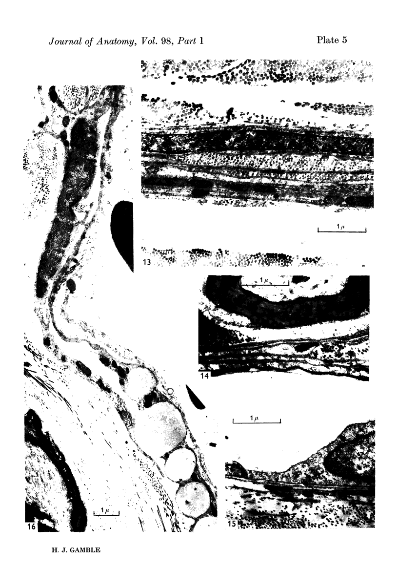
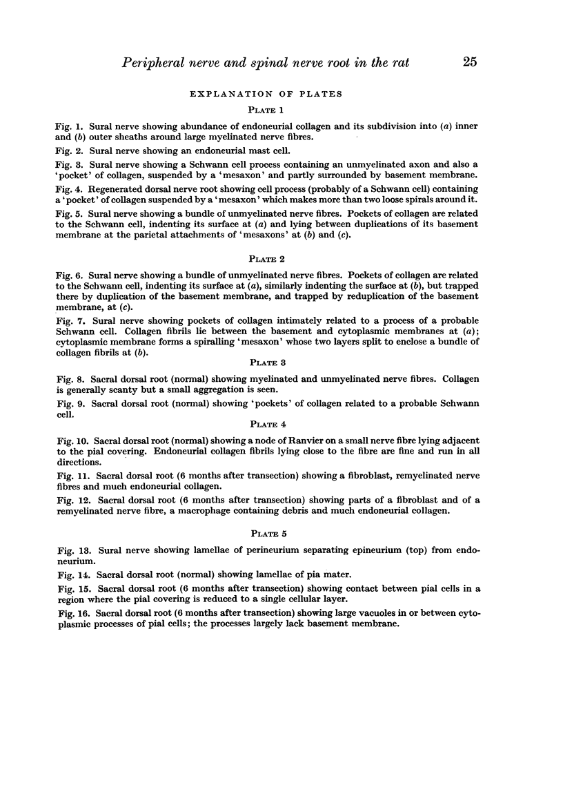
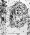
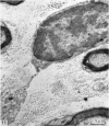
Full text
PDF
Images in this article
Selected References
These references are in PubMed. This may not be the complete list of references from this article.
- BARTON A. A. An electron microscope study of degeneration and regeneration of nerve. Brain. 1962 Dec;85:799–808. doi: 10.1093/brain/85.4.799. [DOI] [PubMed] [Google Scholar]
- CAUSEY G., BARTON A. A. The cellular content of the endoneurium of peripheral nerve. Brain. 1959 Dec;82:594–598. doi: 10.1093/brain/82.4.594. [DOI] [PubMed] [Google Scholar]
- CHAPMAN J. A. Fibroblasts and collagen. Br Med Bull. 1962 Sep;18:233–237. doi: 10.1093/oxfordjournals.bmb.a069985. [DOI] [PubMed] [Google Scholar]
- CRESCITELLI F. Nerve sheath as a barrier to the action of certain substances. Am J Physiol. 1951 Aug;166(2):229–240. doi: 10.1152/ajplegacy.1951.166.2.229. [DOI] [PubMed] [Google Scholar]
- GAMBLE H. J., GOLDBY S. Mast cells in peripheral nerve trunks. Nature. 1961 Mar 4;189:766–767. doi: 10.1038/189766a0. [DOI] [PubMed] [Google Scholar]
- GASSER H. S. Properties of dorsal root unmedullated fibers on the two sides of the ganglion. J Gen Physiol. 1955 May 20;38(5):709–728. doi: 10.1085/jgp.38.5.709. [DOI] [PMC free article] [PubMed] [Google Scholar]
- HARKNESS R. D. Biological functions of collagen. Biol Rev Camb Philos Soc. 1961 Nov;36:399–463. doi: 10.1111/j.1469-185x.1961.tb01596.x. [DOI] [PubMed] [Google Scholar]
- HESS A. The fine structure and morphological organization of non-myelinated nerve fibres. Proc R Soc Lond B Biol Sci. 1956 Mar 13;144(917):496–506. doi: 10.1098/rspb.1956.0006. [DOI] [PubMed] [Google Scholar]
- HUXLEY A. F., STAMPFLI R. Effect of potassium and sodium on resting and action potentials of single myelinated nerve fibers. J Physiol. 1951 Feb;112(3-4):496–508. doi: 10.1113/jphysiol.1951.sp004546. [DOI] [PMC free article] [PubMed] [Google Scholar]
- Laidlaw G. F. Silver Staining of the Endoneurial Fibers of the Cerebrospinal Nerves. Am J Pathol. 1930 Jul;6(4):435–444.3. [PMC free article] [PubMed] [Google Scholar]
- Sanders F. K., Young J. Z. The role of the peripheral stump in the control of fibre diameter in regenerating nerves. J Physiol. 1944 Jun 15;103(1):119–136.2. doi: 10.1113/jphysiol.1944.sp004066. [DOI] [PMC free article] [PubMed] [Google Scholar]
- Simpson S. A., Young J. Z. Regeneration of fibre diameter after cross-unions of visceral and somatic nerves. J Anat. 1945 Apr;79(Pt 2):48–65. [PMC free article] [PubMed] [Google Scholar]
- THOMAS P. K. The connective tissue of peripheral nerve: an electron microscope study. J Anat. 1963 Jan;97:35–44. [PMC free article] [PubMed] [Google Scholar]