

Abstract
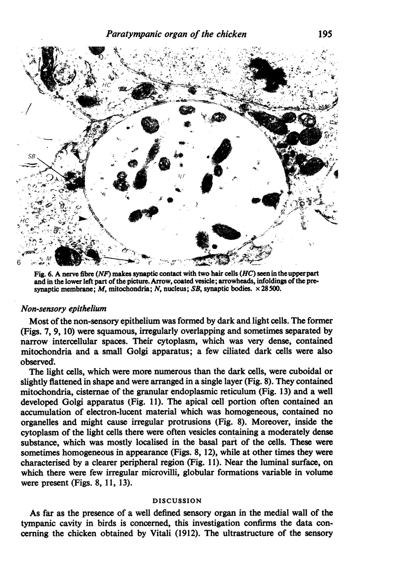
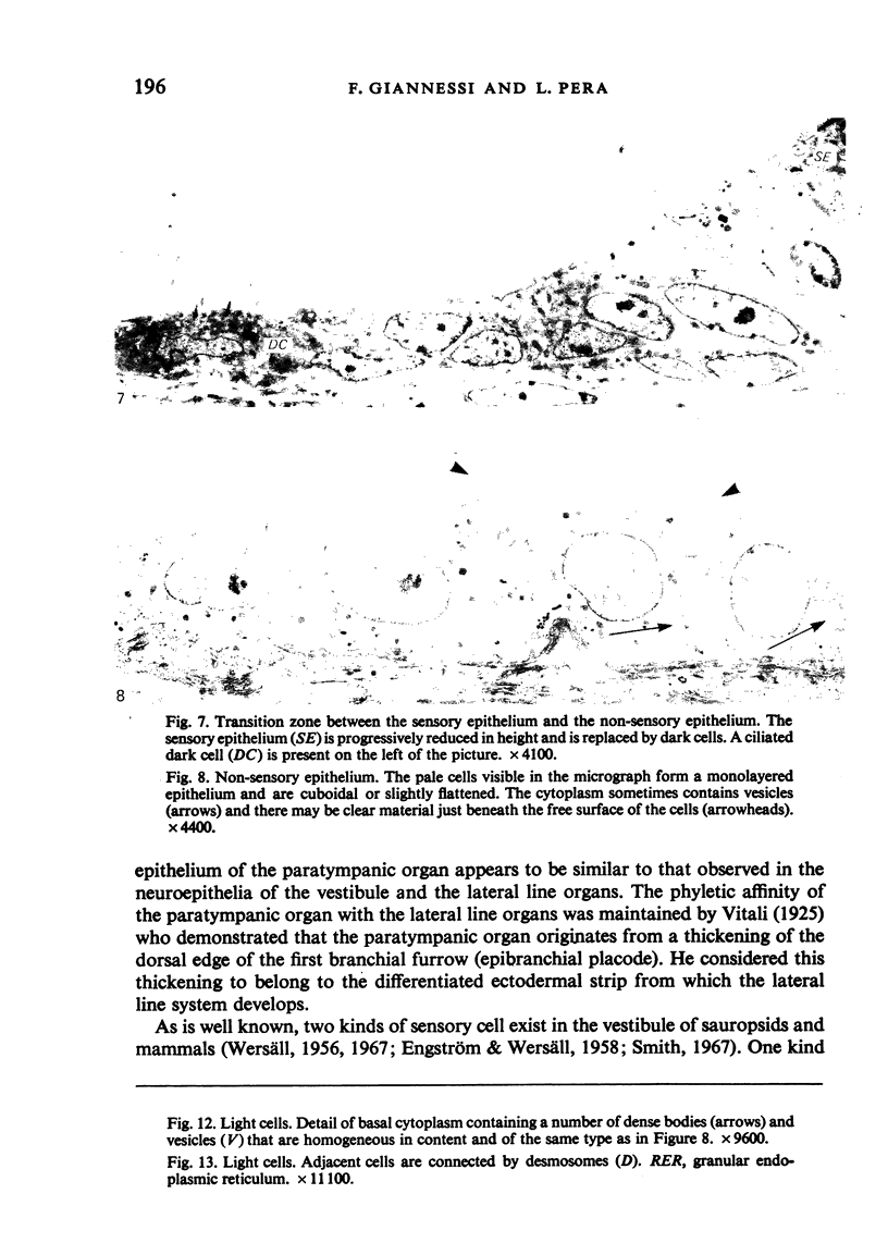
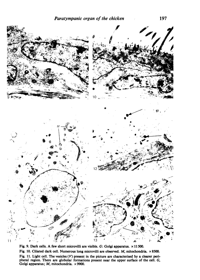
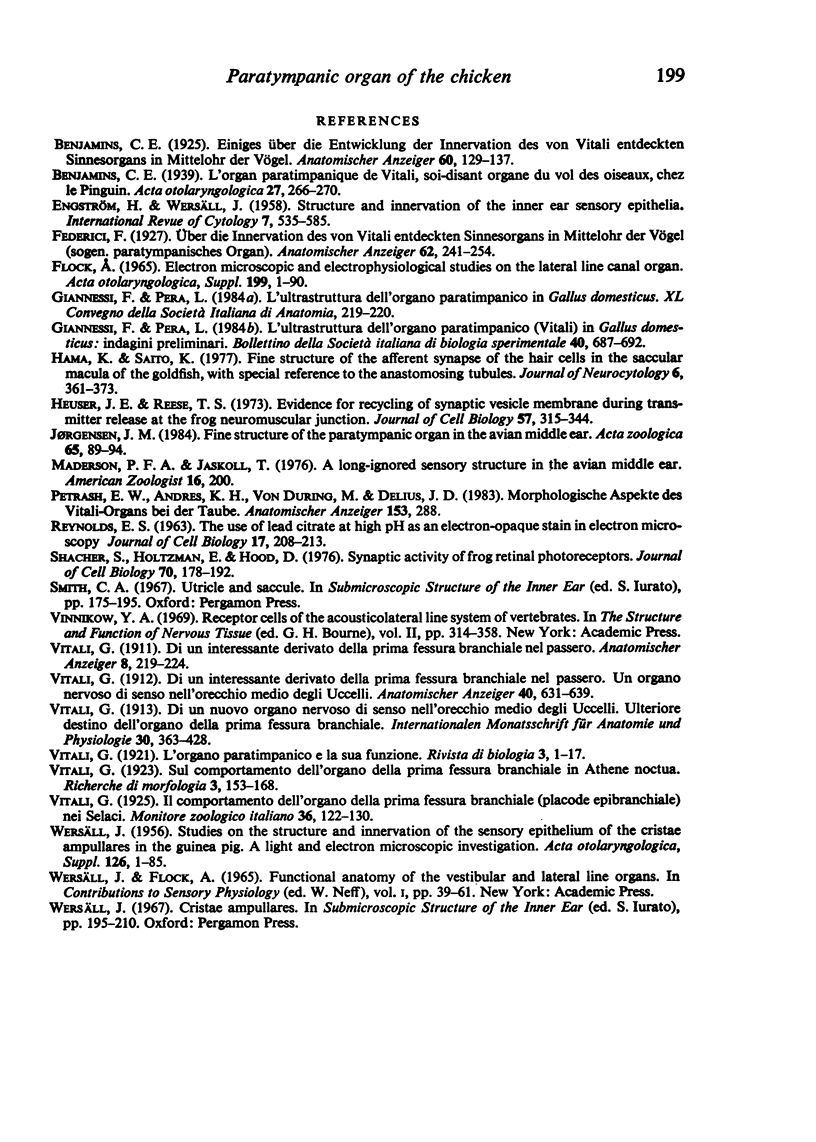
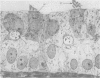
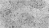
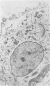
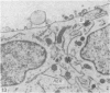
The structure of the paratympanic organ in chickens was investigated by means of the transmission electron microscope. The epithelium lining the lumen of the paratympanic organ consists of sensory and non-sensory components. The sensory epithelium is composed of supporting and hair cells. The hair cells are similar to the type II receptor cells present in the neuroepithelia of the vestibule and of the lateral line organs. The afferent synapses at the bases of the hair cells are also described. The non-sensory epithelium is made up of cells with a clear cytoplasm and arranged in a single layer. It also contains dark, flattened cells which sometimes possess motile cilia. Special emphasis is given to the fact that the results agree with Vitali's theory that the paratympanic organ and the lateral line organs are homologous. It is concluded, therefore, that present knowledge about this structure is not yet sufficient to allow a definitive functional interpretation.
Full text
PDF
Images in this article
Selected References
These references are in PubMed. This may not be the complete list of references from this article.
- Hama K., Saito K. Fine structure of the afferent synapse of the hair cells in the saccular macula of the goldfish, with special reference to the anastomosing tubules. J Neurocytol. 1977 Aug;6(4):361–373. doi: 10.1007/BF01178223. [DOI] [PubMed] [Google Scholar]
- Heuser J. E., Reese T. S. Evidence for recycling of synaptic vesicle membrane during transmitter release at the frog neuromuscular junction. J Cell Biol. 1973 May;57(2):315–344. doi: 10.1083/jcb.57.2.315. [DOI] [PMC free article] [PubMed] [Google Scholar]
- Schacher S., Holtzman E., Hood D. C. Synaptic activity of frog retinal photoreceptors. A peroxidase uptake study. J Cell Biol. 1976 Jul;70(1):178–192. doi: 10.1083/jcb.70.1.178. [DOI] [PMC free article] [PubMed] [Google Scholar]
- WERSAELL J., FLOCK A. FUNCTIONAL ANATOMY OF THE VESTIBULAR AND LATERAL LINE ORGANS. Contrib Sens Physiol. 1965;14:39–61. doi: 10.1016/b978-1-4831-6746-6.50007-9. [DOI] [PubMed] [Google Scholar]
- WERSALL J. Studies on the structure and innervation of the sensory epithelium of the cristae ampulares in the guinea pig; a light and electron microscopic investigation. Acta Otolaryngol Suppl. 1956;126:1–85. [PubMed] [Google Scholar]