

Abstract
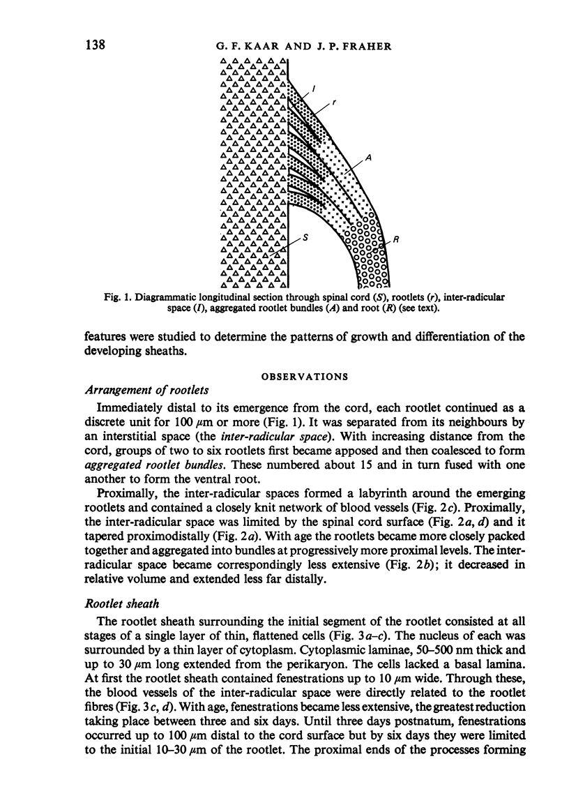
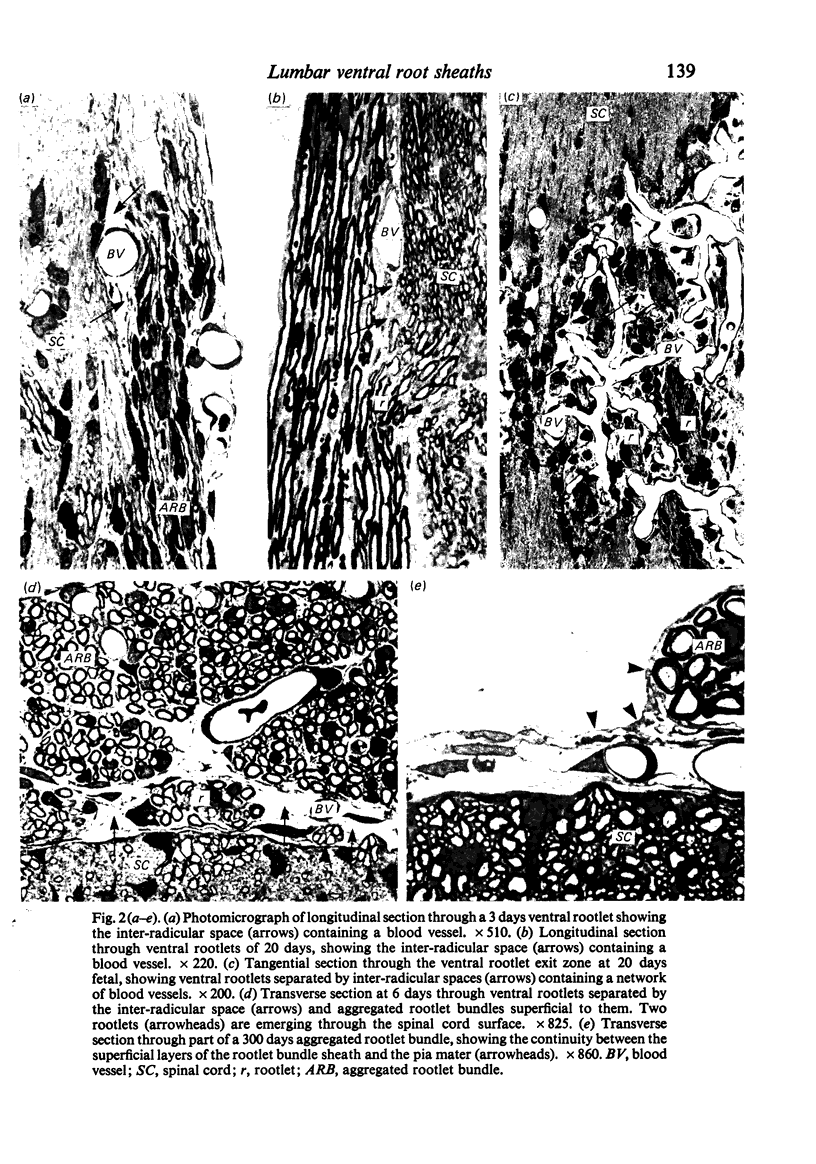
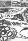
The fifth lumbar ventral spinal nerve rootlets join to form a number of aggregated rootlet bundles. These in turn fuse to form the ventral root. Each rootlet is surrounded by a sheath which consists of a single fenestrated layer of cells and their attenuated cytoplasmic processes and which is open ended proximally. Immediately superficial to the spinal cord surface, rootlets are separated from one another by a labyrinth of inter-radicular spaces containing small blood vessels. Between adjacent rootlets the inter-radicular space tapers distally to an apex. The endoneurial space of the rootlet communicates with the subpial and inter-radicular spaces. Each aggregated rootlet bundle is surrounded by a multilayered sheath. Proximally, the outer layers of this sheath are continuous with the superficial layers of the pia mater. Both of these, as well as the root sheath with which the rootlet bundle sheaths are continuous distally, are complete and lack fenestrations. Accordingly, the endoneurial space, though continuous with the inter-radicular and subpial spaces, is isolated from the subarachnoid space.
Full text
PDF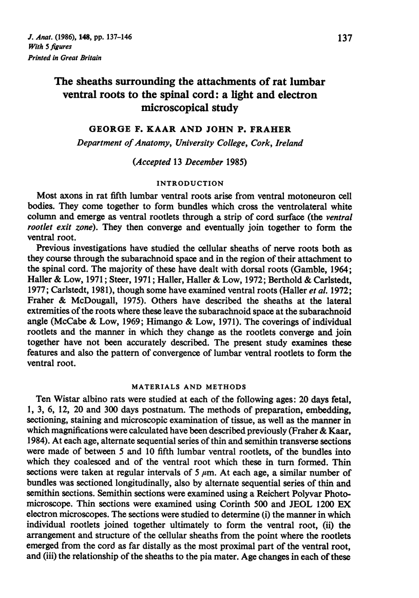
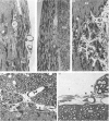
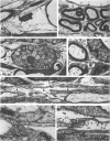
Images in this article
Selected References
These references are in PubMed. This may not be the complete list of references from this article.
- Berthold C. H., Carlstedt T. Observations on the morphology at the transition between the peripheral and the central nervous system in the cat. II. General organization of the transitional region in S1 dorsal rootlets. Acta Physiol Scand Suppl. 1977;446:23–42. [PubMed] [Google Scholar]
- Carlstedt T. An electron-microscopical study of the developing transitional region in feline S1 dorsal rootlets. J Neurol Sci. 1981 Jun;50(3):357–372. doi: 10.1016/0022-510x(81)90148-9. [DOI] [PubMed] [Google Scholar]
- Fraher J. P., Kaar G. F. The transitional node of Ranvier at the junction of the central and peripheral nervous systems: an ultrastructural study of its development and mature form. J Anat. 1984 Sep;139(Pt 2):215–238. [PMC free article] [PubMed] [Google Scholar]
- Fraher J. P., McDougall R. D. Macrophages related to leptomeninges and ventral nerve roots. An ultrastructural study. J Anat. 1975 Dec;120(Pt 3):537–549. [PMC free article] [PubMed] [Google Scholar]
- GAMBLE H. J. COMPARATIVE ELECTRON-MICROSCOPIC OBSERVATIONS ON THE CONNECTIVE TISSUES OF A PERIPHERAL NERVE AND A SPINAL NERVE ROOT IN THE RAT. J Anat. 1964 Jan;98:17–26. [PMC free article] [PubMed] [Google Scholar]
- Haller F. R., Haller C., Low F. N. The fine structure of cellular layers and connective tissue space at spinal nerve root attachments in the rat. Am J Anat. 1972 Jan;133(1):109–123. doi: 10.1002/aja.1001330107. [DOI] [PubMed] [Google Scholar]
- Haller F. R., Low F. N. The fine structure of the peripheral nerve root sheath in the subarachnoid space in the rat and other laboratory animals. Am J Anat. 1971 May;131(1):1–19. doi: 10.1002/aja.1001310102. [DOI] [PubMed] [Google Scholar]
- Himango W. A., Low F. N. The fine structure of a lateral recess of the subarachnoid space in the rat. Anat Rec. 1971 Sep;171(1):1–19. doi: 10.1002/ar.1091710102. [DOI] [PubMed] [Google Scholar]
- McCabe J. S., Low F. N. The subarachnoid angle: an area of transition in peripheral nerve. Anat Rec. 1969 May;164(1):15–33. doi: 10.1002/ar.1091640102. [DOI] [PubMed] [Google Scholar]
- Morse D. E., Low F. N. The fine structure of the pia mater of the rat. Am J Anat. 1972 Mar;133(3):349–367. doi: 10.1002/aja.1001330309. [DOI] [PubMed] [Google Scholar]
- NELSON E., BLINZINGER K., HAGER H. Electron microscopic observations on subarachnoid and perivascular spaces of the Syrian hamster brain. Neurology. 1961 Apr;11(4):285–295. doi: 10.1212/wnl.11.4.285. [DOI] [PubMed] [Google Scholar]
- Nabeshima S., Reese T. S., Landis D. M., Brightman M. W. Junctions in the meninges and marginal glia. J Comp Neurol. 1975 Nov 15;164(2):127–169. doi: 10.1002/cne.901640202. [DOI] [PubMed] [Google Scholar]
- PEASE D. C., SCHULTZ R. L. Electron microscopy of rat cranial meninges. Am J Anat. 1958 Mar;102(2):301–321. doi: 10.1002/aja.1001020207. [DOI] [PubMed] [Google Scholar]
- Rovainen C. M., Lemcoe G. E., Peterson A. Structure and chemistry of glucose-producing cells in meningeal tissue of the lamprey. Brain Res. 1971 Jul 9;30(1):99–118. doi: 10.1016/0006-8993(71)90008-4. [DOI] [PubMed] [Google Scholar]
- Steer J. M. Some observations on the fine structure of rat dorsal spinal nerve roots. J Anat. 1971 Sep;109(Pt 3):467–485. [PMC free article] [PubMed] [Google Scholar]
- Waggener J. D., Beggs J. The membranous coverings of neural tissues: an electron microscopy study. J Neuropathol Exp Neurol. 1967 Jul;26(3):412–426. doi: 10.1097/00005072-196707000-00005. [DOI] [PubMed] [Google Scholar]