

Abstract
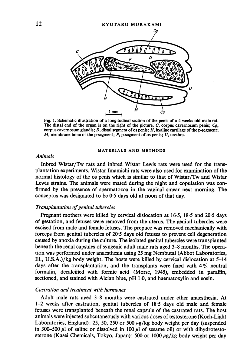
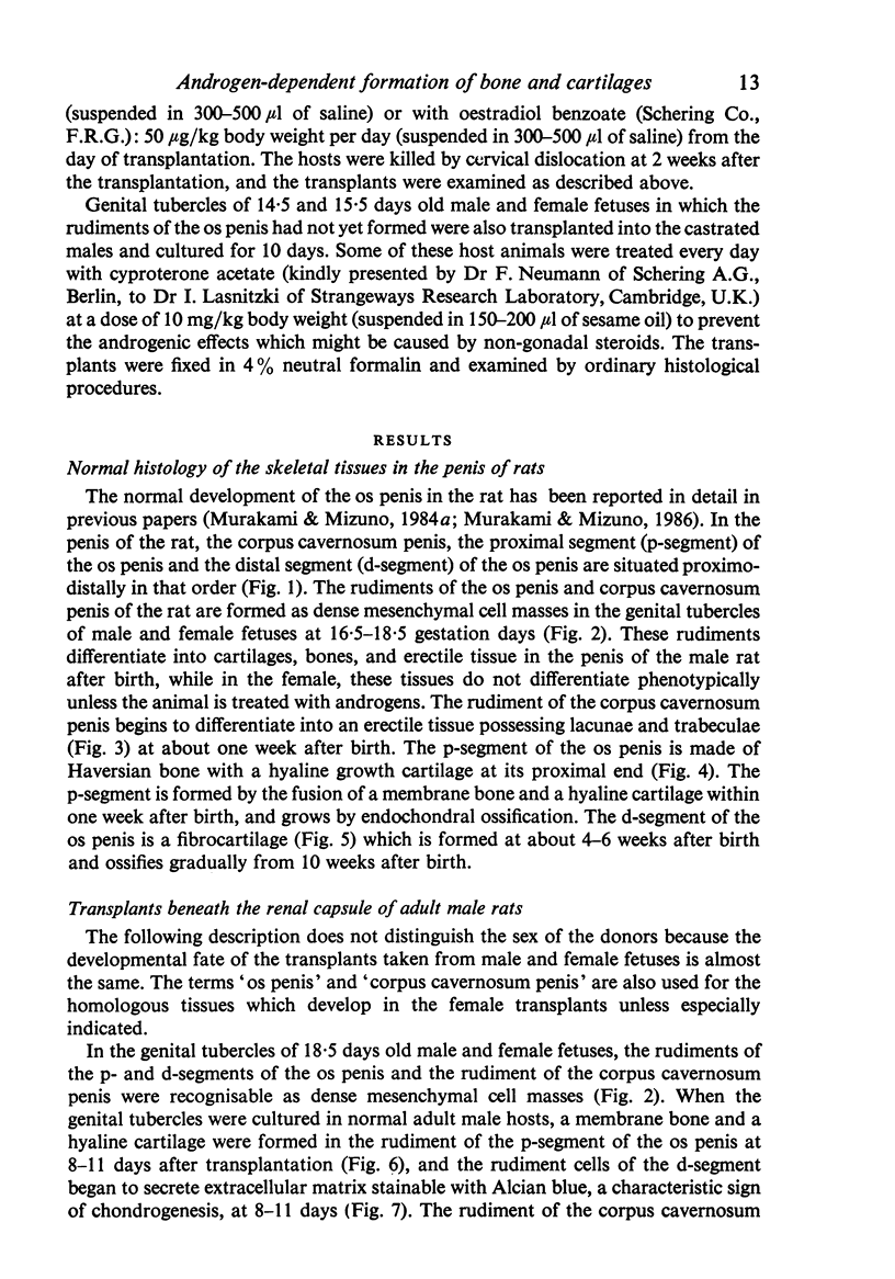
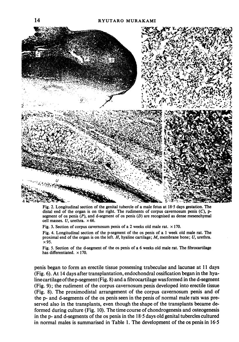
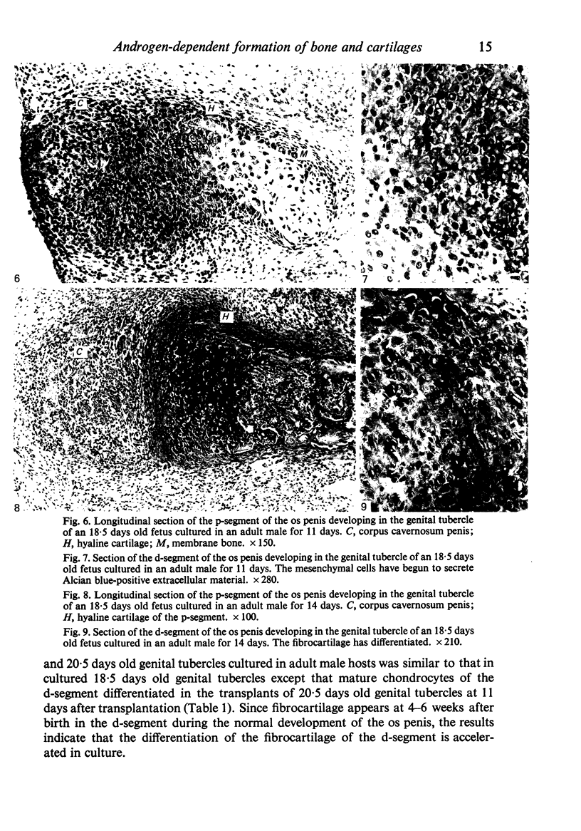
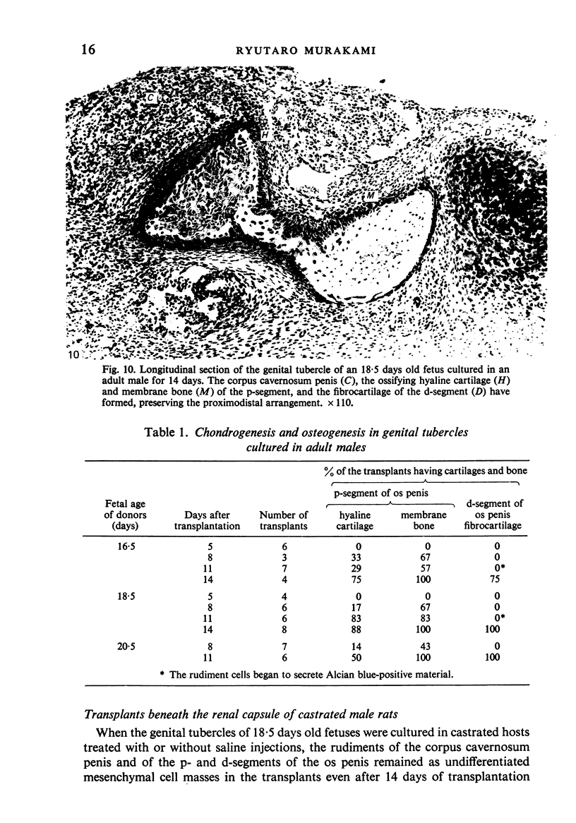
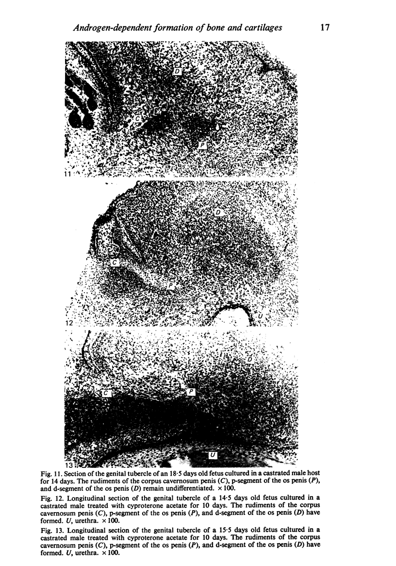
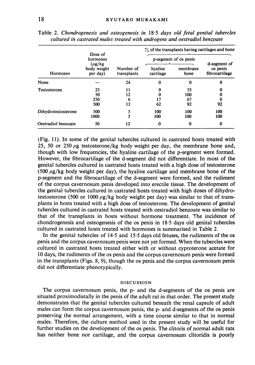
Genital tubercles of male and female rats were cultured beneath the renal capsule of castrated and intact adult male rats treated with androgens, oestrogen, or anti-androgen, and the development of the os penis in the transplants was studied. When the genital tubercles were cultured in normal male hosts, a membrane bone and a hyaline cartilage of the proximal segment of the os penis were formed 8-11 days after transplantation, and a fibrocartilage of the distal segment of the os penis at 11-14 days. In genital tubercles cultured in castrated males, the rudiments of both the proximal and distal segments remained as undifferentiated mesenchymal cell masses. However, similarly cultured genital tubercles were found to develop cartilages and bone when the hosts were treated with high doses of androgens. The potency of androgen-dependent chondrogenesis and osteogenesis was equivalent in the male and female genital tubercles. Chondrogenesis and osteogenesis of the os penis were caused by androgens, while the rudiments of the os penis were formed independently of androgens. The overt differentiation of the corpus cavernosum penis was also caused by androgens.
Full text
PDF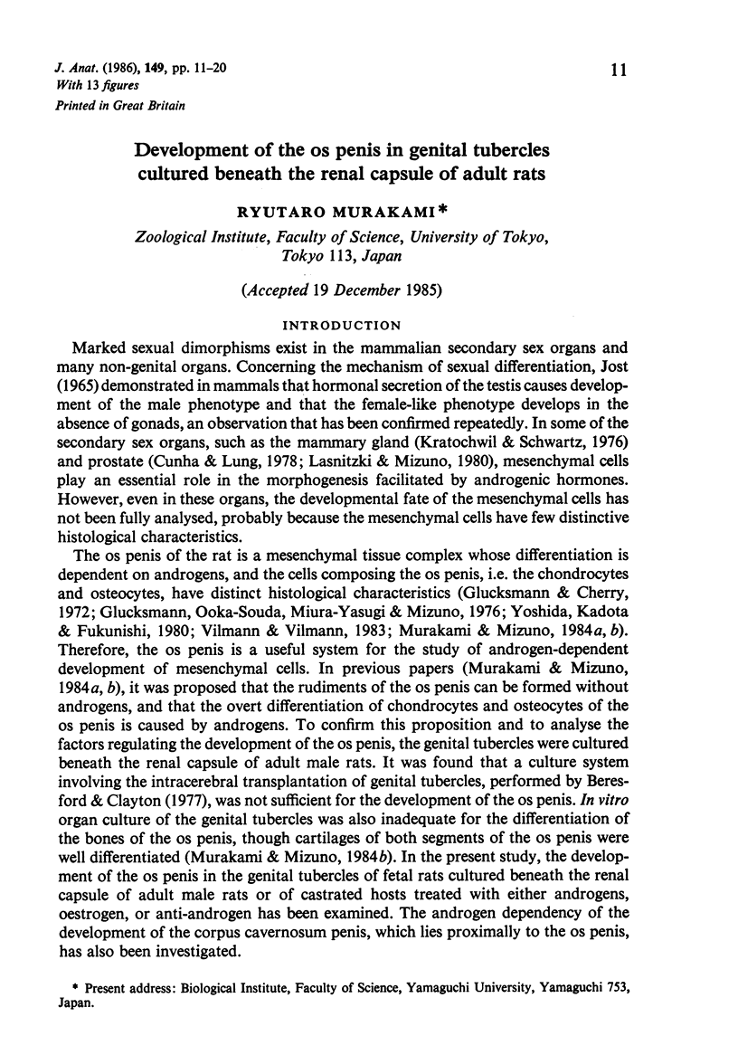
Images in this article
Selected References
These references are in PubMed. This may not be the complete list of references from this article.
- Beresford W. A., Clayton S. P. Intracerebral transplantation of the genital tubercle in the rat: the fate of the penile bone and cartilages. J Anat. 1977 Apr;123(Pt 2):297–311. [PMC free article] [PubMed] [Google Scholar]
- Cihák R., Gutmann E., Hanzlíková V. Involution and hormone-induced persistence of the M. sphincter (levator) ani in female rats. J Anat. 1970 Jan;106(Pt 1):93–110. [PMC free article] [PubMed] [Google Scholar]
- Corpéchot C., Baulieu E. E., Robel P. Testosterone, dihydrotestosterone and androstanediols in plasma, testes and prostates of rats during development. Acta Endocrinol (Copenh) 1981 Jan;96(1):127–135. doi: 10.1530/acta.0.0960127. [DOI] [PubMed] [Google Scholar]
- Cunha G. R., Lung B. The possible influence of temporal factors in androgenic responsiveness of urogenital tissue recombinants from wild-type and androgen-insensitive (Tfm) mice. J Exp Zool. 1978 Aug;205(2):181–193. doi: 10.1002/jez.1402050203. [DOI] [PubMed] [Google Scholar]
- Glucksmann A., Cherry C. P. The hormonal induction of an os clitoridis in the neonatal and adult rat. J Anat. 1972 Jul;112(Pt 2):223–231. [PMC free article] [PubMed] [Google Scholar]
- Glucksmann A., Ooka-Souda S., Miura-Yasugi E., Mizuno T. The effect of neonatal treatment of male mice with antiandrogens and of females with androgens on the development of the os penis and os clitoridis. J Anat. 1976 Apr;121(Pt 2):363–370. [PMC free article] [PubMed] [Google Scholar]
- Habert R., Picon R. Testosterone, dihydrotestosterone and estradiol-17 beta levels in maternal and fetal plasma and in fetal testes in the rat. J Steroid Biochem. 1984 Aug;21(2):193–198. doi: 10.1016/0022-4731(84)90383-2. [DOI] [PubMed] [Google Scholar]
- Kratochwil K., Schwartz P. Tissue interaction in androgen response of embryonic mammary rudiment of mouse: identification of target tissue for testosterone. Proc Natl Acad Sci U S A. 1976 Nov;73(11):4041–4044. doi: 10.1073/pnas.73.11.4041. [DOI] [PMC free article] [PubMed] [Google Scholar]
- Lasnitzki I., Mizuno T. Prostatic induction: interaction of epithelium and mesenchyme from normal wild-type mice and androgen-insensitive mice with testicular feminization. J Endocrinol. 1980 Jun;85(3):423–428. doi: 10.1677/joe.0.0850423. [DOI] [PubMed] [Google Scholar]
- Murakami R., Mizuno T. Culture organotypique du tubercule génital de foetus de rat: induction de l'os pénien par testostérone. C R Seances Soc Biol Fil. 1984;178(5):576–579. [PubMed] [Google Scholar]
- Murakami R., Mizuno T. Proximal-distal sequence of development of the skeletal tissues in the penis of rat and the inductive effect of epithelium. J Embryol Exp Morphol. 1986 Mar;92:133–143. [PubMed] [Google Scholar]
- Picon R. Testosterone secretion by foetal rat testes in vitro. J Endocrinol. 1976 Nov;71(2):231–238. doi: 10.1677/joe.0.0710231. [DOI] [PubMed] [Google Scholar]
- Vilmann A., Vilmann H. Os penis of the rat. IV. The proximal growth cartilage. Acta Anat (Basel) 1983;117(2):136–144. doi: 10.1159/000145779. [DOI] [PubMed] [Google Scholar]
- Warren D. W., Haltmeyer G. C., Eik-Nes K. B. Testosterone in the fetal rat testis. Biol Reprod. 1973 Jun;8(5):560–565. doi: 10.1093/biolreprod/8.5.560. [DOI] [PubMed] [Google Scholar]