Abstract
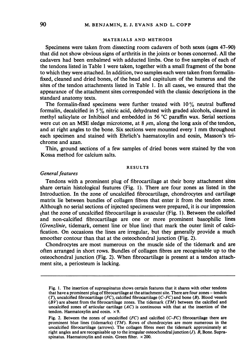
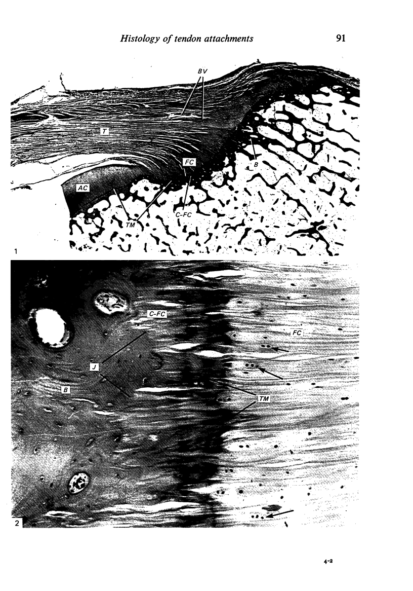
Based on a parallel study of a wide range of human tendons from embalmed dissecting room subjects and from a study of dried bones, an explanation is offered for the well known similarity in gross appearance between the markings left by certain tendons (e.g. those of the rotator cuff) and by articular surfaces on dried bones. Epiphyseal tendons leave markings on bones that look like those left by articular surfaces. These tendons have a prominent zone of fibrocartilage at their attachment site and the deepest part of this is calcified, just as the deepest part of articular hyaline cartilage is calcified. After maceration of the soft tissues, the calcified (fibro) cartilage is left attached to the bone at articular surfaces and at the sites of tendon attachment. In all cases, the tissues separate at the basophilic tidemark between the calcified and uncalcified regions. This tidemark is smooth where there is much overlying uncalcified (fibro) cartilage and it is the smoothness that gives the typical appearance of the dried bone. Blood vessels do not generally traverse the tendon fibrocartilage plugs. Hence the areas are devoid of vascular foramina. The functional significance of tendon fibrocartilage is discussed with particular reference to supraspinatus. It is suggested that the uncalcified fibrocartilage ensures that the tendon fibres do not bend, splay out or become compressed at a hard tissue interface, and are thereby offered some protection from wear and tear. It is also suggested that the fibrocartilage plug of supraspinatus prevents the tendon from rubbing on the head of the humerus.
Full text
PDF
Images in this article
Selected References
These references are in PubMed. This may not be the complete list of references from this article.
- BIERMANN H. Die Knochenbildung im Bereich periostaler-diaphysärer Sehnenund Bandanstäze. Z Zellforsch Mikrosk Anat. 1957;46(6):635–671. [PubMed] [Google Scholar]
- Haines R. W., Mohuiddin A. Metaplastic bone. J Anat. 1968 Nov;103(Pt 3):527–538. [PMC free article] [PubMed] [Google Scholar]
- KNESE K. H., BIERMANN H. Die Knochenbildung an Sehnen- und Bandansătzen im Bereich ursprünglich chondraier Apophysen. Z Zellforsch Mikrosk Anat. 1958;49(2):142–187. [PubMed] [Google Scholar]
- Meachim G., Bentley G. Horizontal splitting in patellar articular cartilage. Arthritis Rheum. 1978 Jul-Aug;21(6):669–674. doi: 10.1002/art.1780210610. [DOI] [PubMed] [Google Scholar]
- SCHNEIDER H. Zur Struktur der Sehnenansatzzonen. Z Anat Entwicklungsgesch. 1956;119(5):431–456. [PubMed] [Google Scholar]


