

Abstract
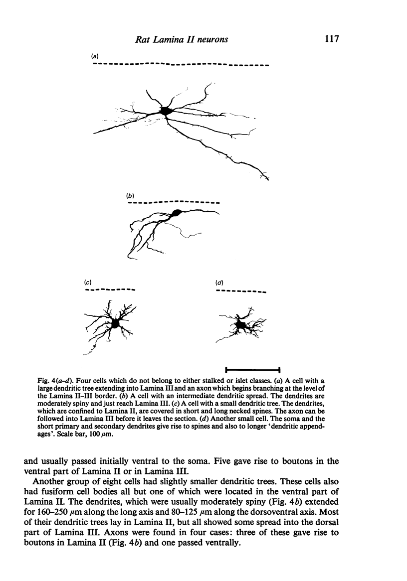
Golgi-stained neurons in Lamina II of the rat spinal cord were examined by light microscopy. Stalked and islet cells similar to those seen in other species were found. Stalked cells were present in large numbers in the dorsal part of the lamina where they made up nearly half the population of stained cells. Islet cells were found throughout the lamina and constituted about one third of the total population. In the ventral part of the lamina half of the stained cells did not fall into either category, but could be divided into groups on the basis of dendritic spread. The axons of many of these cells either remained in Lamina II or passed ventrally into Lamina III. Some of these cells may correspond to the stellate or the II-III border cells which have been seen in human spinal cord and cat medulla respectively.
Full text
PDF
Selected References
These references are in PubMed. This may not be the complete list of references from this article.
- Beal J. A., Cooper M. H. The neurons in the gelatinosal complex (Laminae II and III) of the monkey (Macaca mulatta): a Golgi study. J Comp Neurol. 1978 May 1;179(1):89–121. doi: 10.1002/cne.901790107. [DOI] [PubMed] [Google Scholar]
- Bennett G. J., Abdelmoumene M., Hayashi H., Dubner R. Physiology and morphology of substantia gelatinosa neurons intracellularly stained with horseradish peroxidase. J Comp Neurol. 1980 Dec 15;194(4):809–827. doi: 10.1002/cne.901940407. [DOI] [PubMed] [Google Scholar]
- Gobel S. Golgi studies in the substantia gelatinosa neurons in the spinal trigeminal nucleus. J Comp Neurol. 1975 Aug 1;162(3):397–415. doi: 10.1002/cne.901620308. [DOI] [PubMed] [Google Scholar]
- Gobel S. Golgi studies of the neurons in layer II of the dorsal horn of the medulla (trigeminal nucleus caudalis). J Comp Neurol. 1978 Jul 15;180(2):395–413. doi: 10.1002/cne.901800213. [DOI] [PubMed] [Google Scholar]
- Ribeiro-da-Silva A., Coimbra A. Two types of synaptic glomeruli and their distribution in laminae I-III of the rat spinal cord. J Comp Neurol. 1982 Aug 1;209(2):176–186. doi: 10.1002/cne.902090205. [DOI] [PubMed] [Google Scholar]
- Schoenen J. The dendritic organization of the human spinal cord: the dorsal horn. Neuroscience. 1982;7(9):2057–2087. doi: 10.1016/0306-4522(82)90120-8. [DOI] [PubMed] [Google Scholar]
- Somogyi P., Hodgson A. J., Smith A. D. An approach to tracing neuron networks in the cerebral cortex and basal ganglia. Combination of Golgi staining, retrograde transport of horseradish peroxidase and anterograde degeneration of synaptic boutons in the same material. Neuroscience. 1979;4(12):1805–1852. doi: 10.1016/0306-4522(79)90059-9. [DOI] [PubMed] [Google Scholar]
- Woolf C. J., Fitzgerald M. The properties of neurones recorded in the superficial dorsal horn of the rat spinal cord. J Comp Neurol. 1983 Dec 10;221(3):313–328. doi: 10.1002/cne.902210307. [DOI] [PubMed] [Google Scholar]