

Abstract
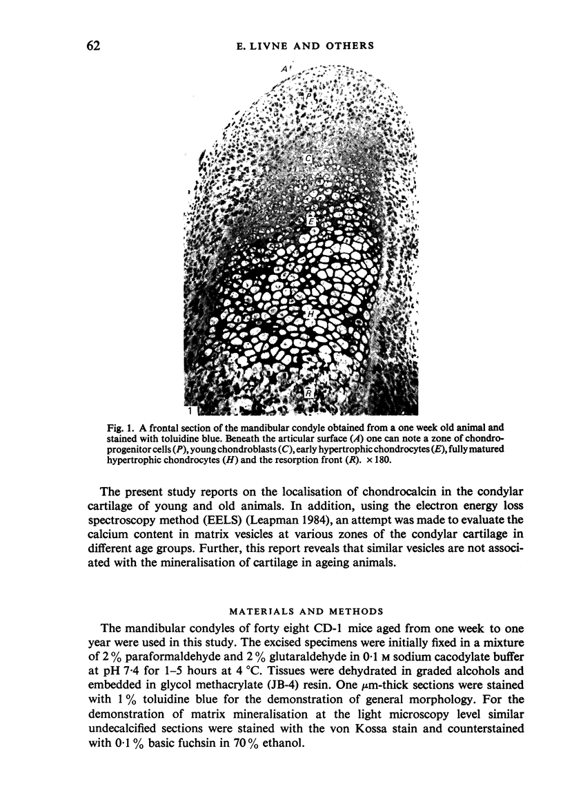
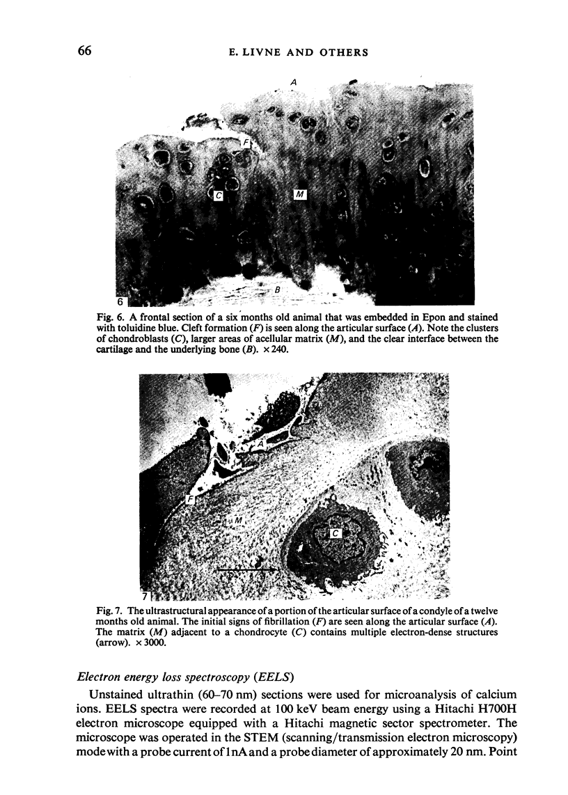
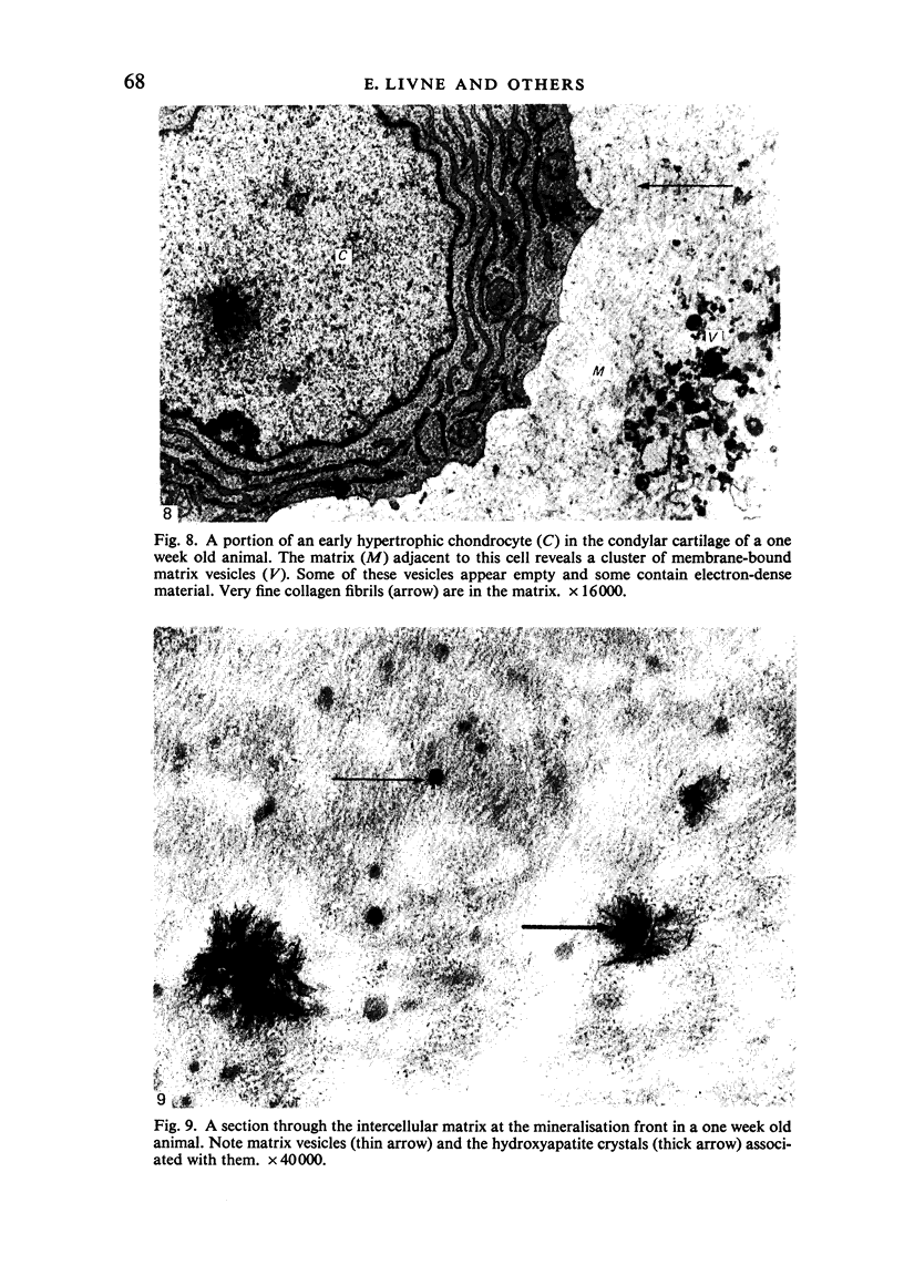
A combined approach of light microscopy, immunofluorescence, transmission electron microscopy and electron energy loss spectroscopy (EELS) was used to study age-related changes in the condylar cartilage in mice. Chondrocalcin, a cartilage matrix calcium-binding protein, was demonstrated by indirect immunofluorescence microscopy using monospecific antibodies. In one week old animals the most intense staining was observed in the matrix around the hypertrophic cells in the mineralising zone, to a lesser degree around the cells in the zone of chondroblasts, while no staining was noted in the zone of chondroprogenitor cells and in the matrix around the early hypertrophic cells. In the mineralisation zone the distribution of chondrocalcin correlated with that of mineral deposits as revealed by the von Kossa stain. The matrix between the early hypertrophic cells as shown by transmission electron microscopy revealed the presence of matrix vesicles and demonstrated a gradual accumulation of hydroxyapatite in the mineralising zone. In one month old animals chondrocalcin localisation was mainly confined to the lower hypertrophic zone which also demonstrated positive von Kossa staining was seen along the articular surface. In older animals multiple electron-dense structure that resembled matrix vesicles were observed in the non-mineralising portions of the condylar cartilage. Use of the EELS method confirmed the almost complete lack of calcium ions in these structures. In contrast, with the use of the same method, detectable amounts of calcium were recorded in vesicles in the mineralising zones of all age groups. Hence what appear ultrastructurally as structures similar to matrix vesicles represent atypical vesicles that might characterise an ageing and degenerative articular cartilage and are not necessarily associated with the mineralisation process.
Full text
PDF


Images in this article


Selected References
These references are in PubMed. This may not be the complete list of references from this article.
- Ali S. Y. Analysis of matrix vesicles and their role in the calcification of epiphyseal cartilage. Fed Proc. 1976 Feb;35(2):135–142. [PubMed] [Google Scholar]
- Anderson H. C. Vesicles associated with calcification in the matrix of epiphyseal cartilage. J Cell Biol. 1969 Apr;41(1):59–72. doi: 10.1083/jcb.41.1.59. [DOI] [PMC free article] [PubMed] [Google Scholar]
- Appleton J., Morris D. C. The use of the potassium pyroantimonate-osmium method as a means of identifying and localizing calcium at the ultrastructural level in the cells of calcifying systems. J Histochem Cytochem. 1979 Feb;27(2):676–680. doi: 10.1177/27.2.448059. [DOI] [PubMed] [Google Scholar]
- Arsenault A. L., Ottensmeyer F. P. Quantitative spatial distributions of calcium, phosphorus, and sulfur in calcifying epiphysis by high resolution electron spectroscopic imaging. Proc Natl Acad Sci U S A. 1983 Mar;80(5):1322–1326. doi: 10.1073/pnas.80.5.1322. [DOI] [PMC free article] [PubMed] [Google Scholar]
- Bonucci E. Fine structure of early cartilage calcification. J Ultrastruct Res. 1967 Sep;20(1):33–50. doi: 10.1016/s0022-5320(67)80034-0. [DOI] [PubMed] [Google Scholar]
- Choi H. U., Tang L. H., Johnson T. L., Pal S., Rosenberg L. C., Reiner A., Poole A. R. Isolation and characterization of a 35,000 molecular weight subunit fetal cartilage matrix protein. J Biol Chem. 1983 Jan 10;258(1):655–661. [PubMed] [Google Scholar]
- Dearden L. C., Bonucci E., Cuicchio M. An investigation of ageing in human costal cartilage. Cell Tissue Res. 1974;152(3):305–337. doi: 10.1007/BF00223953. [DOI] [PubMed] [Google Scholar]
- Dougherty W. J. Ca-enriched amorphous mineral deposits associated with the plasma membranes of chondrocytes and matrix vesicles of rat epiphyseal cartilage. Calcif Tissue Int. 1983 Jul;35(4-5):486–495. doi: 10.1007/BF02405082. [DOI] [PubMed] [Google Scholar]
- Koski K. Cartilage in the face. Birth Defects Orig Artic Ser. 1975;11(7):231–254. [PubMed] [Google Scholar]
- Landis W. J., Glimcher M. J. Electron optical and analytical observations of rat growth plate cartilage prepared by ultracryomicrotomy: the failure to detect a mineral phase in matrix vesicles and the identification of heterodispersed particles as the initial solid phase of calcium phosphate deposited in the extracellular matrix. J Ultrastruct Res. 1982 Mar;78(3):227–268. doi: 10.1016/s0022-5320(82)80001-4. [DOI] [PubMed] [Google Scholar]
- Livne E., Silbermann M. Changes in the structure and chemical composition of the mandibular condylar cartilage of the neonatal mouse. Arch Oral Biol. 1983;28(9):805–812. doi: 10.1016/0003-9969(83)90036-5. [DOI] [PubMed] [Google Scholar]
- Livne E., von der Mark K., Silbermann M. Morphologic and cytochemical changes in maturing and osteoarthritic articular cartilage in the temporomandibular joint of mice. Arthritis Rheum. 1985 Sep;28(9):1027–1038. doi: 10.1002/art.1780280910. [DOI] [PubMed] [Google Scholar]
- Matsuzawa T., Anderson H. C. Phosphatases of epiphyseal cartilage studied by electron microscopic cytochemical methods. J Histochem Cytochem. 1971 Dec;19(12):801–808. doi: 10.1177/19.12.801. [DOI] [PubMed] [Google Scholar]
- Meachim G. Age changes in articular cartilage. Clin Orthop Relat Res. 1969 May-Jun;64:33–44. [PubMed] [Google Scholar]
- Meikle M. C. The mineralization of condylar cartilage in the rat mandible: an electron microscopic enzyme histochemical study. Arch Oral Biol. 1976;21(1):33–43. doi: 10.1016/0003-9969(76)90157-6. [DOI] [PubMed] [Google Scholar]
- Mitchell N., Shepard N. The ultrastructure of articular cartilage in rheumatoid arthritis. A preliminary report. J Bone Joint Surg Am. 1970 Oct;52(7):1405–1423. [PubMed] [Google Scholar]
- Poole A. R., Pidoux I., Reiner A., Choi H., Rosenberg L. C. Association of an extracellular protein (chondrocalcin) with the calcification of cartilage in endochondral bone formation. J Cell Biol. 1984 Jan;98(1):54–65. doi: 10.1083/jcb.98.1.54. [DOI] [PMC free article] [PubMed] [Google Scholar]
- Poole A. R., Pidoux I., Rosenberg L. Role of proteoglycans in endochondral ossification: immunofluorescent localization of link protein and proteoglycan monomer in bovine fetal epiphyseal growth plate. J Cell Biol. 1982 Feb;92(2):249–260. doi: 10.1083/jcb.92.2.249. [DOI] [PMC free article] [PubMed] [Google Scholar]
- Silbermann M., Lewinson D. An electron microscopic study of the premineralizing zone of the condylar cartilage of the mouse mandible. J Anat. 1978 Jan;125(Pt 1):55–70. [PMC free article] [PubMed] [Google Scholar]
- Silbermann M., Livne E. Skeletal changes in the condylar cartilage of the neonate mouse mandible. Biol Neonate. 1979;35(1-2):95–105. doi: 10.1159/000241159. [DOI] [PubMed] [Google Scholar]