

Abstract
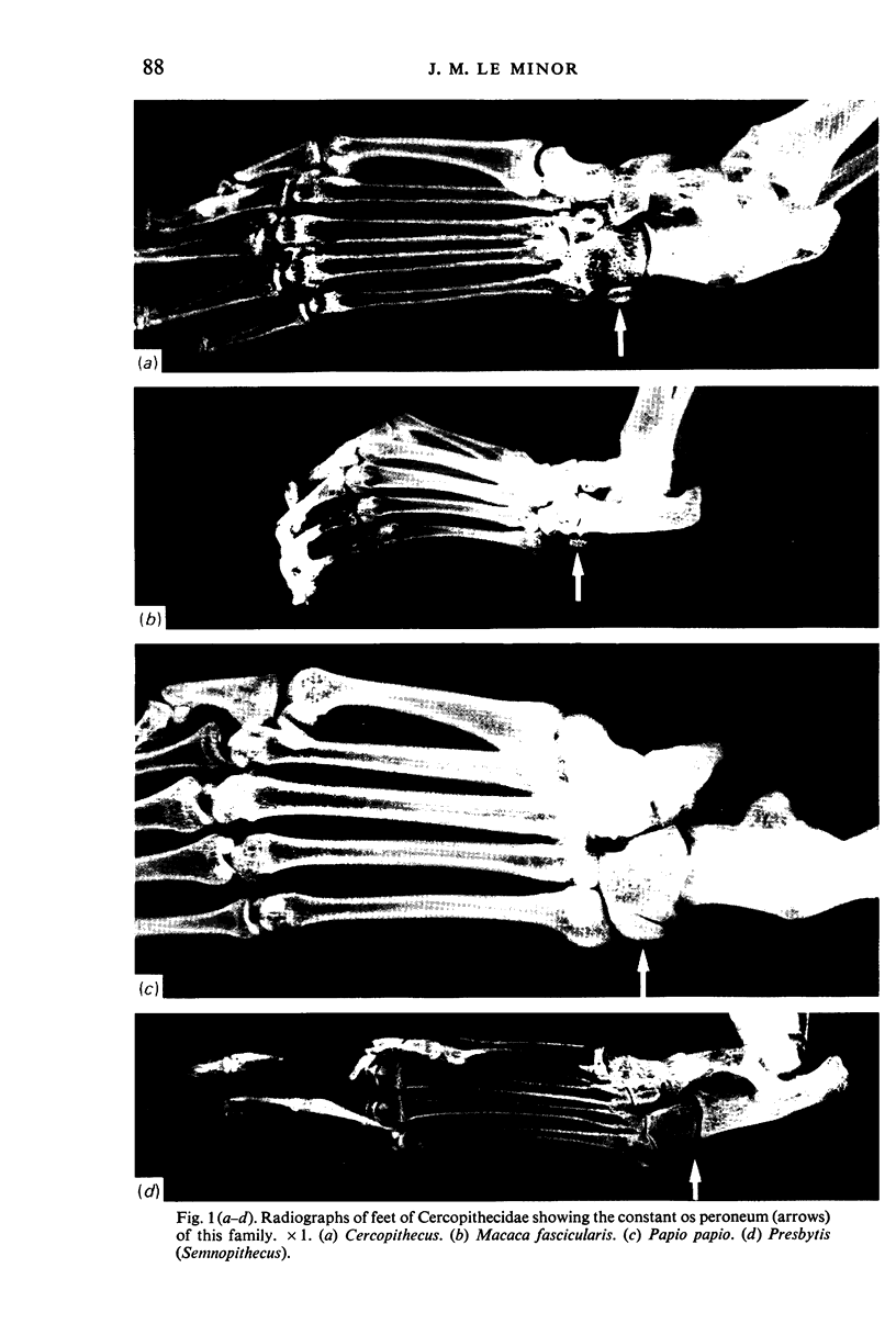
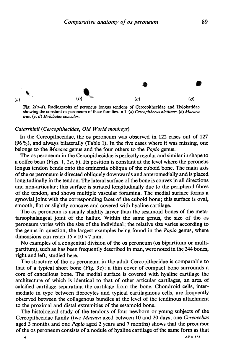
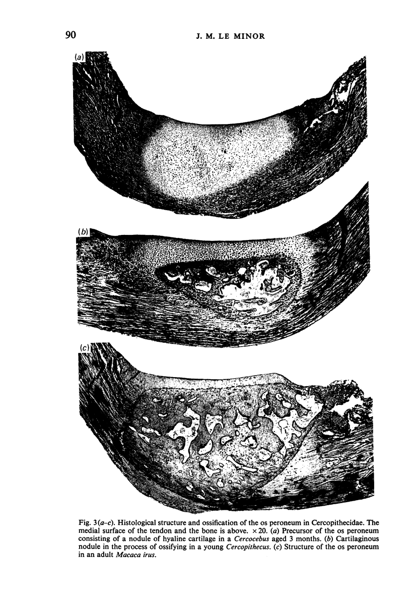
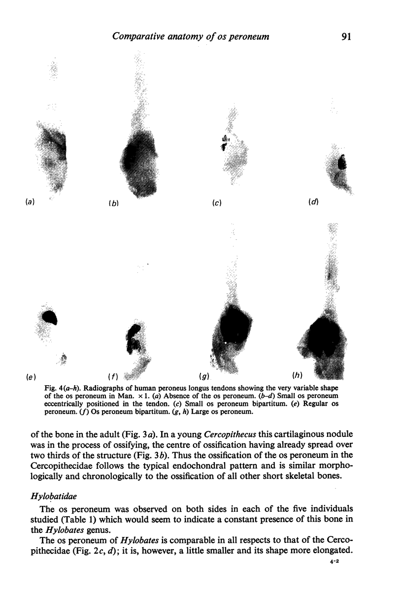
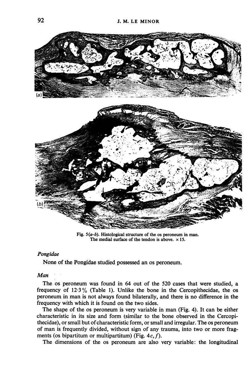
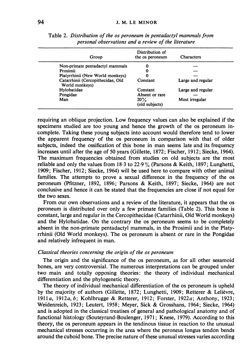
The os peroneum is found in only a few primate families and seems to be completely absent in the non-primate pentadactyl mammals, in the Prosimii and in the Platyrrhinii (New World monkeys). In the Cercopithecidae (Catarrhinii, Old World monkeys) and the Hylobatidae, the os peroneum is a coffee bean-shaped constant, large and regular bone. The lateral surface of the bone is convex in all directions and non-articular. The medial surface is covered with hyaline cartilage and articulates by means of a synovial joint with the corresponding facet of the cuboid bone. The histological structure and the mode of ossification of the os peroneum are identical to that of other short bones of the skeleton. The os peroneum of the Cercopithecidae and Hylobatidae is an example of a new skeletal element that has appeared in a tendon subject to unusual mechanical stress. In the case of the peroneus longus tendon the stress is due to repetitive friction because of the functional importance of this muscle in the adduction and pseudo-opposability of the hallux. This osseous element is genetically fixed and hereditarily transmitted. Its mode of appearance is analogous to that accepted for the origin of the patella. In the Pongidae, the os peroneum is absent or rare. In man, this bone is relatively infrequent (approx 20% of mature individuals) and its shape is most irregular. In this case, the os peroneum appears as a regressive form of the typical bone observed in the above families, which is in the process of disappearing. Besides fundamental genetical factors, this regression is probably in relation to the disappearance of the functional importance of the peroneus longus muscle to the loss of the hallux opposability. Thus the mechanical factors cannot be dissociated from the genetic and phylogenetic factors in explaining the appearance and the regression of the os peroneum.
Full text
PDF
Images in this article
Selected References
These references are in PubMed. This may not be the complete list of references from this article.
- BARNETT C. H., LEWIS O. J. The evolution of some traction epiphyses in birds and mammals. J Anat. 1958 Oct;92(4):593–601. [PMC free article] [PubMed] [Google Scholar]
- Haines R. W. Note on the independence of sesamoid and epiphysial centres of ossification. J Anat. 1940 Oct;75(Pt 1):101–105. [PMC free article] [PubMed] [Google Scholar]
- LEUTERT G. Uber den Bau der Sehne des Musculus fibularis longus im Bereich des äusseren Fussrandes. Z Mikrosk Anat Forsch. 1955;61(4):512–532. [PubMed] [Google Scholar]
- Mains D. B., Sullivan R. C. Fracture of the os peroneum. A case report. J Bone Joint Surg Am. 1973 Oct;55(7):1529–1530. [PubMed] [Google Scholar]
- Manners-Smith T. A Study of the Cuboid and Os Peroneum in the Primate Foot. J Anat Physiol. 1908 Jul;42(Pt 4):397–414. [PMC free article] [PubMed] [Google Scholar]
- Parsons F G. Further Remarks on Traction Epiphyses. J Anat Physiol. 1908 Jul;42(Pt 4):388–396. [PMC free article] [PubMed] [Google Scholar]
- Parsons F G, Keith A. Seventh Report of the Committee of Collective Investigation of the Anatomical Society of Great Britain and Ireland, 1896-97. J Anat Physiol. 1897 Oct;32(Pt 1):164–186. [PMC free article] [PubMed] [Google Scholar]
- Parsons F G. Observations on Traction Epiphyses. J Anat Physiol. 1904 Apr;38(Pt 3):248–258. [PMC free article] [PubMed] [Google Scholar]
- SICK H. L'ADAPTATION DES TENDONS 'A LA R'EFLEXION. Arch Anat Histol Embryol. 1964;47:369–446. [PubMed] [Google Scholar]
- SIECKE H. BEITRAG ZUR GENESE DES OS PERONEUM. (BEOBACHTUNGEN AN 250 ROENTGENOLOGISCH FESTGESTELLTEN OSSA PERONEA) Z Orthop Ihre Grenzgeb. 1964 Apr;98:358–370. [PubMed] [Google Scholar]
- WELTI H. [Contribution to the study of the morphology and function of the tibialis anterior and peroneus longus muscles. Study of comparative anatomy]. Arch Anat Histol Embryol. 1961;44:59–100. [PubMed] [Google Scholar]
- WIRTSCHAFTER Z. T., TSUJIMURA J. K. The sesamoid bones in Long-Evans strain rats. Anat Rec. 1961 Nov;141:195–204. doi: 10.1002/ar.1091410304. [DOI] [PubMed] [Google Scholar]
- WIRTSCHAFTER Z. T., TSUJIMURA J. K. The sesamoid bones in the C3H mouse. Anat Rec. 1961 Mar;139:399–408. doi: 10.1002/ar.1091390308. [DOI] [PubMed] [Google Scholar]
- Wrobel K. H. Funktionelle Anpassungserscheinungen der Muskulatur des Lorisidenfusses. Gegenbaurs Morphol Jahrb. 1966;109(3):448–469. [PubMed] [Google Scholar]
- Wütschke J. Patella partita und Patella duplex (kritische Betrachtung zur Differentialdiagnose) Fortschr Geb Rontgenstr Nuklearmed. 1966 Feb;104(2):260–263. [PubMed] [Google Scholar]