Abstract
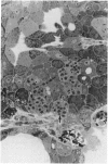
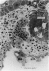
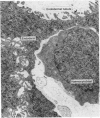
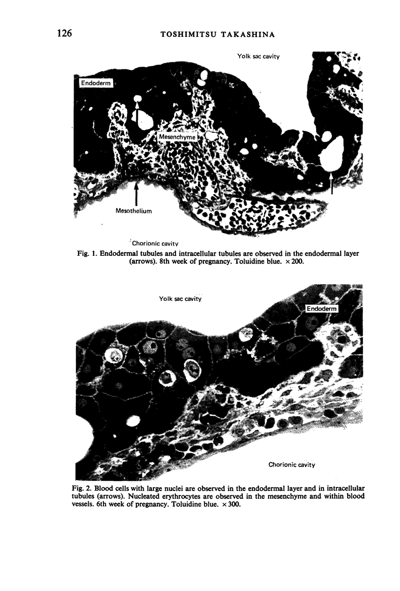
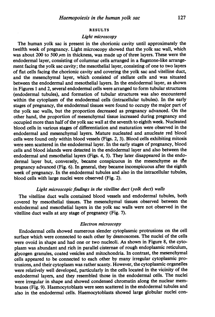
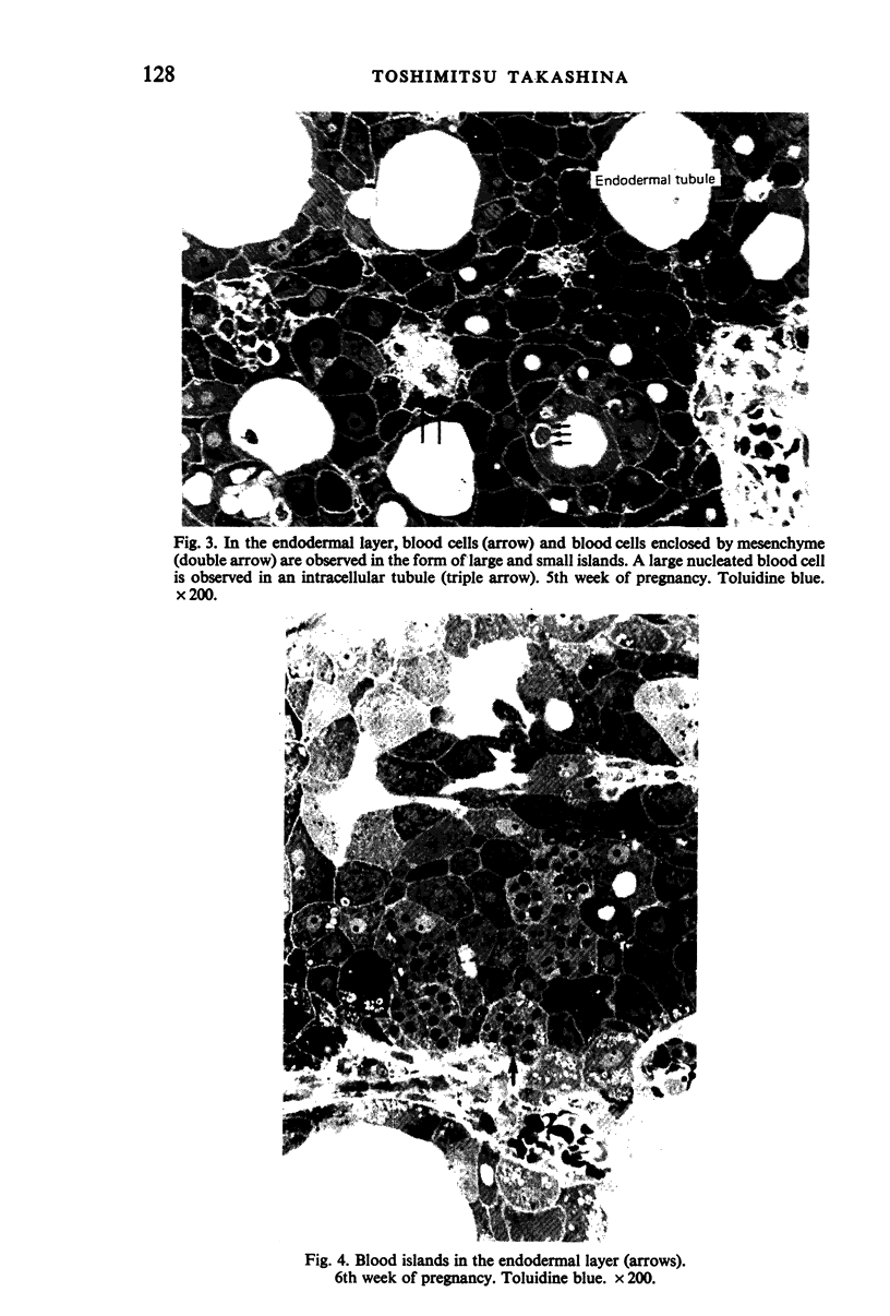
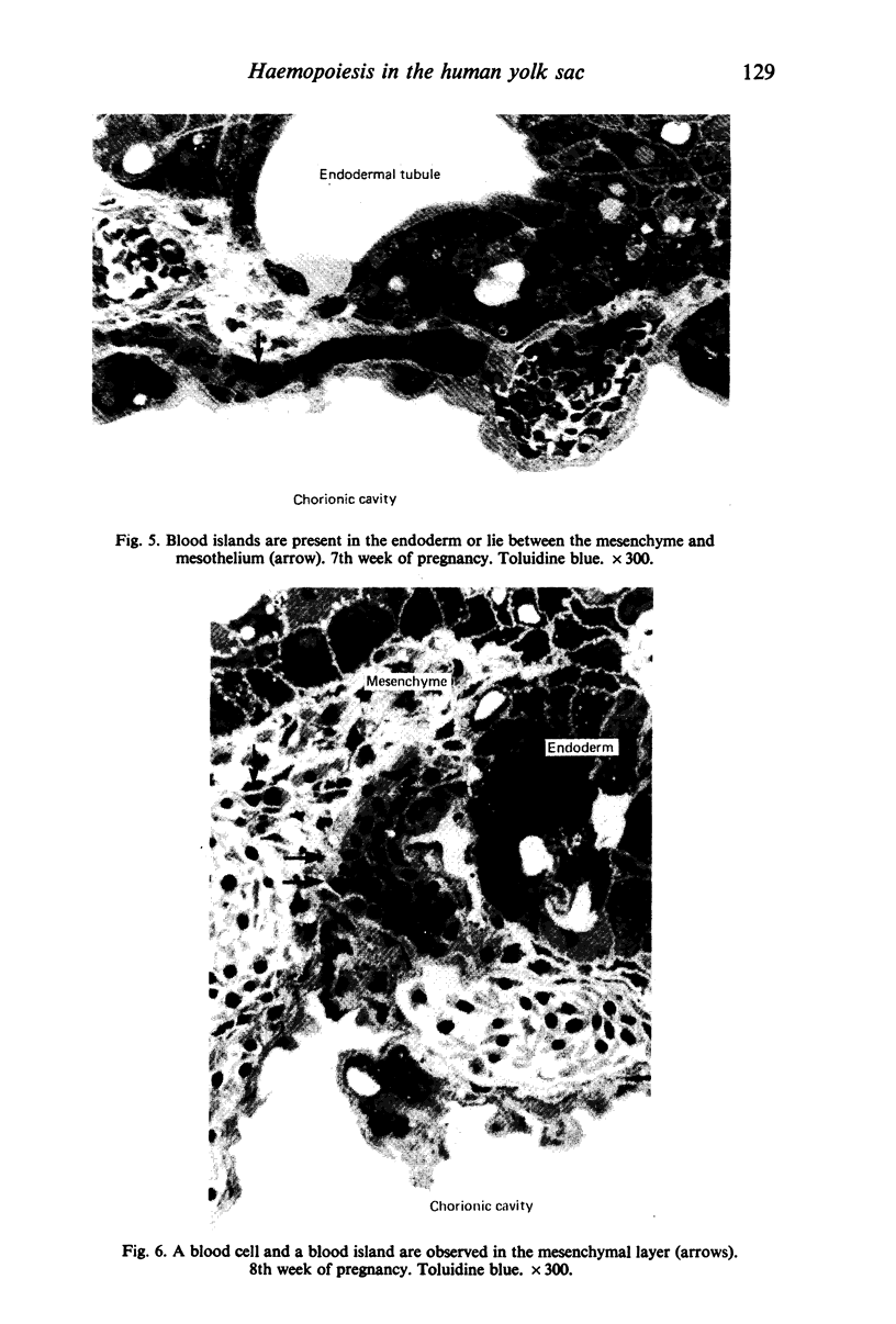
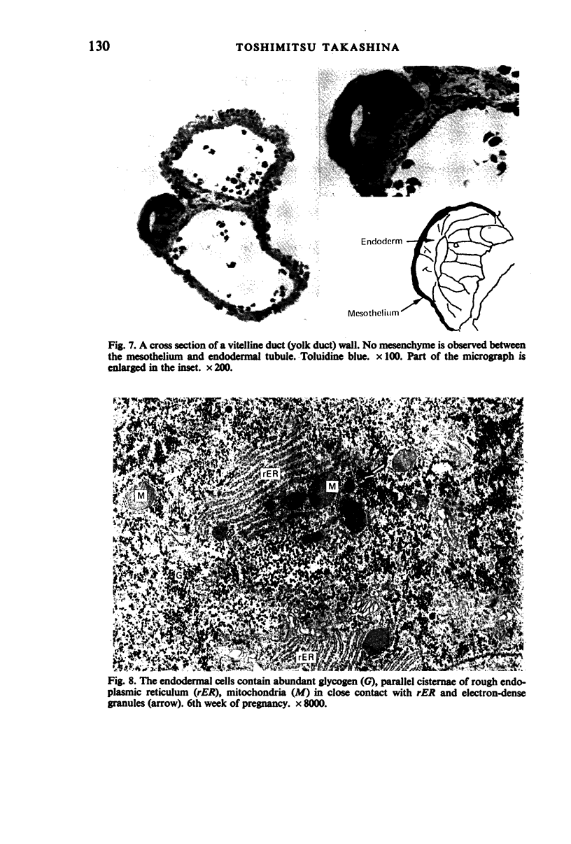
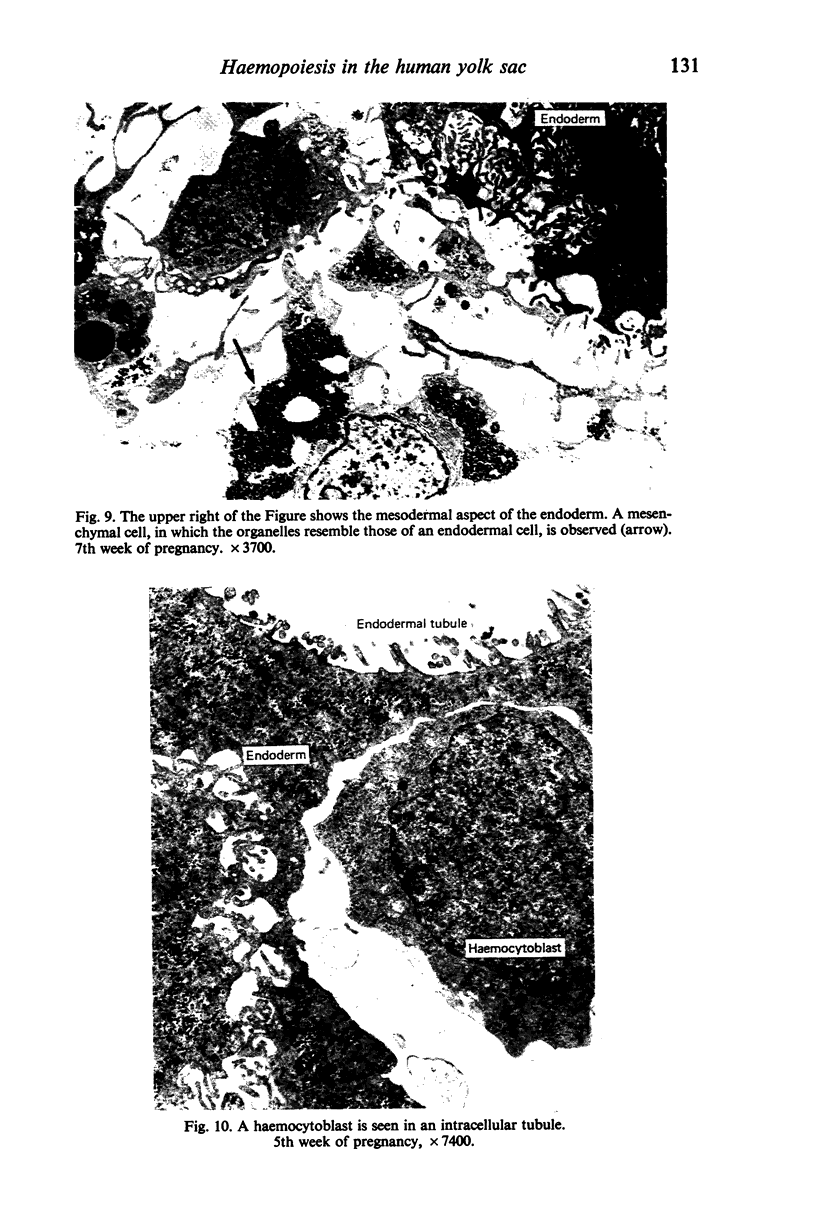
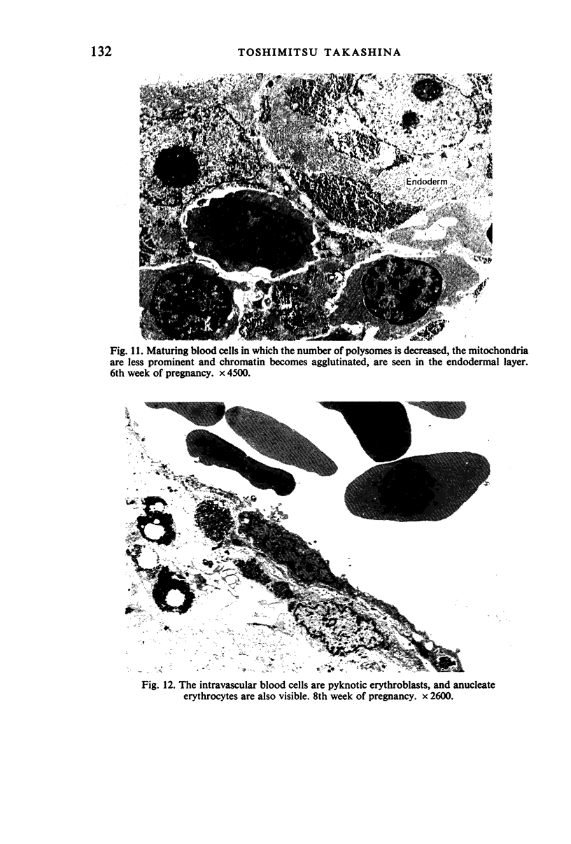
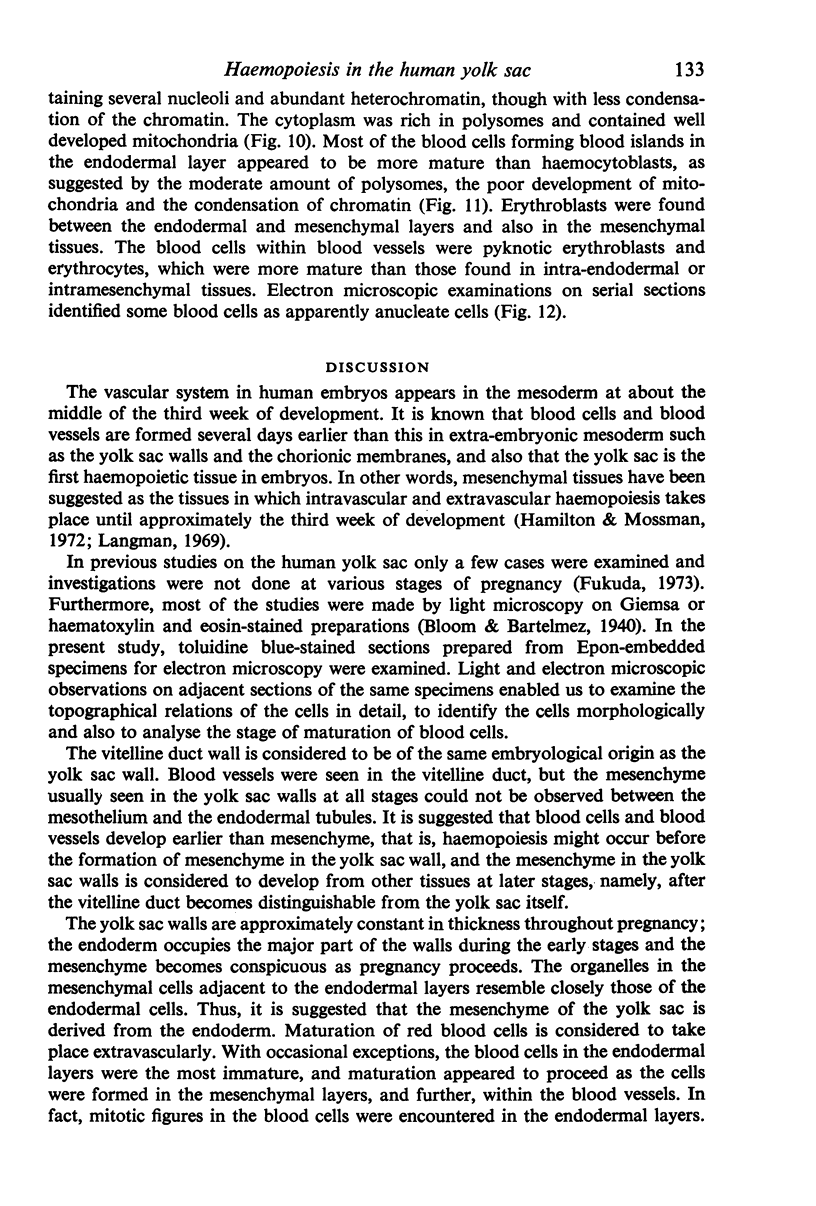
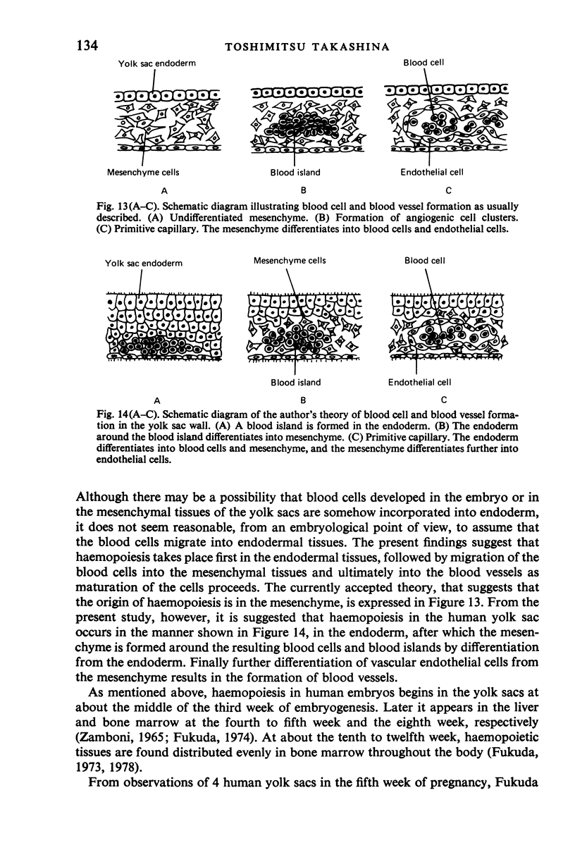
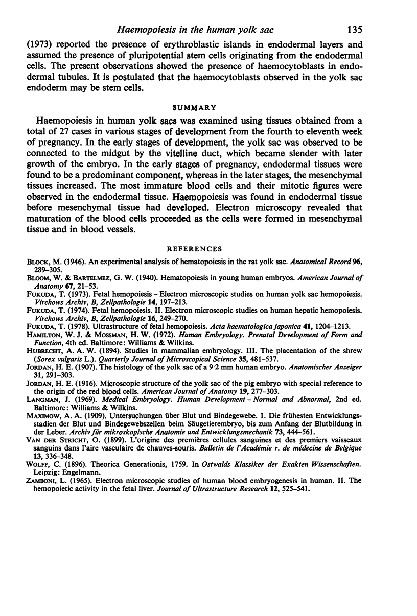
Haemopoiesis in human yolk sacs was examined using tissues obtained from a total of 27 cases in various stages of development from the fourth to eleventh week of pregnancy. In the early stages of development, the yolk sac was observed to be connected to the midgut by the vitelline duct, which became slender with later growth of the embryo. In the early stages of pregnancy, endodermal tissues were found to be a predominant component, whereas in the later stages, the mesenchymal tissues increased. The most immature blood cells and their mitotic figures were observed in the endodermal tissue. Haemopoiesis was found in endodermal tissue before mesenchymal tissue had developed. Electron microscopy revealed that maturation of the blood cells proceeded as the cells were formed in mesenchymal tissue and in blood vessels.
Full text
PDF
Images in this article
Selected References
These references are in PubMed. This may not be the complete list of references from this article.
- Fukuda T. Fetal hemopoiesis. I. Electron microscopic studies on human yolk sac hemopoiesis. Virchows Arch B Cell Pathol. 1973 Dec 7;14(3):197–213. [PubMed] [Google Scholar]
- Fukuda T. Fetal hemopoiesis. II. Electron microscopic studies on human hepatic hemopoiesis. Virchows Arch B Cell Pathol. 1974;16(3):249–270. [PubMed] [Google Scholar]
- Fukuda T. Ultrastructure of fetal hemopoiesis. Nihon Ketsueki Gakkai Zasshi. 1978 Dec;41(6):1204–1213. [PubMed] [Google Scholar]
- Zamboni L. Electron microscopic studies of blood embryogenesis in humans. II. The hemopoietic activity in the fetal liver. J Ultrastruct Res. 1965 Jun;12(5):525–541. doi: 10.1016/s0022-5320(65)80045-4. [DOI] [PubMed] [Google Scholar]


