

Abstract
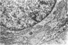
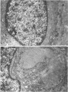
Articular cartilage of eight young NZW rabbits was investigated by electron microscopy. A simple and rapid stereological method was designed for quantifying the synthetic activity of the chondrocytes in the different zones of uncalcified articular cartilage by measuring the amount of rough endoplasmic reticulum (RER) on electron micrographs. The surface area of RER per unit volume of chondrocyte cytoplasm, of cartilage, and per chondrocyte, were determined. In addition the volume density, and mean diameter, of RER cisternae were computed. The surface area of RER was least in the superficial zone (402 micron 2) and largest in the deep zone (879 micron 2) chondrocytes. The RER surface area per unit volume of tissue was, however, significantly (P less than 0.05) greater in the superficial zone (12.8 X 10(-2) micron2/micron3) than in the deep zone (8.5 X 10(-2) micron2/micron3) of the articular cartilage. Percentages of chondrocytes displaying masses of intracytoplasmic fine filaments were also registered. 8.5% of the chondrocytes in the superficial, 48.8% in the middle, and 53.5% in the deep zone contained intracytoplasmic fine filaments. Chondrocytes containing filaments are probably degenerating cells. They contained diminished amounts of RER. The altered nutritional status in the deeper zones of the cartilage might have caused these changes in the cells. The formation of the calcified zone of the cartilage in the young rabbits, contributing to the cessation of the nutritional supply of chondrocytes from the subchondral bone marrow spaces, may have affected the process. Since the proportion of RER can be regarded as indicating the synthetic activity, and that of filaments as indicating the degree of degeneration, these parameters can be used in the evaluation of the functional status of the uncalcified articular cartilage chondrocytes.
Full text
PDF


Images in this article
Selected References
These references are in PubMed. This may not be the complete list of references from this article.
- BARNETT C. H., COCHRANE W., PALFREY A. J. AGE CHANGES IN ARTICULAR CARTILAGE OF RABBITS. Ann Rheum Dis. 1963 Nov;22:389–400. doi: 10.1136/ard.22.6.389. [DOI] [PMC free article] [PubMed] [Google Scholar]
- Brighton C. T., Kitajima T., Hunt R. M. Zonal analysis of cytoplasmic components of articular cartilage chondrocytes. Arthritis Rheum. 1984 Nov;27(11):1290–1299. doi: 10.1002/art.1780271112. [DOI] [PubMed] [Google Scholar]
- Cruz-Orive L. M., Weibel E. R. Sampling designs for stereology. J Microsc. 1981 Jun;122(Pt 3):235–257. doi: 10.1111/j.1365-2818.1981.tb01265.x. [DOI] [PubMed] [Google Scholar]
- DAVIES D. V., BARNETT C. H., COCHRANE W., PALFREY A. J. Electron microscopy of articular cartilage in the young adult rabbit. Ann Rheum Dis. 1962 Mar;21:11–22. doi: 10.1136/ard.21.1.11. [DOI] [PMC free article] [PubMed] [Google Scholar]
- Meachim G., Roy S. Intracytoplasmic filaments in the cells of adult human articular cartilage. Ann Rheum Dis. 1967 Jan;26(1):50–58. doi: 10.1136/ard.26.1.50. [DOI] [PMC free article] [PubMed] [Google Scholar]
- Müller A. E., Cruz-Orive L. M., Gehr P., Weibel E. R. Comparison of two subsampling methods for electron microscopic morphometry. J Microsc. 1981 Jul;123(Pt 1):35–49. doi: 10.1111/j.1365-2818.1981.tb01278.x. [DOI] [PubMed] [Google Scholar]
- Paukkonen K., Selkäinaho K., Jurvelin J., Helminen H. J. Morphometry of articular cartilage: a stereological method using light microscopy. Anat Rec. 1984 Dec;210(4):675–682. doi: 10.1002/ar.1092100415. [DOI] [PubMed] [Google Scholar]
- Roy S., Meachim G. Chondrocyte ultrastructure in adult human articular cartilage. Ann Rheum Dis. 1968 Nov;27(6):544–558. doi: 10.1136/ard.27.6.544. [DOI] [PMC free article] [PubMed] [Google Scholar]
- SILBERBERG R., SILBEREBERG M., FEIR D. LIFE CYCLE OF ARTICULAR CARTILAGE CELLS: AN ELECTRON MICROSCOPE STUDY OF THE HIP JOINT OF THE MOUSE. Am J Anat. 1964 Jan;114:17–47. doi: 10.1002/aja.1001140103. [DOI] [PubMed] [Google Scholar]
- Weiss C., Rosenberg L., Helfet A. J. An ultrastructural study of normal young adult human articular cartilage. J Bone Joint Surg Am. 1968 Jun;50(4):663–674. doi: 10.2106/00004623-196850040-00002. [DOI] [PubMed] [Google Scholar]