Abstract
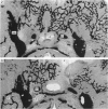
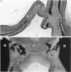
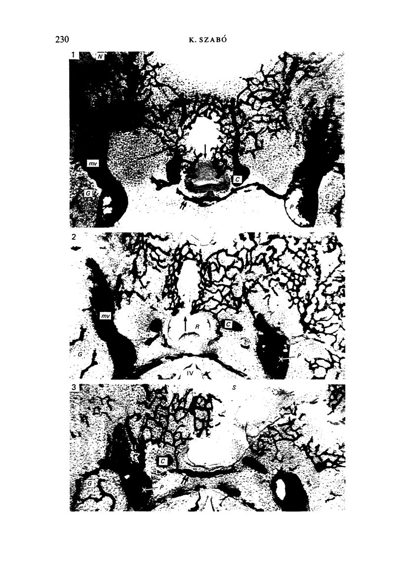
The origin of the portal vessels and the adjacent structures are described in rat embryos (gestational Days 11, 12 and 13) injected with India ink via the umbilical veins. Without the interposition of mesoderm Rathke's pouch comes into contact with the prosencephalic vesicle and its diverticulum, the processus infundibularis. Developmental changes in the shape of the prosencephalic vesicle influence the formation of Rathke's pouch. Before separation of the latter from the diencephalon and the processus infundibularis (Day 12), branches start growing around Rathke's pouch from the capillary network covering the prospective hypothalamus, and these vessels may be now considered as the forerunners of the primary portal vessels. After the separation of the anterior wall of Rathke's pouch from the diencephalon (Day 13) the primary portal veins are present and the hypothalamo-adenohypophyseal portal circulation is ready to function. The hypothalamic plexus is supplied by the primitive maxillary artery and by some small branches of the internal carotid arteries. The venous outflow leads into the intercavernous sinus and into the primitive maxillary veins. No arterial vessels in the primitive pharyngeal wall could be observed before the appearance of the primary portal vessels. The present observations clearly demonstrate that the adenohypophyseal vessels are derived only from the vascular network of the diencephalon, without contribution from the pharyngeal vessels, and that the portal vascular bed develops as a single entity.
Full text
PDF
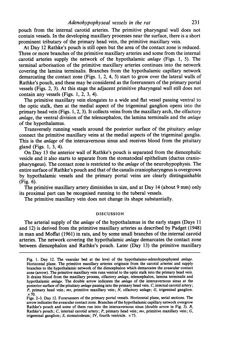
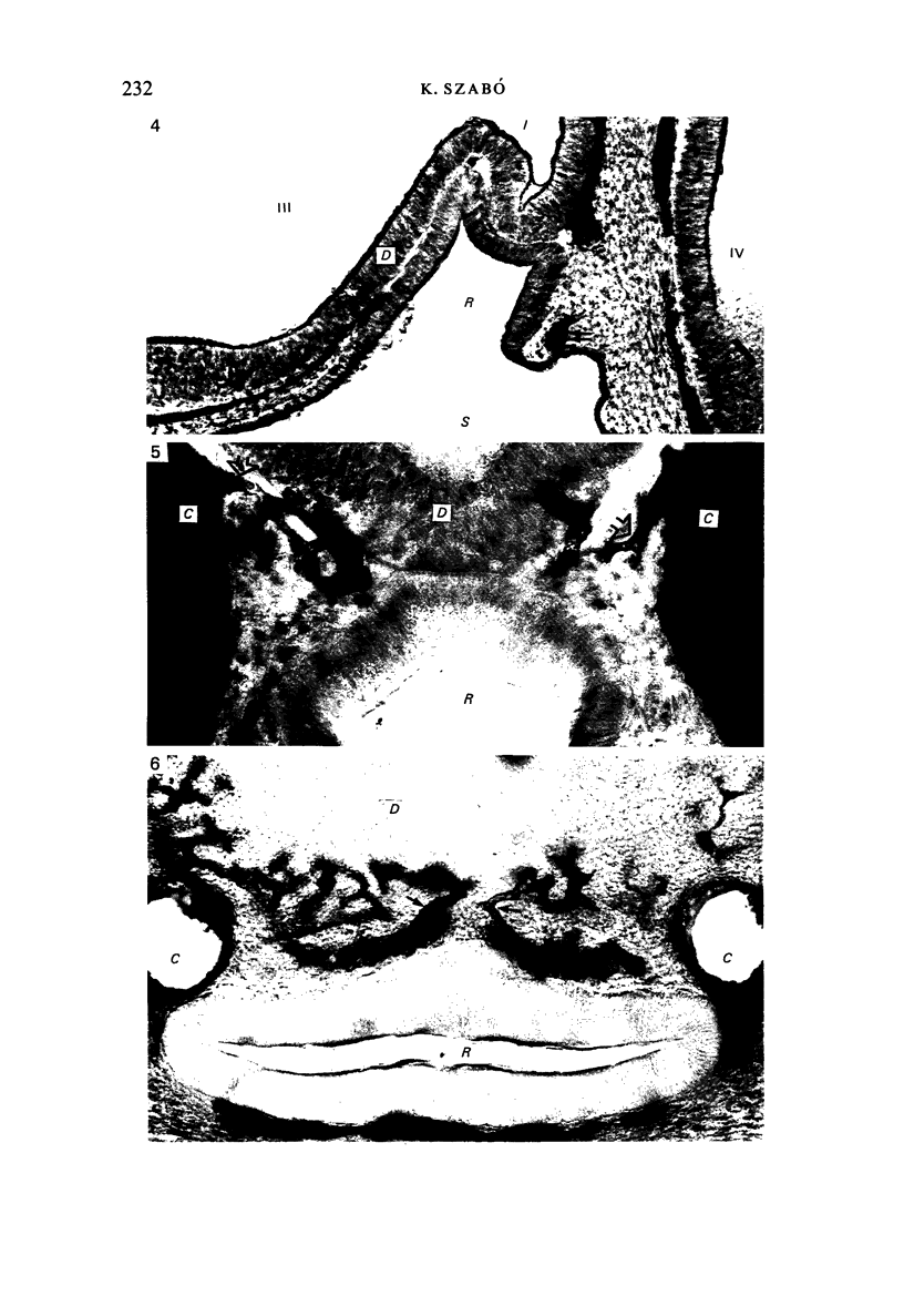
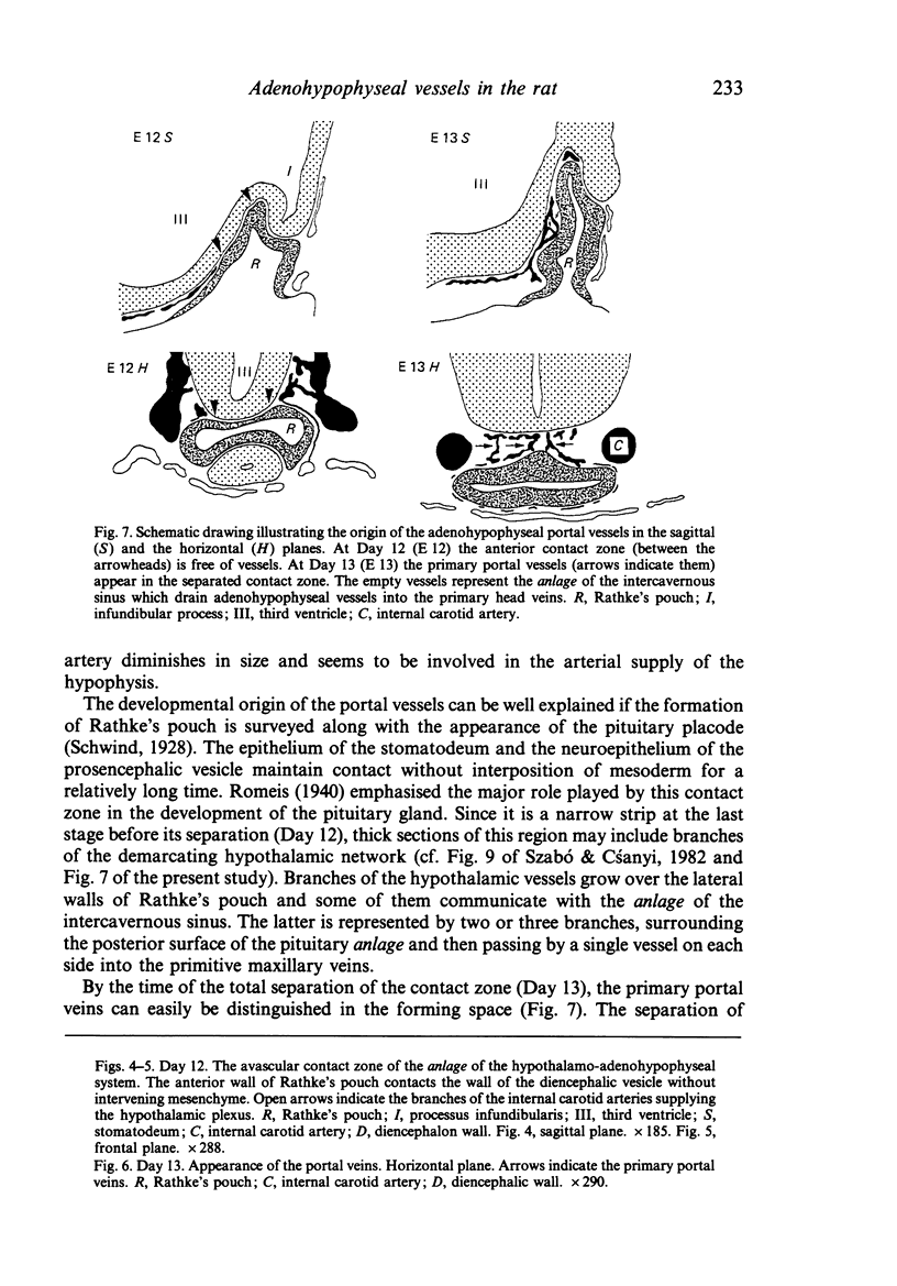
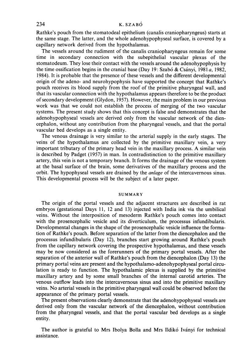
Images in this article
Selected References
These references are in PubMed. This may not be the complete list of references from this article.
- GLYDON R. S. The development of the blood supply of the pituitary in the albino rat, with special reference to the portal vessels. J Anat. 1957 Apr;91(2):237–244. [PMC free article] [PubMed] [Google Scholar]
- Szabó K., Csányi K. Eine modifizierte Tuscheinjektionsmethode zur Darstellung embryonaler Gefässe in der Ratte. Mikroskopie. 1981 Dec;38(11-12):319–324. [PubMed] [Google Scholar]


