Abstract
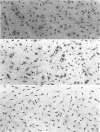
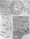
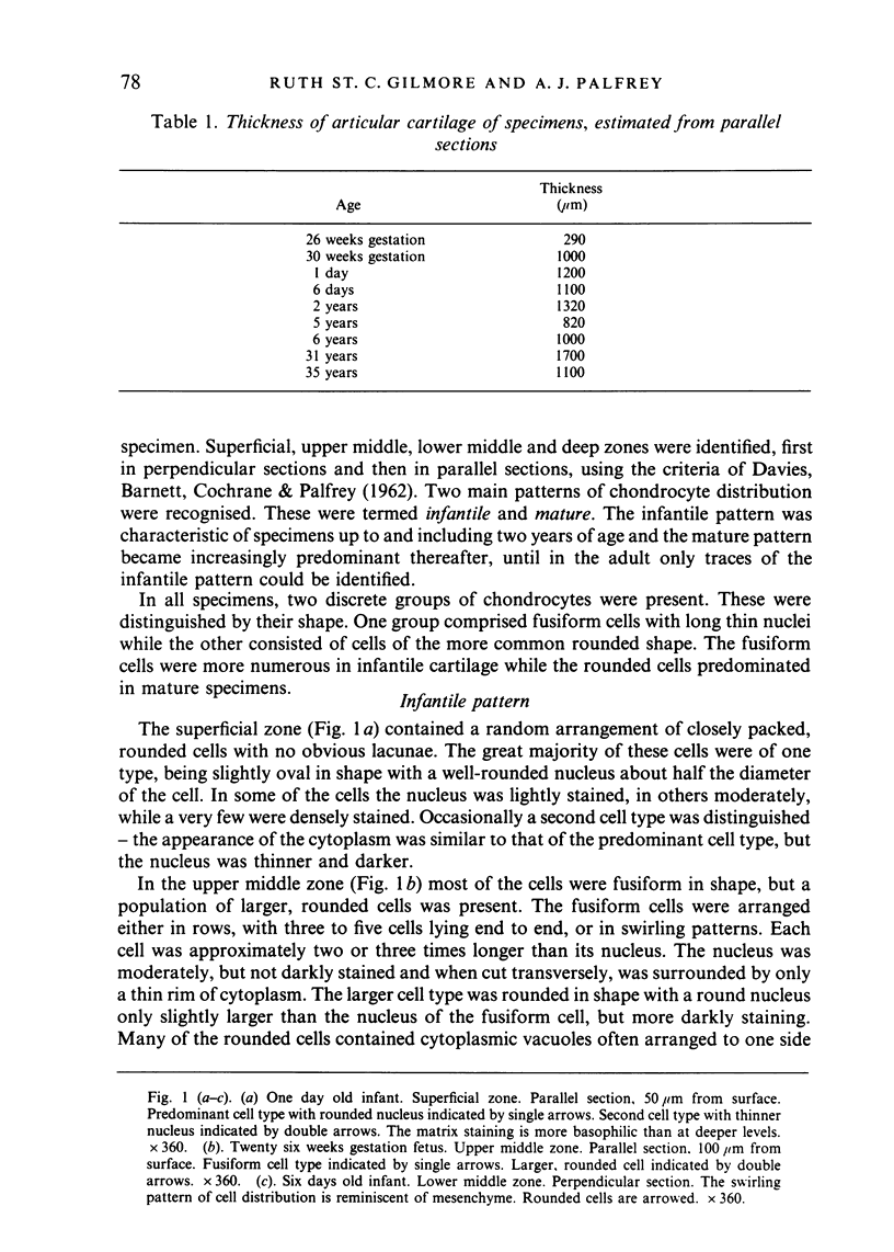
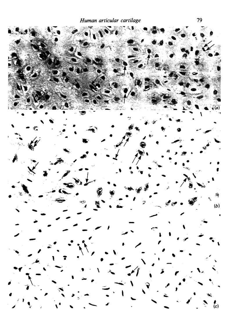
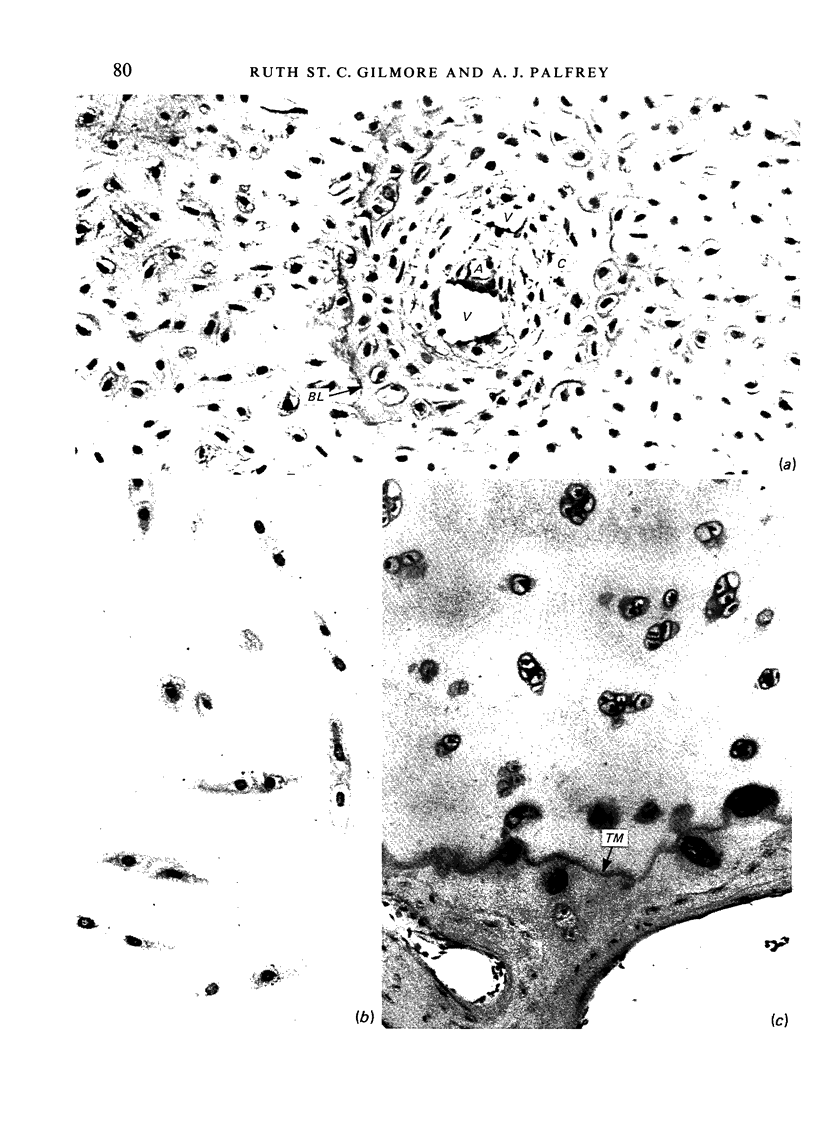
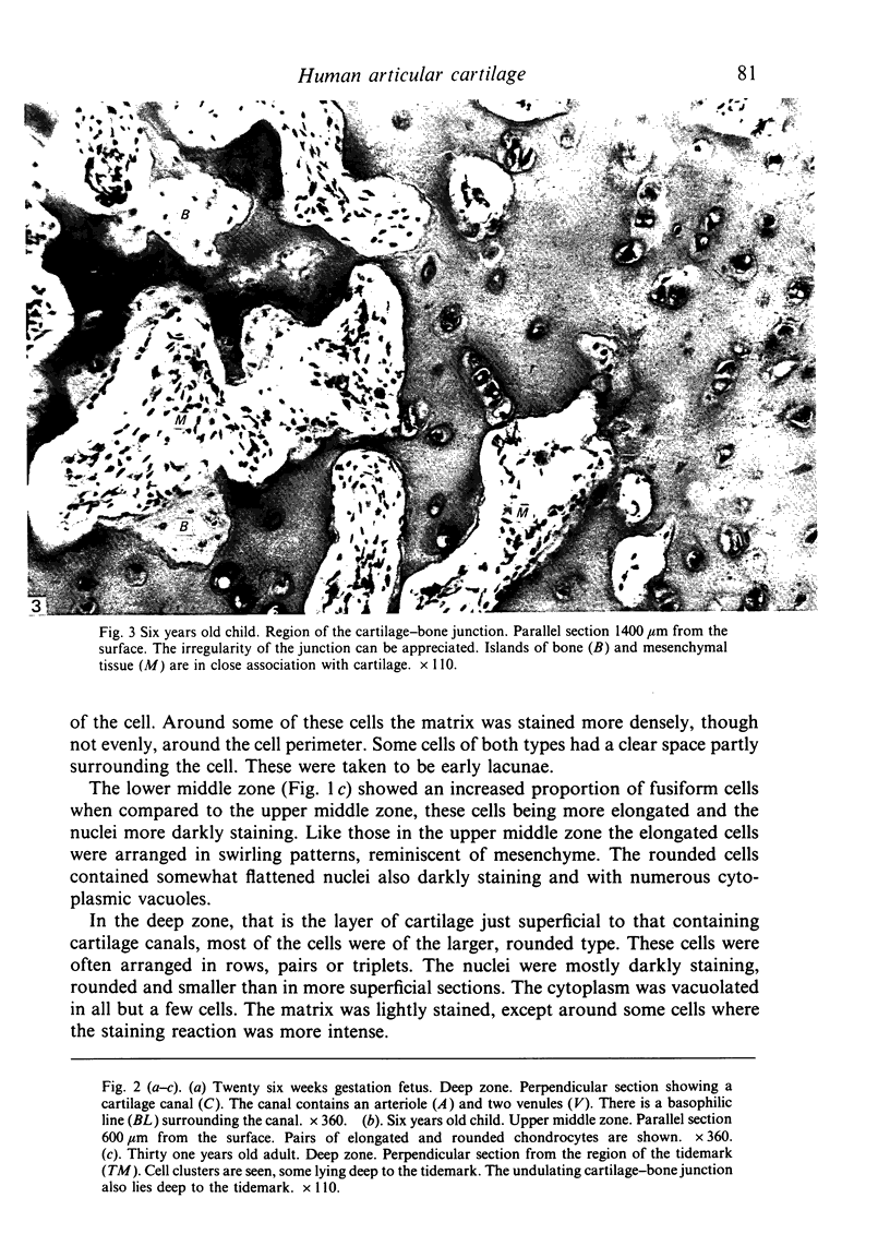
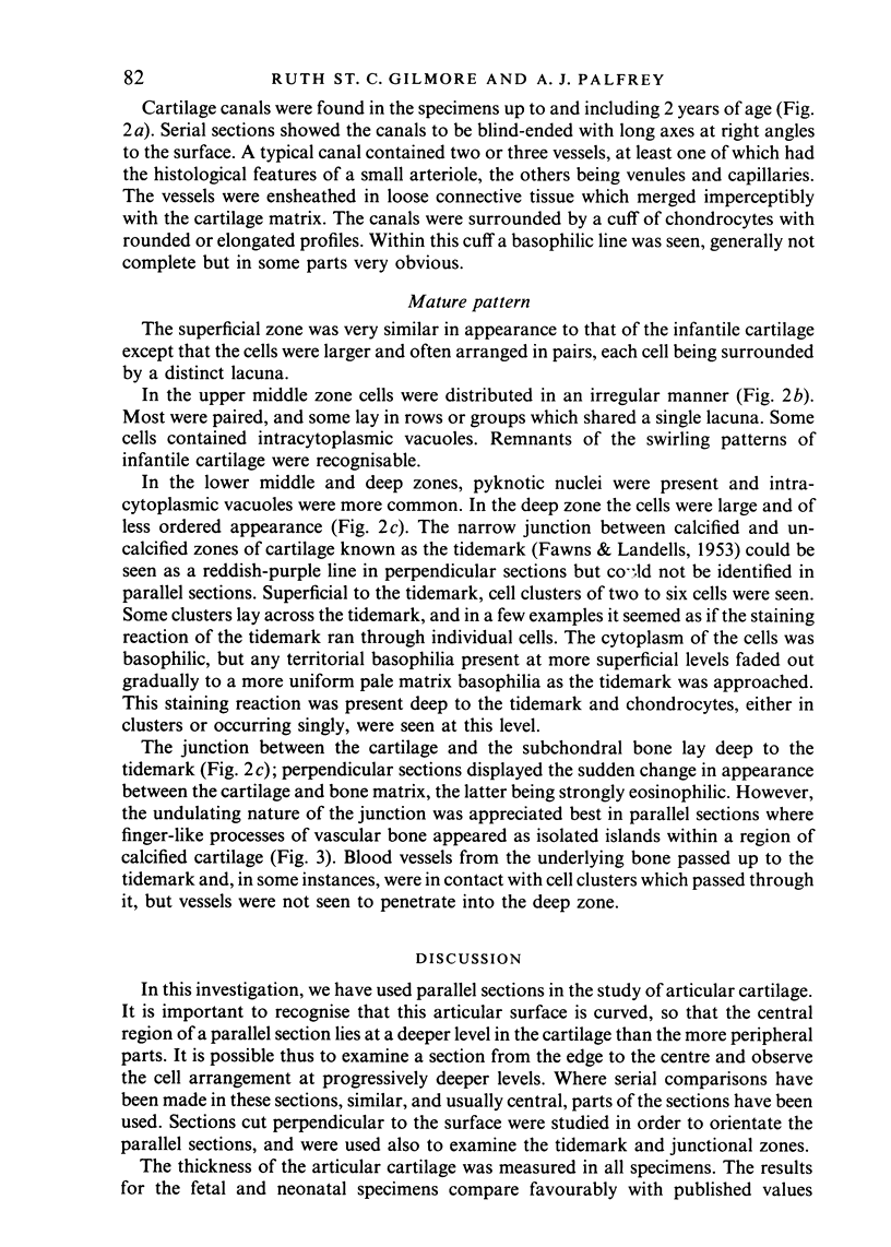
Paraffin sections cut both parallel to and perpendicular to the surface were used to study the histological structure of the articular cartilage of the lateral femoral condyle of infants, children and adults. Two main cell types were present--fusiform chondrocytes lying in swirling patterns were the predominant cell type of the cartilage up to and including two years, while a more rounded cell, randomly arranged, was commoner in the older specimens. Evidence suggests that these cells represent two distinct populations of chondrocyte, the round cells being derived from the earlier fusiform cells. Cartilage canals were a feature of the deeper regions of the presumptive articular cartilage in young specimens in which the epiphysis was still cartilaginous. The basophilic tidemark which marks the junction between the calcified and uncalcified cartilage in perpendicular sections was not seen in parallel sections. The calcified cartilage layer contained numerous processes of vascular bone which extended up from the subchondral bone and were a characteristic feature of the cartilage-bone interface. This layer of the articular cartilage cannot therefore be considered to be truly avascular.
Full text
PDF
Images in this article
Selected References
These references are in PubMed. This may not be the complete list of references from this article.
- DAVIES D. V., BARNETT C. H., COCHRANE W., PALFREY A. J. Electron microscopy of articular cartilage in the young adult rabbit. Ann Rheum Dis. 1962 Mar;21:11–22. doi: 10.1136/ard.21.1.11. [DOI] [PMC free article] [PubMed] [Google Scholar]
- FAWNS H. T., LANDELLS J. W. Histochemical studies of rheumatic conditions. I. Observations on the fine structures of the matrix of normal bone and cartilage. Ann Rheum Dis. 1953 Jun;12(2):105–113. doi: 10.1136/ard.12.2.105. [DOI] [PMC free article] [PubMed] [Google Scholar]
- Haines R. W. Cartilage Canals. J Anat. 1933 Oct;68(Pt 1):45–64. [PMC free article] [PubMed] [Google Scholar]
- Haines R. W. Growth of Cartilage Canals in the Patella. J Anat. 1937 Jul;71(Pt 4):471–479. [PMC free article] [PubMed] [Google Scholar]
- Hurrell D. J. The Vascularisation of Cartilage. J Anat. 1934 Oct;69(Pt 1):47–61. [PMC free article] [PubMed] [Google Scholar]
- Maroudas A., Bullough P., Swanson S. A., Freeman M. A. The permeability of articular cartilage. J Bone Joint Surg Br. 1968 Feb;50(1):166–177. [PubMed] [Google Scholar]
- Mital M. A., Millington P. F. Osseous pathway of nutrition to articular cartilage of the human femoral head. Lancet. 1970 Apr 18;1(7651):842–842. doi: 10.1016/s0140-6736(70)92443-8. [DOI] [PubMed] [Google Scholar]
- Redler I., Mow V. C., Zimny M. L., Mansell J. The ultrastructure and biomechanical significance of the tidemark of articular cartilage. Clin Orthop Relat Res. 1975 Oct;(112):357–362. [PubMed] [Google Scholar]
- Stockwell R. A. The interrelationship of cell density and cartilage thickness in mammalian articular cartilage. J Anat. 1971 Sep;109(Pt 3):411–421. [PMC free article] [PubMed] [Google Scholar]
- Wilsman N. J., Van Sickle D. C. Cartilage canals, their morphology and distribution. Anat Rec. 1972 May;173(1):79–93. doi: 10.1002/ar.1091730107. [DOI] [PubMed] [Google Scholar]


