Abstract
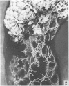
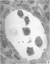
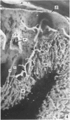
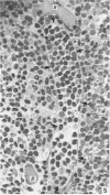
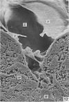
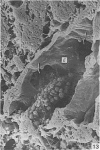
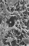
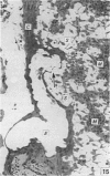
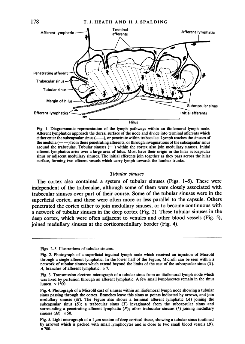
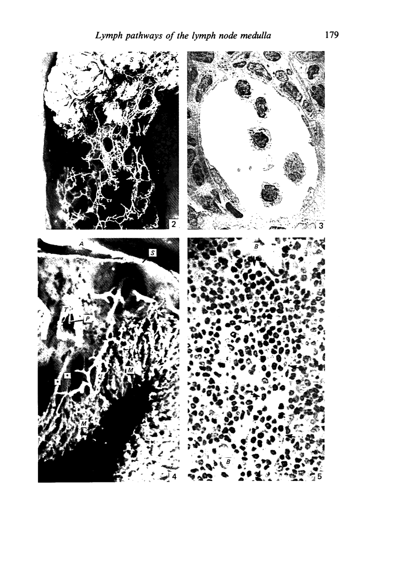
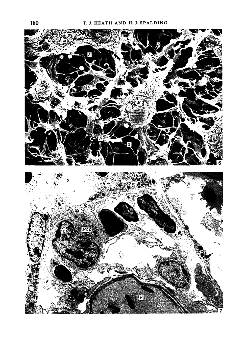
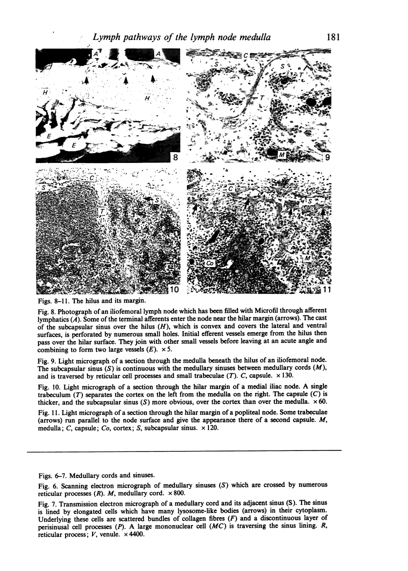
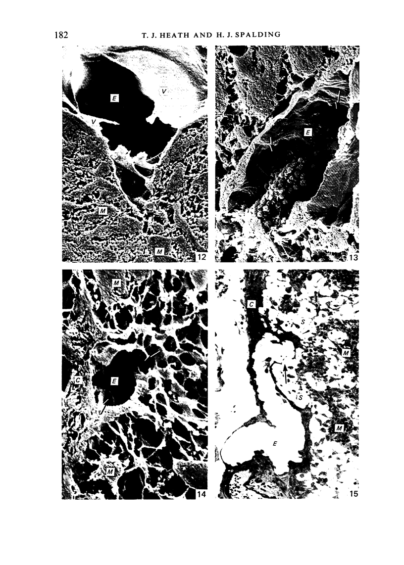
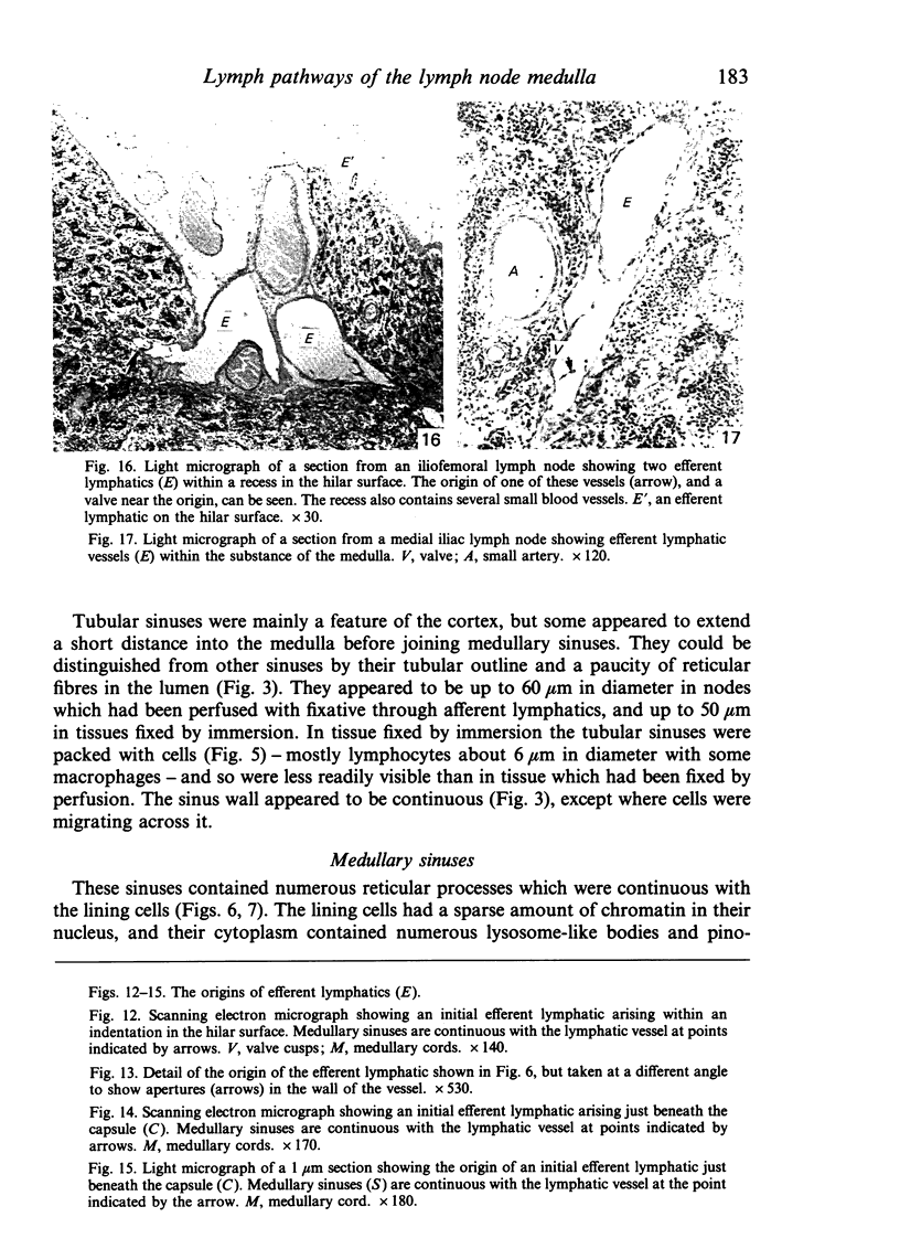
Medullary sinuses are continuous with penetrating afferent lymphatics, and with the trabecular and tubular sinuses which penetrate through the cortex. Tubular sinuses are often associated with blood vessels, especially in the deep cortex, and they appear to be important in the transport of lymphocytes. The subcapsular sinus is continuous over the cortex and the medulla, although trabeculae and reticular processes appear to restrict the flow of afferent lymph to the subcapsular sinus over the medulla. Lymph leaves the medulla through up to 100 or more initial efferent lymphatics, some only 60 micron across. Almost all of these arise from sinuses adjacent to the capsule lining the hilus. Some efferents remain associated with the capsule for a short distance whereas others, especially in nodes with a deep hilar depression, leave immediately at an angle of 30-90 degrees.
Full text
PDF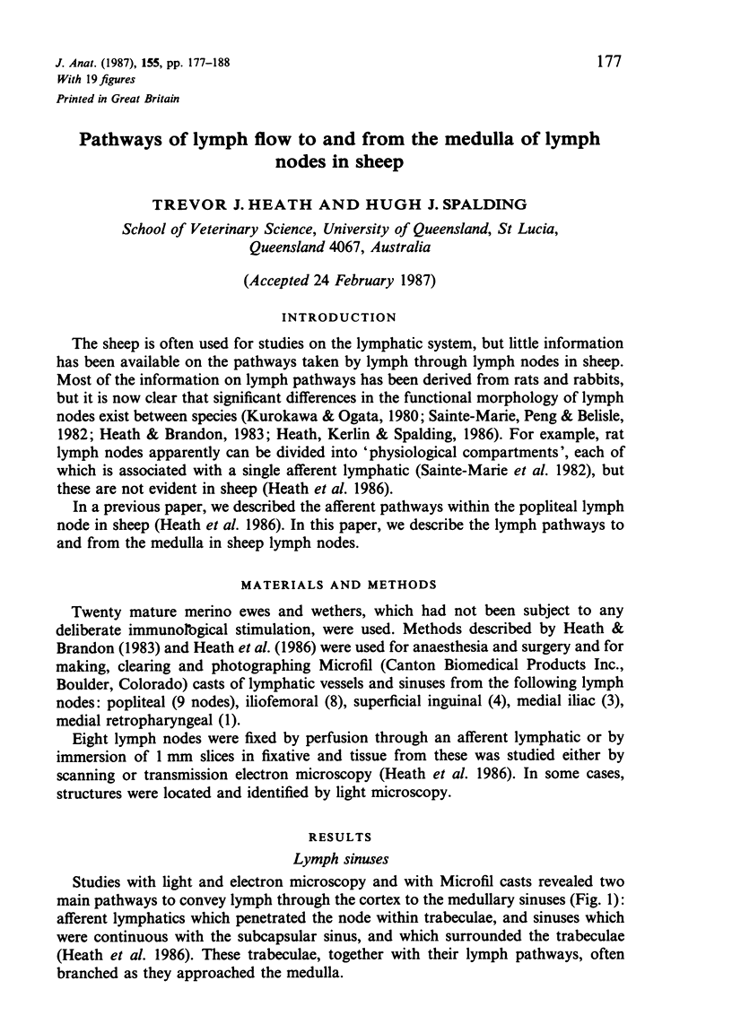
Images in this article
Selected References
These references are in PubMed. This may not be the complete list of references from this article.
- Farr A. G., Cho Y., De Bruyn P. P. The structure of the sinus wall of the lymph node relative to its endocytic properties and transmural cell passage. Am J Anat. 1980 Mar;157(3):265–284. doi: 10.1002/aja.1001570304. [DOI] [PubMed] [Google Scholar]
- Forkert P. G., Thliveris J. A., Bertalanffy F. D. Structure of sinuses in the human lymph node. Cell Tissue Res. 1977 Sep 14;183(1):115–130. doi: 10.1007/BF00219996. [DOI] [PubMed] [Google Scholar]
- Heath T. J., Kerlin R. L. Lymph drainage from the mammary gland in sheep. J Anat. 1986 Feb;144:61–70. [PMC free article] [PubMed] [Google Scholar]
- Heath T. J., Kerlin R. L., Spalding H. J. Afferent pathways of lymph flow within the popliteal node in sheep. J Anat. 1986 Dec;149:65–75. [PMC free article] [PubMed] [Google Scholar]
- Heath T., Brandon R. Lymphatic and blood vessels of the popliteal node in sheep. Anat Rec. 1983 Nov;207(3):461–472. doi: 10.1002/ar.1092070308. [DOI] [PubMed] [Google Scholar]
- Kelly R. H. Functional anatomy of lymph nodes. I. The paracortical cords. Int Arch Allergy Appl Immunol. 1975;48(6):836–849. doi: 10.1159/000231371. [DOI] [PubMed] [Google Scholar]
- Kurokawa T., Ogata T. A scanning electron microscopic study on the lymphatic microcirculation of the rabbit mesenteric lymph node. A corrosion cast study. Acta Anat (Basel) 1980;107(4):439–466. doi: 10.1159/000145272. [DOI] [PubMed] [Google Scholar]
- Sainte-Marie G., Peng F. S., Bélisle C. Overall architecture and pattern of lymph flow in the rat lymph node. Am J Anat. 1982 Aug;164(4):275–309. doi: 10.1002/aja.1001640402. [DOI] [PubMed] [Google Scholar]


