

Abstract
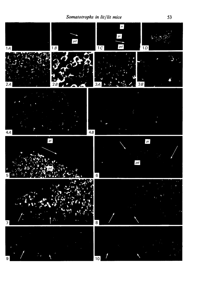
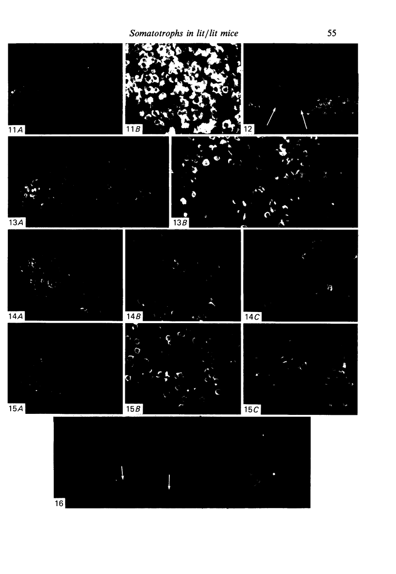
Regional and sexual patterns in the distribution and density of somatotroph cells exposed to anti-growth hormone serum were analysed by means of immunofluorescence histochemistry in adenohypophyses of normal C57BL mice and abnormal (lit/lit) mutant mice, which exhibit postnatal growth deficits. In adult (3-4 months) lit/lit mice, the regional distribution of somatotrophs both in males and females was normal; however, there was a sparsity of somatotrophs, relative to the normal condition, in the lateral wings of the pars distalis, and sexual differences in the concentration of immunoreactive cells were not as prominent as in the normal mice. In the midline region of the pars distalis a cranioventral zone virtually devoid of somatotrophs occurred in lit/lit as well as in normal mice, especially in the females, though it was not as well defined in lit/lit because of the overall sparsity of somatotrophs. In normal immature mice at 8 and 14 days after birth, the lateral wings did not show the striking sexual differences in density of somatotroph distribution as they did in the normal adults, and at 8 days they were more sparsely populated with somatotrophs than at 14 days. In 14 day lit/lit mice, the lateral wings were less densely populated with somatotrophs than their normal counterparts, but at 8 days these differences were not detectable. In both normal and abnormal 8 day mice, the medial and midline regions of the pars distalis contained less intensely immunoreactive somatotrophs than did the lateral wings.(ABSTRACT TRUNCATED AT 250 WORDS)
Full text
PDF
Images in this article

Selected References
These references are in PubMed. This may not be the complete list of references from this article.
- Baker B. L., Gross D. S. Cytology and distribution of secretory cell types in the mouse hypophysis as demonstrated with immunocytochemistry. Am J Anat. 1978 Oct;153(2):193–215. doi: 10.1002/aja.1001530203. [DOI] [PubMed] [Google Scholar]
- Baker B. L., Midgley A. R., Jr, Gersten B. E., Yu Y. Y. Differentiation of growth hormone- and prolactin-containing acidophils with peroxidase-labeled antibody. Anat Rec. 1969 Jun;164(2):163–171. doi: 10.1002/ar.1091640204. [DOI] [PubMed] [Google Scholar]
- Beamer W. H., Eicher E. M. Stimulation of growth in the little mouse. J Endocrinol. 1976 Oct;71(1):37–45. doi: 10.1677/joe.0.0710037. [DOI] [PubMed] [Google Scholar]
- Cheng T. C., Beamer W. G., Phillips J. A., 3rd, Bartke A., Mallonee R. L., Dowling C. Etiology of growth hormone deficiency in little, Ames, and Snell dwarf mice. Endocrinology. 1983 Nov;113(5):1669–1678. doi: 10.1210/endo-113-5-1669. [DOI] [PubMed] [Google Scholar]
- Christensen E., Wilson D. B. Fine structure of somatotrophs and mammotrophs in the pituitary pars distalis of the little (lit) mutant mouse. Virchows Arch B Cell Pathol Incl Mol Pathol. 1981;37(1):89–96. doi: 10.1007/BF02892558. [DOI] [PubMed] [Google Scholar]
- Dearden N. M., Holmes R. L. Cyto-differentiation and portal vascular development in the mouse adenohypophysis. J Anat. 1976 Jul;121(Pt 3):551–569. [PMC free article] [PubMed] [Google Scholar]
- Eicher E. M., Beamer W. G. Inherited ateliotic dwarfism in mice. Characteristics of the mutation, little, on chromosome 6. J Hered. 1976 Mar-Apr;67(2):87–91. doi: 10.1093/oxfordjournals.jhered.a108682. [DOI] [PubMed] [Google Scholar]
- Jansson J. O., Downs T. R., Beamer W. G., Frohman L. A. Receptor-associated resistance to growth hormone-releasing factor in dwarf "little" mice. Science. 1986 Apr 25;232(4749):511–512. doi: 10.1126/science.3008329. [DOI] [PubMed] [Google Scholar]
- Nakane P. K. Classifications of anterior pituitary cell types with immunoenzyme histochemistry. J Histochem Cytochem. 1970 Jan;18(1):9–20. doi: 10.1177/18.1.9. [DOI] [PubMed] [Google Scholar]
- Poole M. C., Kornegay W. D., 3rd Cellular distribution within the rat adenohypophysis: a morphometric study. Anat Rec. 1982 Sep;204(1):45–53. doi: 10.1002/ar.1092040107. [DOI] [PubMed] [Google Scholar]
- Roux M., Bartke A., Dumont F., Dubois M. P. Immunohistological study of the anterior pituitary gland - pars distalis and pars intermedia - in dwarf mice. Cell Tissue Res. 1982;223(2):415–420. doi: 10.1007/BF01258498. [DOI] [PubMed] [Google Scholar]
- Watanabe Y. G. An immunohistochemical study on the mouse adenohypophysis with reference to the spatial relationship between GH cells and other types of hormone-producing cells. Anat Embryol (Berl) 1985;172(3):277–280. doi: 10.1007/BF00318975. [DOI] [PubMed] [Google Scholar]
- Watanabe Y. G., Daikoku S. An immunohistochemical study on the cytogenesis of adenohypophysial cells in fetal rats. Dev Biol. 1979 Feb;68(2):557–567. doi: 10.1016/0012-1606(79)90226-4. [DOI] [PubMed] [Google Scholar]
- Wilson D. B., Christensen E. Fine structure of somatotrophs and mammotrophs during development of the dwarf (dw) mutant mouse. J Anat. 1981 Oct;133(Pt 3):407–417. [PMC free article] [PubMed] [Google Scholar]
- Wilson D. B., Wyatt D. P. Growth hormone and prolactin immunoreactivity in the pituitary gland of postnatal little (lit) mice. Histol Histopathol. 1986 Oct;1(4):309–313. [PubMed] [Google Scholar]
- YAMADA K., SANO M., ITO T. A postnatal histogenetic study of the anterior pituitary of the mouse. Okajimas Folia Anat Jpn. 1957 Aug;30(2-3):177–195. doi: 10.2535/ofaj1936.30.2-3_177. [DOI] [PubMed] [Google Scholar]