

Abstract
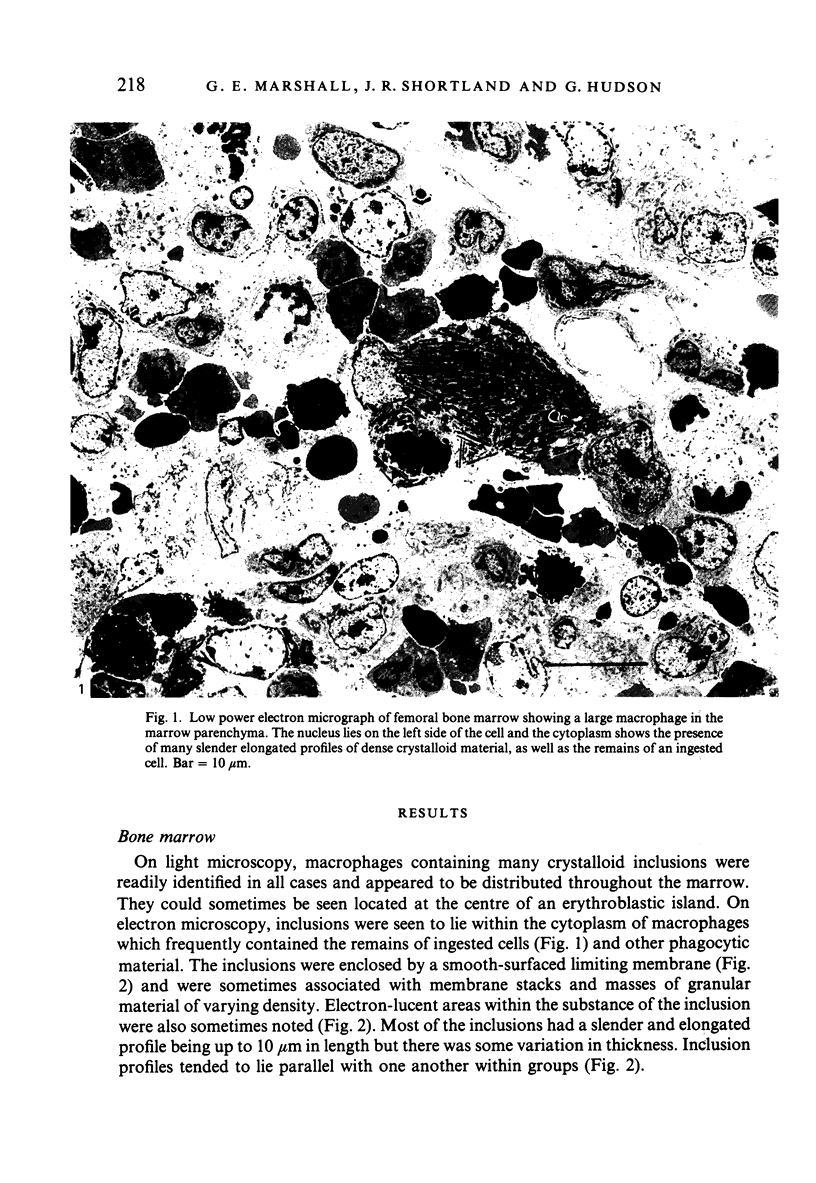
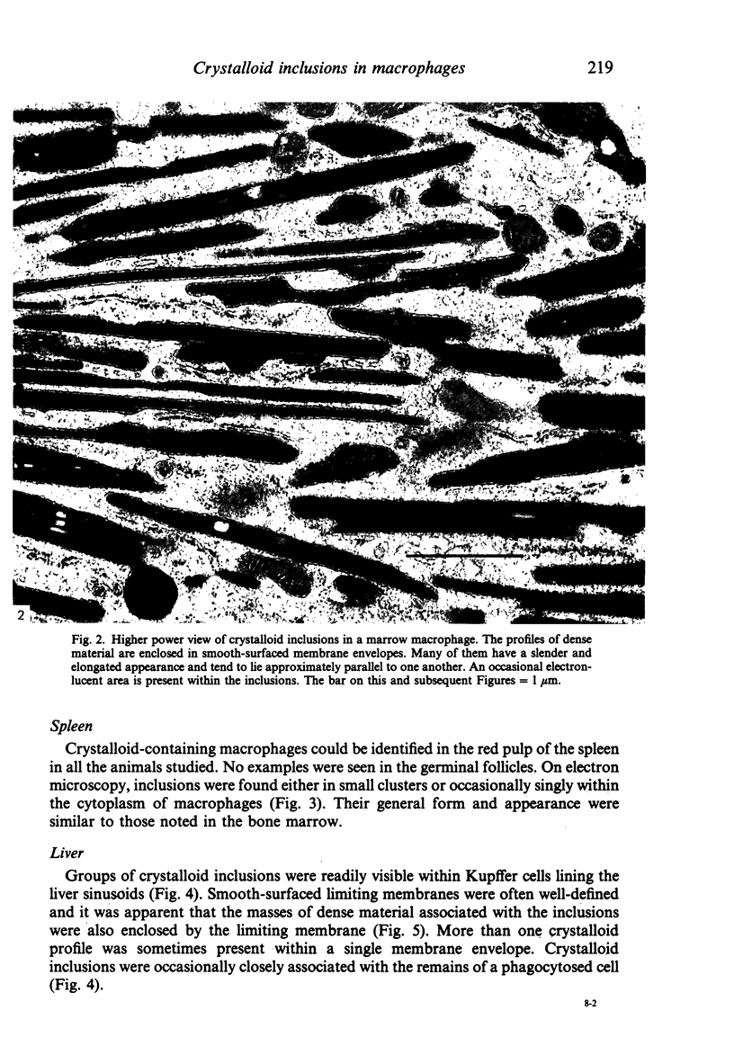
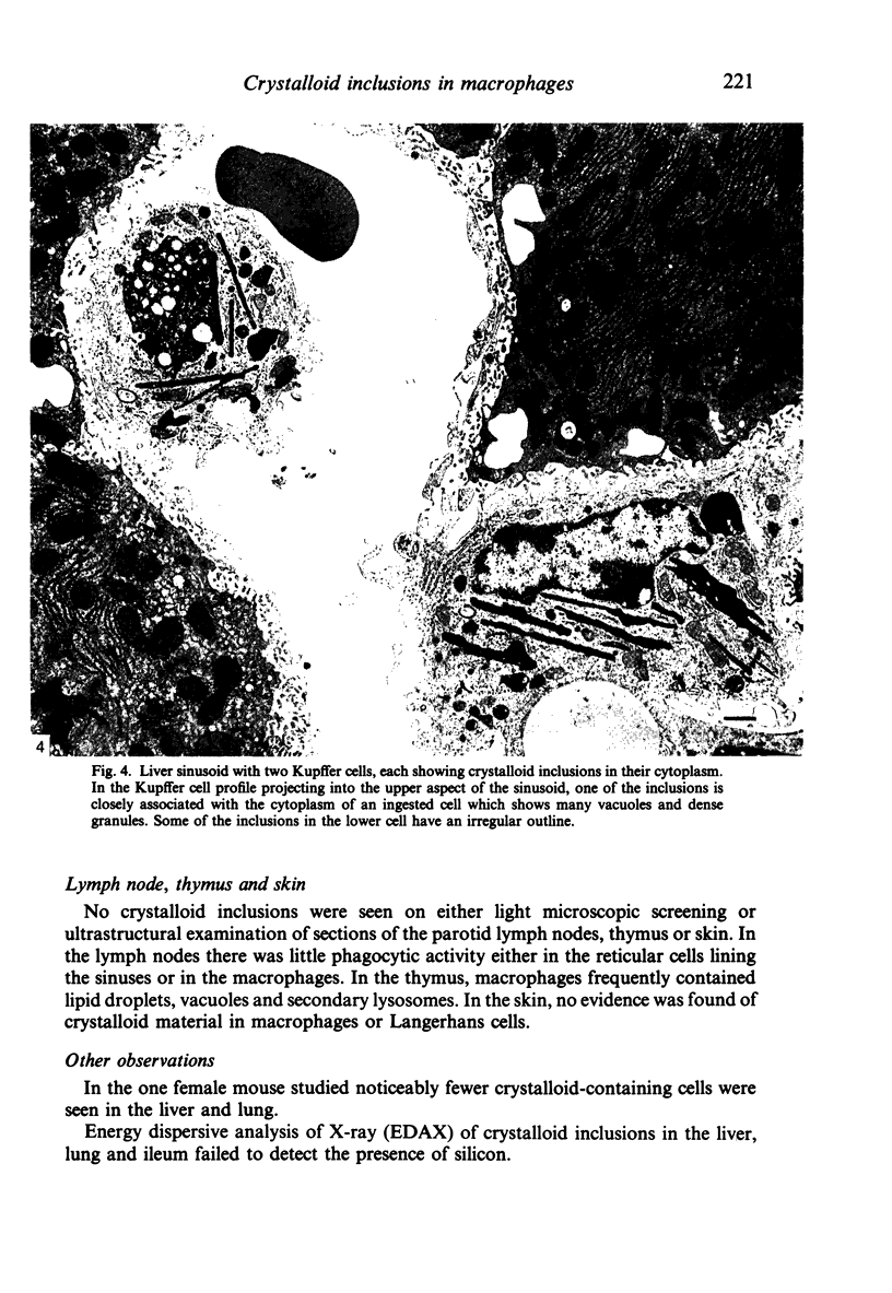
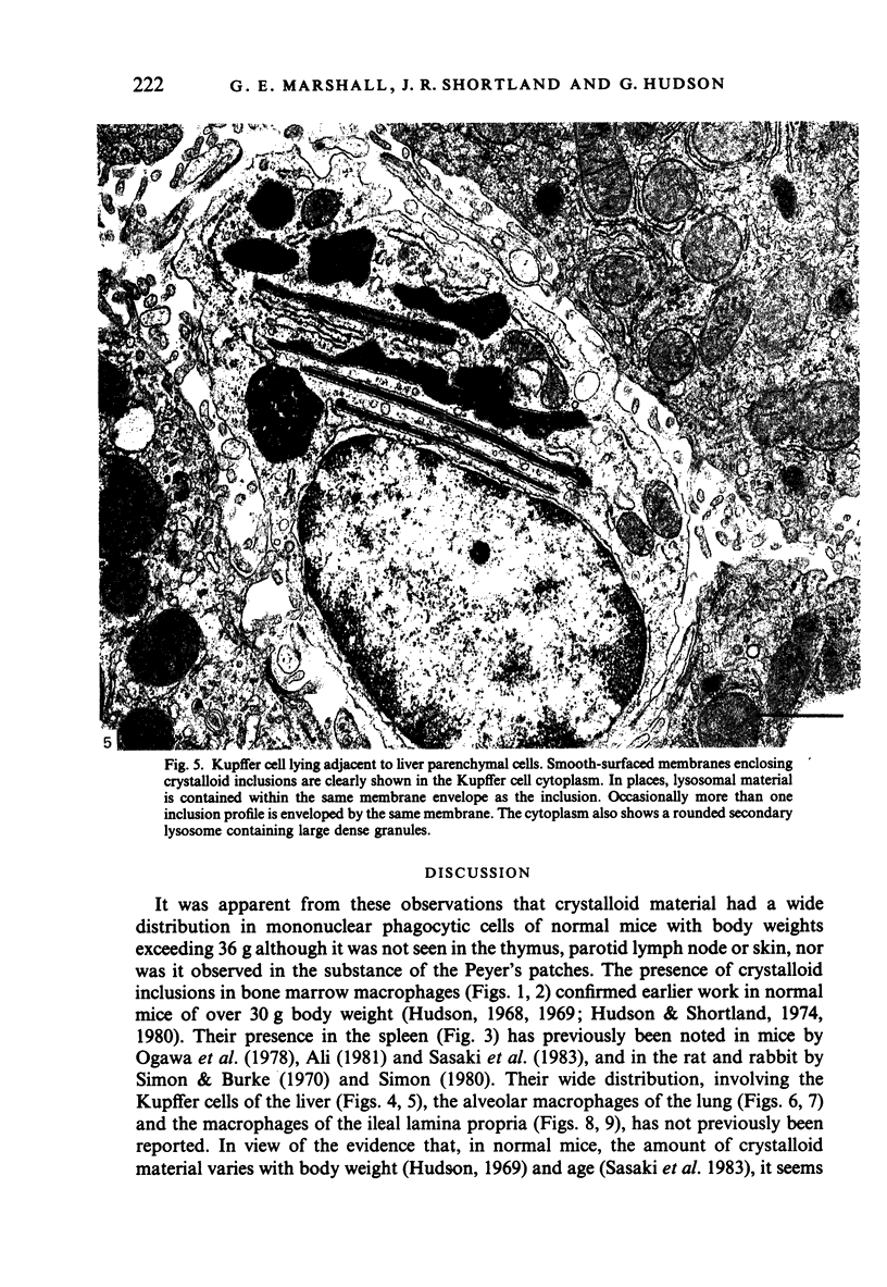
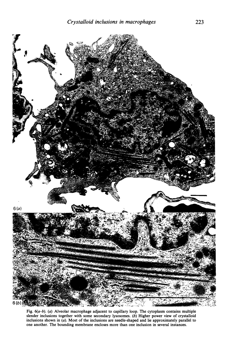
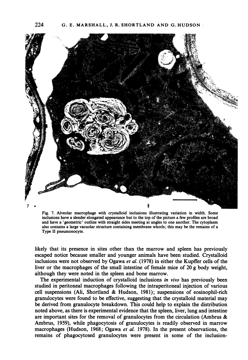
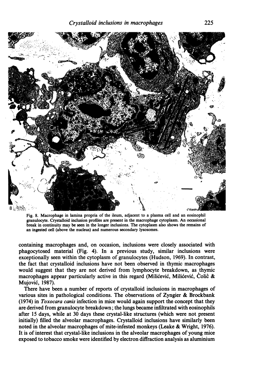
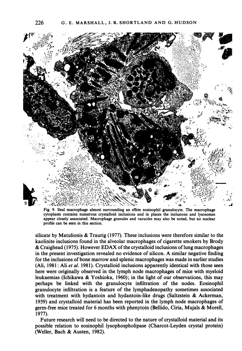
In an attempt to throw further light on the nature and distribution of crystalloid material in macrophages, tissues from a variety of locations in 9 normal adult mice of 36-46 g body weight were studied by light and electron microscopy. Crystalloid inclusions were widely distributed in mononuclear phagocytic cells, being present in macrophages of the bone marrow, spleen, lung and ileum and in the Kupffer cells of the liver. They were not observed in the macrophages of the thymus, parotid lymph node or skin nor were they seen within the lymphatic nodules of Peyer's patches. The observed distribution of crystalloid material seems consistent with its suggested origin from granulocyte breakdown.
Full text
PDF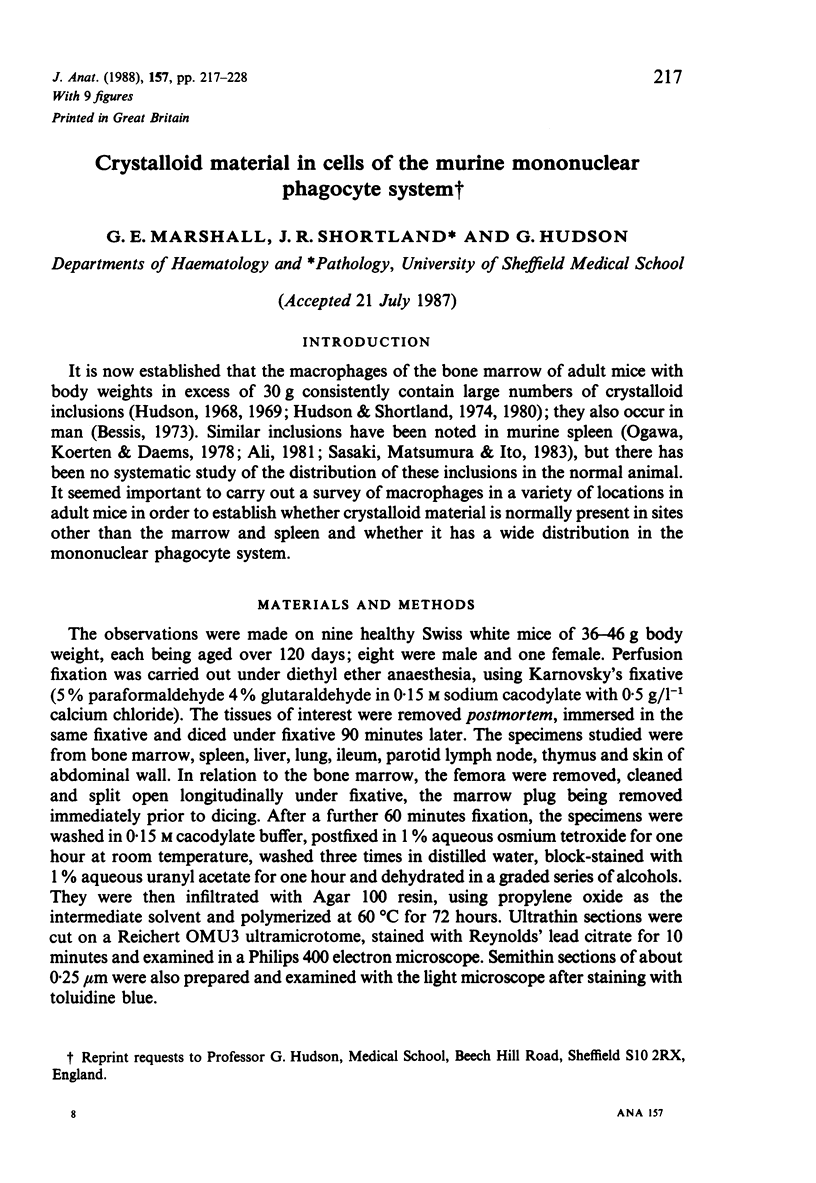
Images in this article
Selected References
These references are in PubMed. This may not be the complete list of references from this article.
- AMBRUS C. M., AMBRUS J. L. Regulation of the leukocyte level. Ann N Y Acad Sci. 1959 Jun 25;77:445–486. doi: 10.1111/j.1749-6632.1959.tb36920.x. [DOI] [PubMed] [Google Scholar]
- Ali B. A., Shortland J. R., Hudson G. Origin of crystalloid inclusions in macrophages II: evidence for derivation from eosinophil granulocyte breakdown. Br J Exp Pathol. 1981 Dec;62(6):662–668. [PMC free article] [PubMed] [Google Scholar]
- Bellido P. D., Ciria M. D., Mujals D. R., Morell A. R. Lymphadenopathies and phenytoin. Lancet. 1977 Jun 25;1(8026):1372–1373. doi: 10.1016/s0140-6736(77)92594-6. [DOI] [PubMed] [Google Scholar]
- Brody A. R., Craighead J. E. Cytoplasmic inclusions in pulmonary macrophages of cigarette smokers. Lab Invest. 1975 Feb;32(2):125–132. [PubMed] [Google Scholar]
- Hudson G. Crystalloid material in macrophages of mouse bone marrow. Acta Anat (Basel) 1968;71(1):100–107. doi: 10.1159/000143176. [DOI] [PubMed] [Google Scholar]
- Hudson G., Shortland J. R. Crystalloid material in marrow macrophages of specific pathogen-free mice. Acta Anat (Basel) 1974;87(3):404–408. doi: 10.1159/000144188. [DOI] [PubMed] [Google Scholar]
- Hudson G. Variations in the amount of crystalloid material in marrow macrophages in mice of different body weights. Acta Anat (Basel) 1969;73(1):136–141. doi: 10.1159/000143290. [DOI] [PubMed] [Google Scholar]
- Leake E. S., Wright M. J. The fine structure of lung macrophages from rhesus and squirrel monkeys, with special reference to the large numbers of mitochondria. Am Rev Respir Dis. 1976 Sep;114(3):581–592. doi: 10.1164/arrd.1976.114.3.581. [DOI] [PubMed] [Google Scholar]
- Matulionis D. H., Traurig H. H. In situ response of lung macrophages and hydrolase activities to cigarette smoke. Lab Invest. 1977 Sep;37(3):314–326. [PubMed] [Google Scholar]
- Milićević N. M., Milićević Z., Colic M., Mujović S. Ultrastructural study of macrophages in the rat thymus, with special reference to the cortico-medullary zone. J Anat. 1987 Feb;150:89–98. [PMC free article] [PubMed] [Google Scholar]
- Ogawa T., Koerten H. K., Daems W. T. Peroxidase activity in monocytes and tissue macrophages of mice. Cell Tissue Res. 1978 Apr 28;188(3):361–373. doi: 10.1007/BF00219778. [DOI] [PubMed] [Google Scholar]
- SALTZSTEIN S. L., ACKERMAN L. V. Lymphadenopathy induced by anticonvulsant drugs and mimicking clinically pathologically malignant lymphomas. Cancer. 1959 Jan-Feb;12(1):164–182. doi: 10.1002/1097-0142(195901/02)12:1<164::aid-cncr2820120122>3.0.co;2-y. [DOI] [PubMed] [Google Scholar]
- Sasaki K., Matsumura G., Ito T. Crystalloid inclusion-containing macrophages in the bone marrow and red pulp of the mouse, with particular relation to age, sex and hydrocortisone administration: qualitative and quantitative electron microscopy. Arch Histol Jpn. 1983 Jun;46(3):381–391. doi: 10.1679/aohc.46.381. [DOI] [PubMed] [Google Scholar]
- Simon G. T., Burke J. S. Electron microscopy of the spleen. 3. Erythro-leukophagocytosis. Am J Pathol. 1970 Mar;58(3):451–470. [PMC free article] [PubMed] [Google Scholar]
- Weller P. F., Bach D., Austen K. F. Human eosinophil lysophospholipase: the sole protein component of Charcot-Leyden crystals. J Immunol. 1982 Mar;128(3):1346–1349. [PubMed] [Google Scholar]
- Zyngier F. R., Brockbank A. Electron microscopy of the lung in experimental Toxocara canis infection. Ann Trop Med Parasitol. 1974 Jun;68(2):229–233. doi: 10.1080/00034983.1974.11686940. [DOI] [PubMed] [Google Scholar]