

Abstract
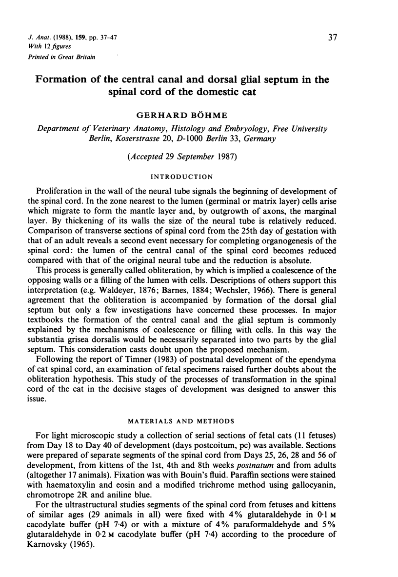
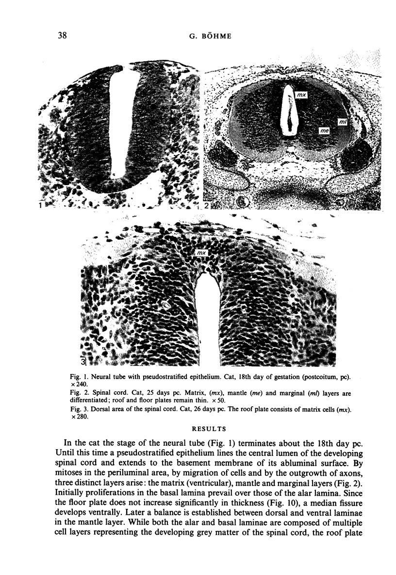
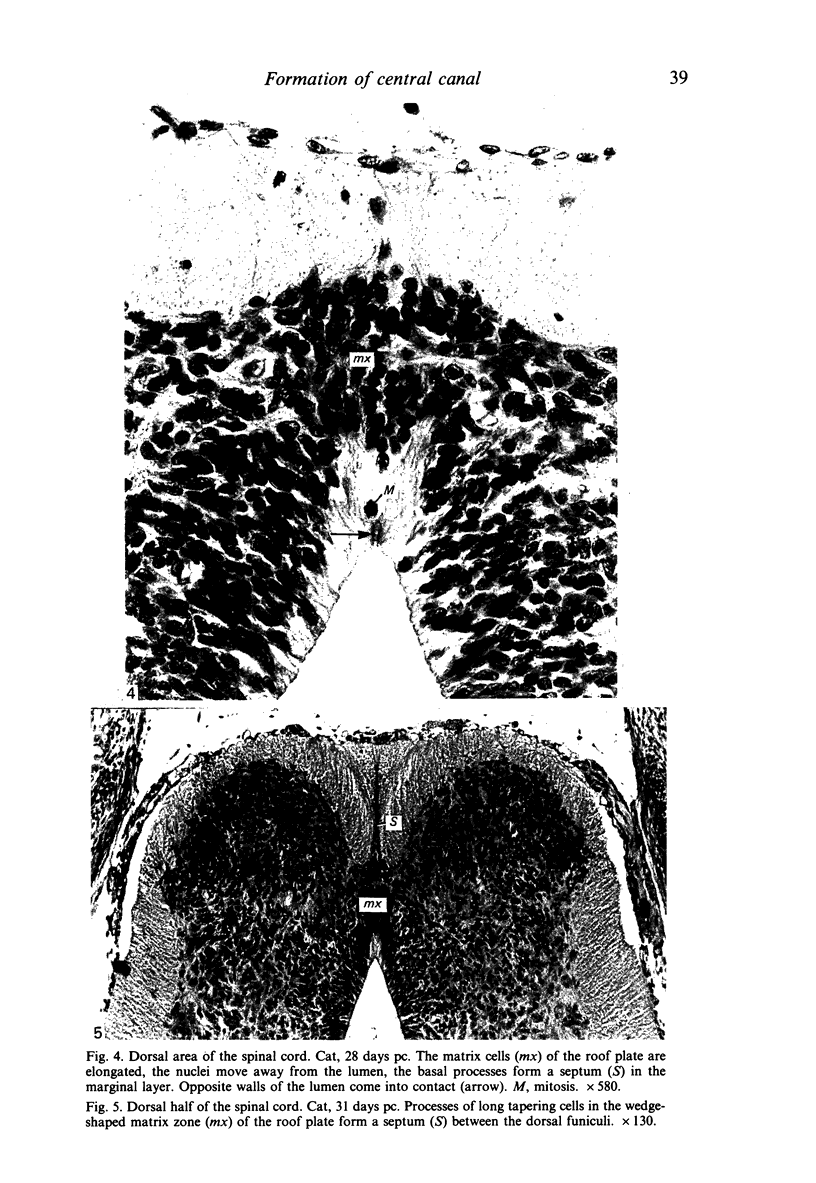
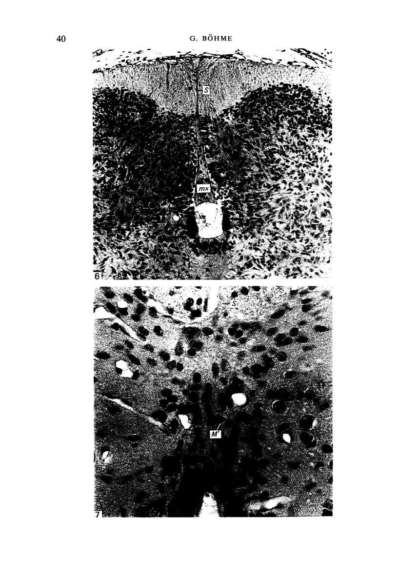
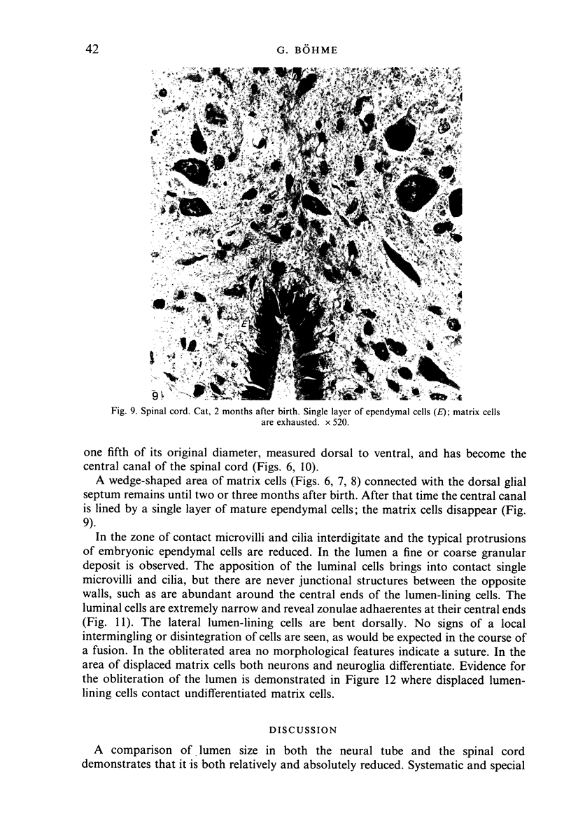
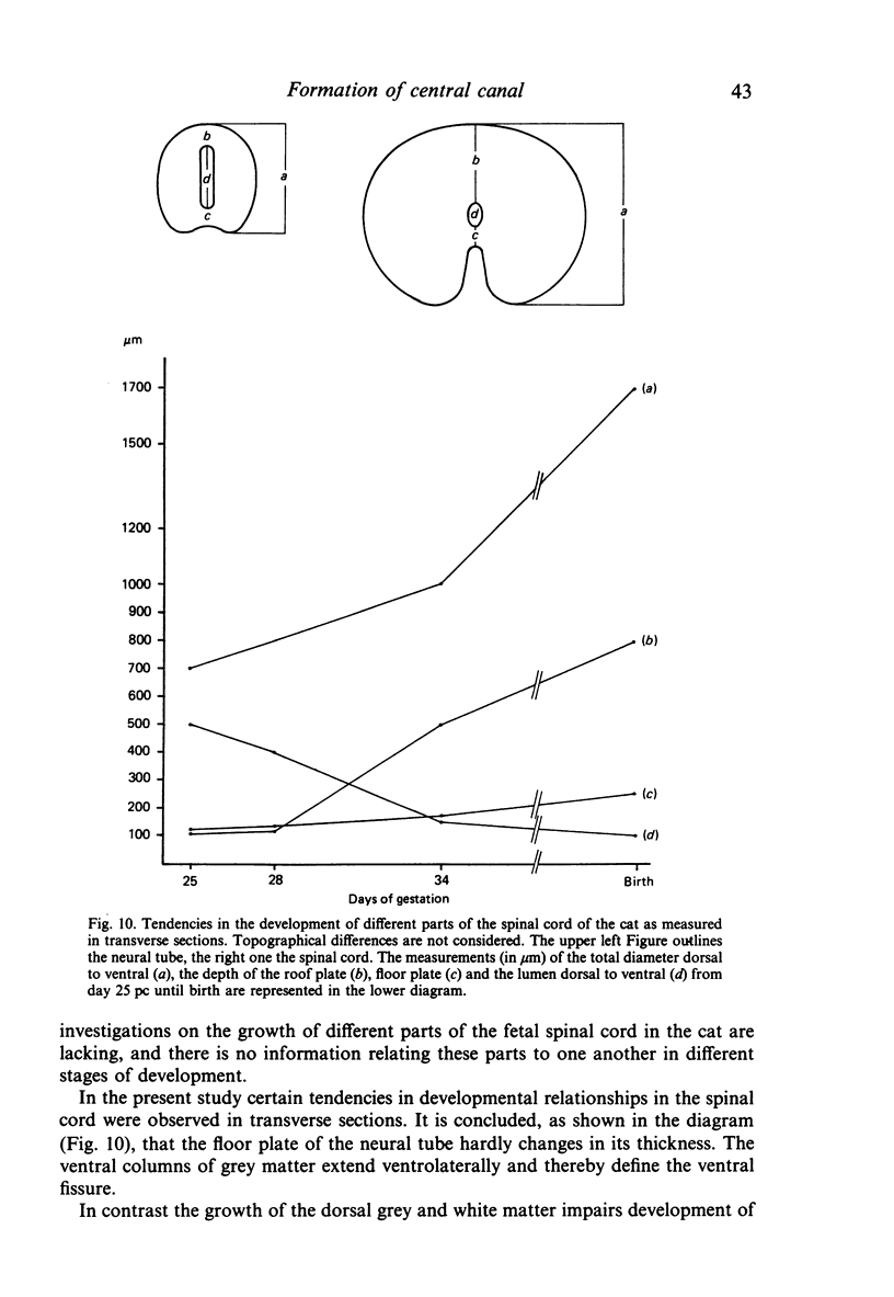
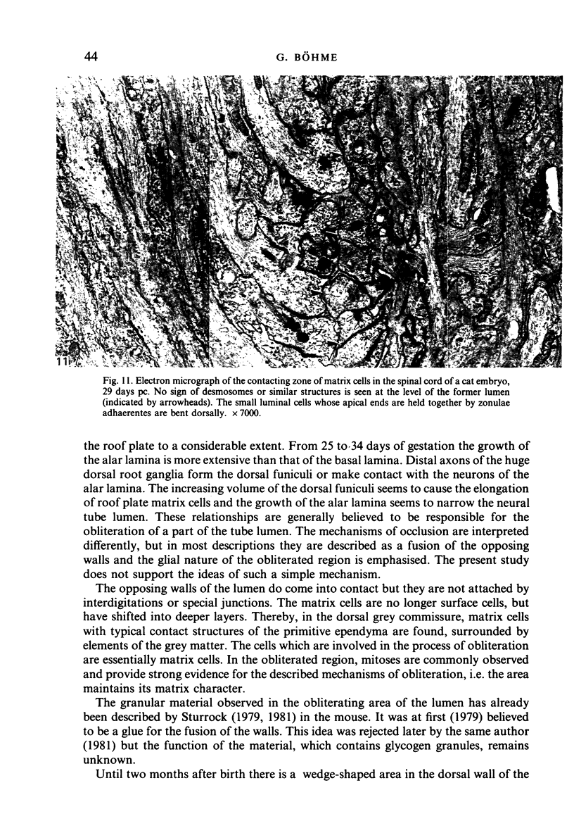
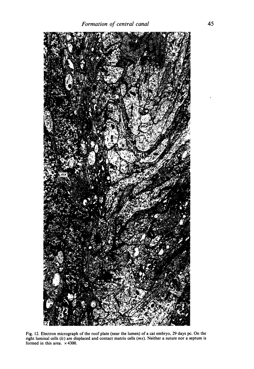
Development of the neural tube results in a relative reduction of its lumen accompanied by an increasing thickness of its wall. The central canal measures only about one fifth of that of the former neural canal. This has been said to be the result of an obliteration or fusion of a part of the lumen. This transformation of the central canal takes place between fetal days 28 and 34 in the cat and is characterised by an elongation and shifting of the dorsal ependymal matrix cells and by an apposition of the lateral walls in the same region. It is suggested that the increase in size of the dorsal funiculi causes the elongation of the ependymal cells, the basal processes of which remain to form the dorsal glial septum. The proliferation of neurons and the resultant growth of the dorsal grey horns is believed to be responsible for the narrowing of the lumen. The lumen-contacting matrix cells are displaced from the former surface. These 'blast' cells develop into neurons or glial cells. Until two or three months after birth there is a small wedge-shaped area in the dorsal wall of the central canal which consists of fetal matrix cells with long tapering basal processes extending into the glial septum. After this date the matrix is exhausted and the ependyma forms the complete lining of the surface of the central canal.
Full text
PDF

Images in this article

Selected References
These references are in PubMed. This may not be the complete list of references from this article.
- Sturrock R. R. A comparison of the processes of ventricular coarctation and choroid and ependymal fusion in the mouse brain. J Anat. 1979 Sep;129(Pt 2):235–242. [PMC free article] [PubMed] [Google Scholar]
- Sturrock R. R. An electron microscopic study of the development of the ependyma of the central canal of the mouse spinal cord. J Anat. 1981 Jan;132(Pt 1):119–136. [PMC free article] [PubMed] [Google Scholar]
- Wechsler W. Elektronenmikroskopischer beitrag zur Differenzierung des Ependyms am Rückenmark von Hühnerembryonen. Z Zellforsch Mikrosk Anat. 1966;74(3):423–442. [PubMed] [Google Scholar]