

Abstract
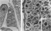
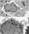
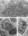
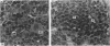
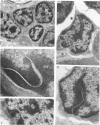
Development of splenic haemopoiesis and morphology of lymphocytes in the embryonic and neonatal mice were examined by light and electron microscopy. At 15 days of gestation, free mononuclear cells were scattered throughout the splenic anlage, and the spleen was prehaemopoietic. At 16 days of gestation, immature erythroid cells and small lymphocytes first appeared and then the spleen became predominantly erythropoietic. In the red pulp, lymphocytes constituted approximately 2% of haemopoietic cells during fetal life and 3.6% at 4 days after birth. Splenic lymphocytes in the embryo had a nucleus 3.5-4.5 microns in diameter, and the majority were small lymphocytes with sparse and dark cytoplasm. Small lymphocytes occasionally had an invagination of the inner nuclear membrane into the nucleoplasm, showing various sectional profiles; in ultrathin sections, the proportion of small lymphocytes having the nuclear membrane invagination comprised 14.3% of the small lymphocytes in the embryonic spleen.
Full text
PDF


Images in this article

Selected References
These references are in PubMed. This may not be the complete list of references from this article.
- Abe K., Sasaki K., Ito T. Comparative ultrastructure and cytometric analysis of small lymphocytes in haemopoietic organs of neonatal mice. J Anat. 1973 Sep;115(Pt 3):393–406. [PMC free article] [PubMed] [Google Scholar]
- BORGHESE E. The present state of research on WW mice. Acta Anat (Basel) 1959;36(3):185–220. doi: 10.1159/000141435. [DOI] [PubMed] [Google Scholar]
- Cline M. J., Moore M. A. Embryonic origin of the mouse macrophage. Blood. 1972 Jun;39(6):842–849. [PubMed] [Google Scholar]
- Djaldetti M., Bessler H., Rifkind R. A. Hematopoiesis in the embryonic mouse spleen: an electron microscopic study. Blood. 1972 Jun;39(6):826–841. [PubMed] [Google Scholar]
- Holyoke E. A., Latta J. S., McLean J. V. A study of the ultrastructure of the developing spleen in the albino rat. J Ultrastruct Res. 1966 Apr;15(1):87–99. doi: 10.1016/s0022-5320(66)80096-5. [DOI] [PubMed] [Google Scholar]
- Levine A. S., Nesbit M. E., White J. G., Yarbro J. W. Effects of fractionated histones on nucleic acid synthesis in 6C3HED mouse ascites tumor cells and in normal spleen cells. Cancer Res. 1968 May;28(5):831–844. [PubMed] [Google Scholar]
- Osmond D. G. The non-thymic origin of lymphocytes. Anat Rec. 1969 Sep;165(1):109–112. doi: 10.1002/ar.1091650114. [DOI] [PubMed] [Google Scholar]
- Parrott D. M., De Sousa M. Thymus-dependent and thymus-independent populations: origin, migratory patterns and lifespan. Clin Exp Immunol. 1971 May;8(5):663–684. [PMC free article] [PubMed] [Google Scholar]
- Sasaki K., Ito T. Effects of estrogen and progesterone on the spleen of the mouse: a light and electron microscopic study. Arch Histol Jpn. 1981 Jul;44(3):203–213. doi: 10.1679/aohc1950.44.203. [DOI] [PubMed] [Google Scholar]
- Sasaki K., Ito T. Effects of gonadectomy and testosterone on lymphocytes in the bone marrow of the mouse: an electron microscopic study. J Anat. 1980 Mar;130(Pt 2):429–438. [PMC free article] [PubMed] [Google Scholar]
- Sasaki K., Ito T. Effects of pregnancy and lactation on lymphocytes in the bone marrow of the mouse: a quantitative electron microscopic study. Arch Histol Jpn. 1980 Aug;43(3):211–219. doi: 10.1679/aohc1950.43.211. [DOI] [PubMed] [Google Scholar]
- Sasaki K., Ito T. Sex-related changes in the population of lymphocytes in the bone marrow of the mouse. An electron microscope study. Arch Histol Jpn. 1978 Nov;41(5):389–400. doi: 10.1679/aohc1950.41.389. [DOI] [PubMed] [Google Scholar]
- Sasaki K., Kendall M. D. The morphology of the haemopoietic cells of the yolk sac in mice with particular reference to nucleolar changes. J Anat. 1985 Mar;140(Pt 2):279–295. [PMC free article] [PubMed] [Google Scholar]
- Sasaki K., Matsumura G. Hemopoietic cells in the liver and spleen of the embryonic and early postnatal mouse: a karyometrical observation. Anat Rec. 1987 Dec;219(4):378–383. doi: 10.1002/ar.1092190408. [DOI] [PubMed] [Google Scholar]
- Sasaki K., Matsumura G., Ito T. Light and electron microscopy of bone marrow hemopoiesis in late embryonal and early postnatal mice: a qualitative and quantitative study. Arch Histol Jpn. 1984 Aug;47(3):239–250. doi: 10.1679/aohc.47.239. [DOI] [PubMed] [Google Scholar]
- Takahashi K., Takahashi H., Naito M., Sato T., Kojima M. Ultrastructural and functional development of macrophages in the dermal tissue of rat fetuses. Cell Tissue Res. 1983;232(3):539–552. doi: 10.1007/BF00216427. [DOI] [PubMed] [Google Scholar]
- Yoffey J. M., Hudson G., Osmond D. G. The lymphocyte in guinea-pig bone marrow. J Anat. 1965 Oct;99(Pt 4):841–860. [PMC free article] [PubMed] [Google Scholar]