Abstract
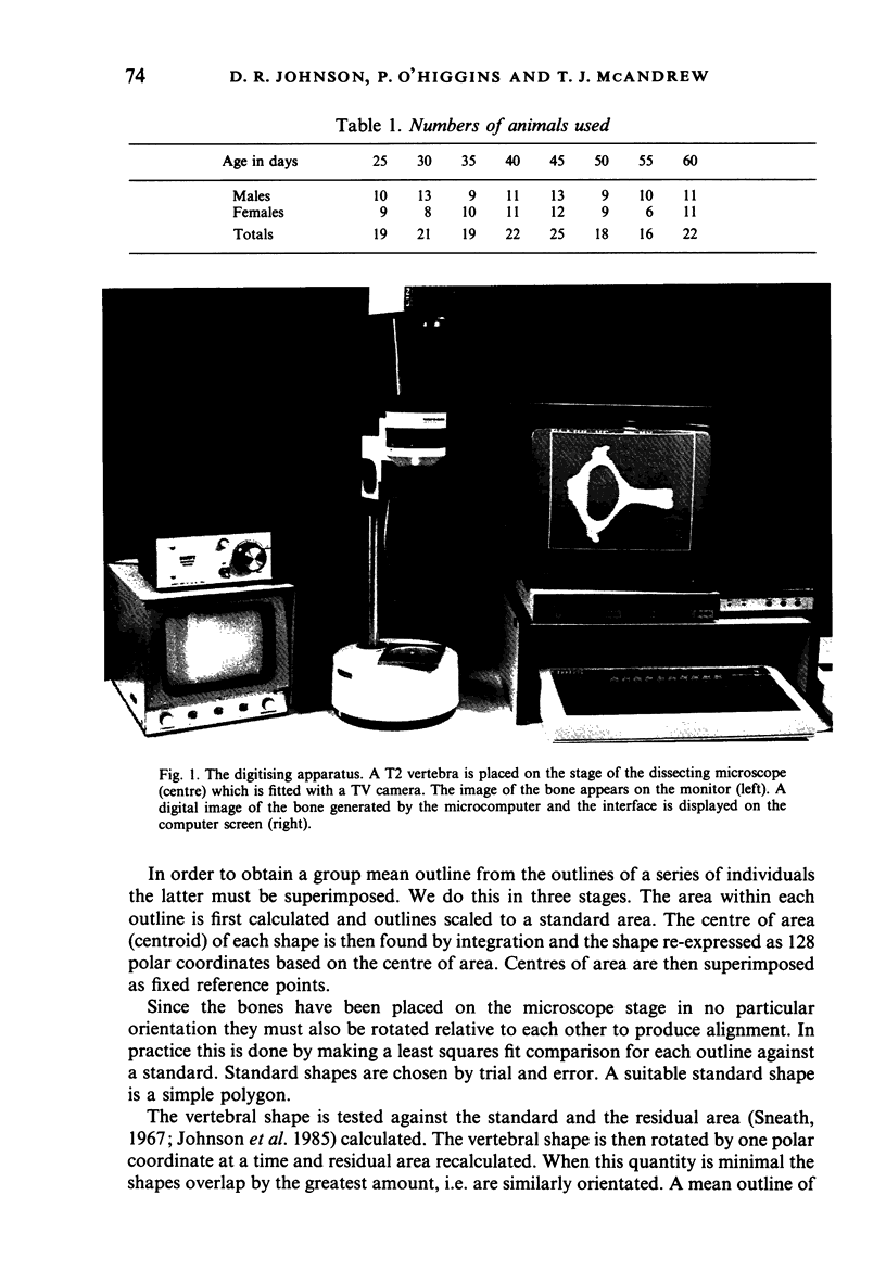
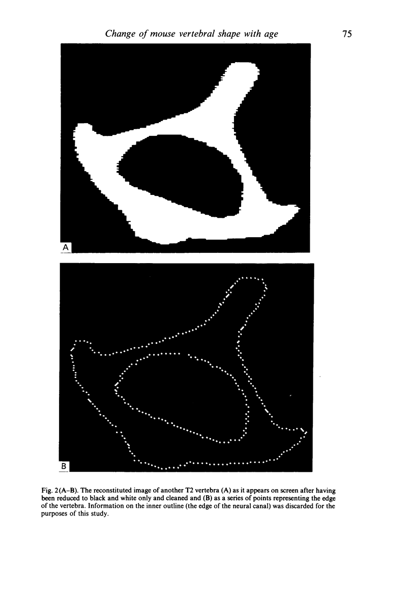
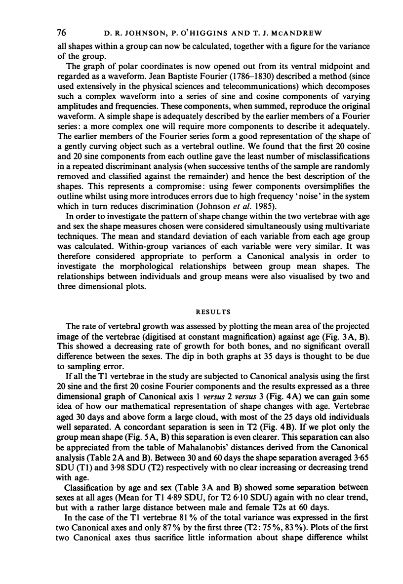
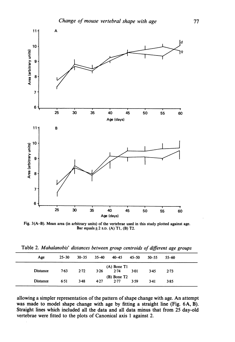
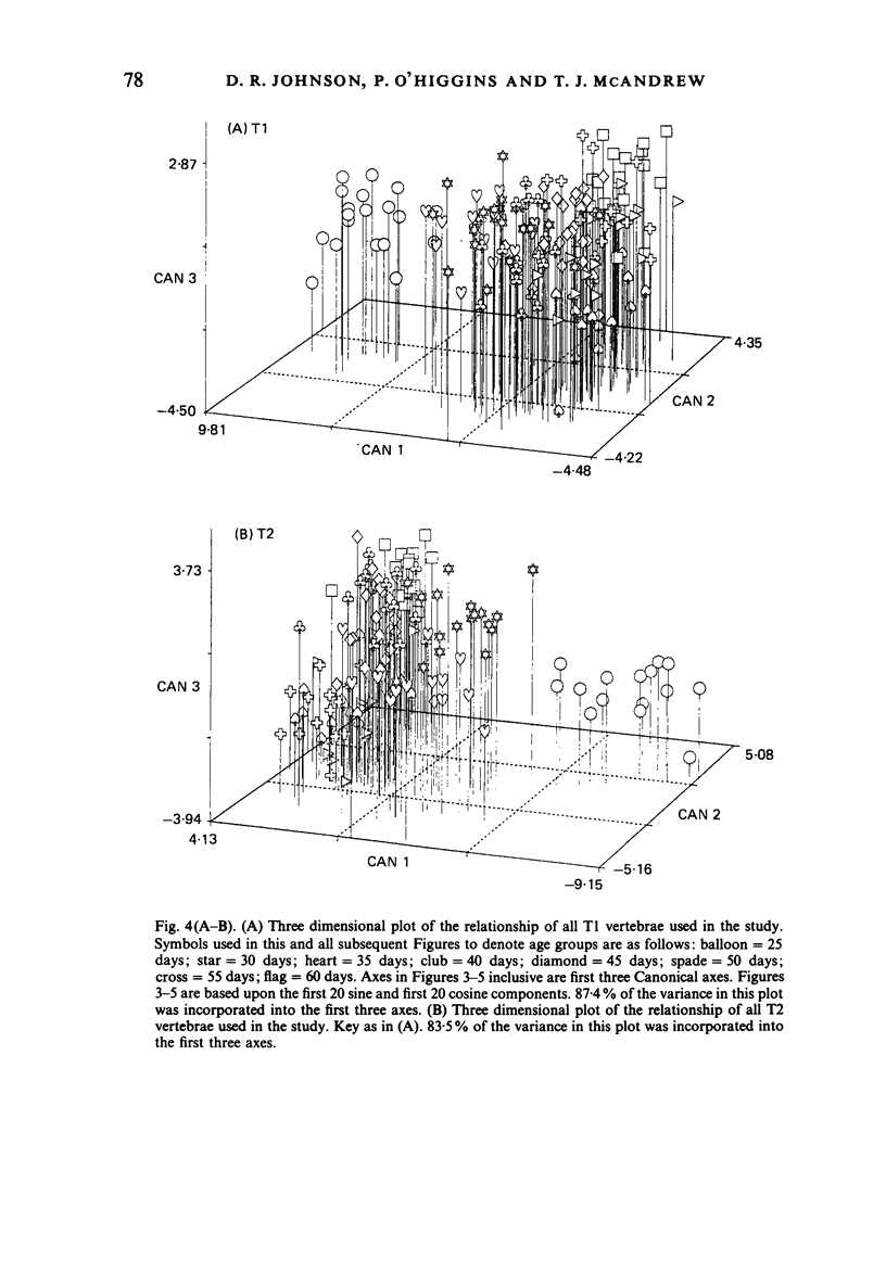
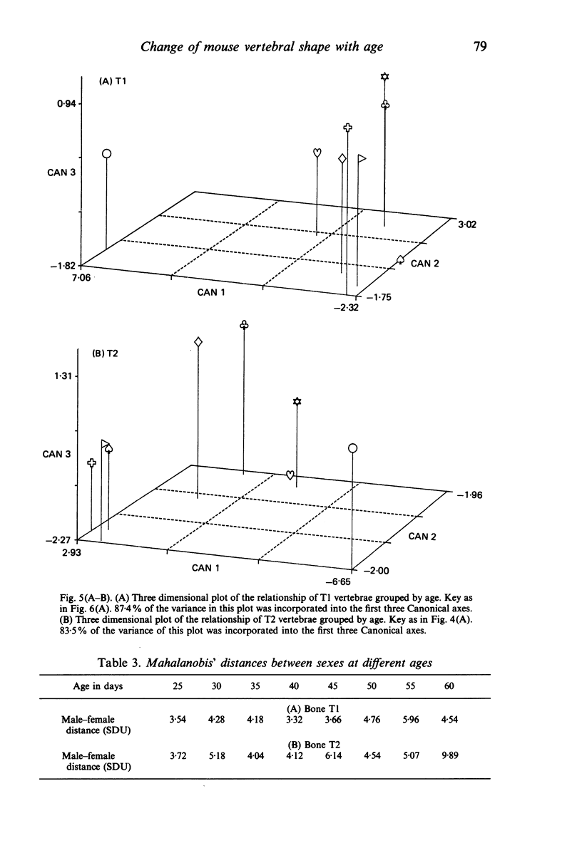
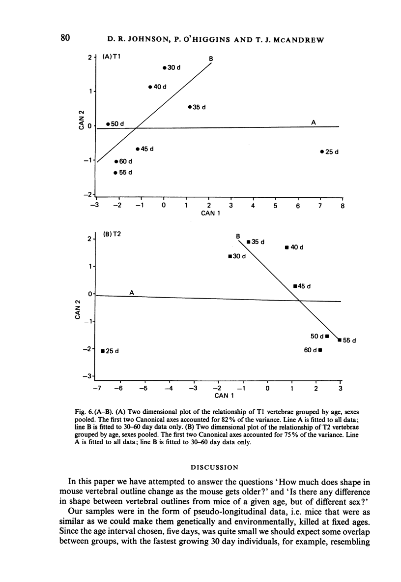
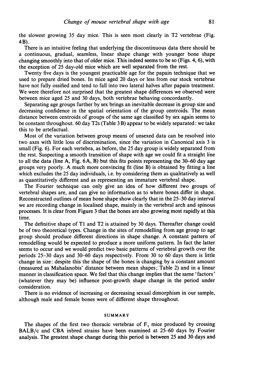
The shapes of the first two thoracic vertebrae of F1 mice produced by crossing BALB/c and CBA inbred strains have been examined at 25-60 days by Fourier analysis. The greatest shape change during this period is between 25 and 30 days and is related to the finalisation of the form of the neural arch and spinous process. Between 30 and 60 days there is continued linear shape change not associated with further increase in vertebral area, and probably due to a constant pattern of localised shape changes. Both vertebrae and both sexes behave similarly.
Full text
PDF
Images in this article
Selected References
These references are in PubMed. This may not be the complete list of references from this article.
- Johnson D. R., O'Higgins P., McAndrew T. J., Adams L. M., Flinn R. M. Measurement of biological shape: a general method applied to mouse vertebrae. J Embryol Exp Morphol. 1985 Dec;90:363–377. [PubMed] [Google Scholar]
- O'Higgins P., Johnson D. R., McAndrew T. J. The clonal model of vertebral column development: a reinvestigation of vertebral shape using Fourier analysis. J Embryol Exp Morphol. 1986 Jul;96:171–182. [PubMed] [Google Scholar]


