Abstract
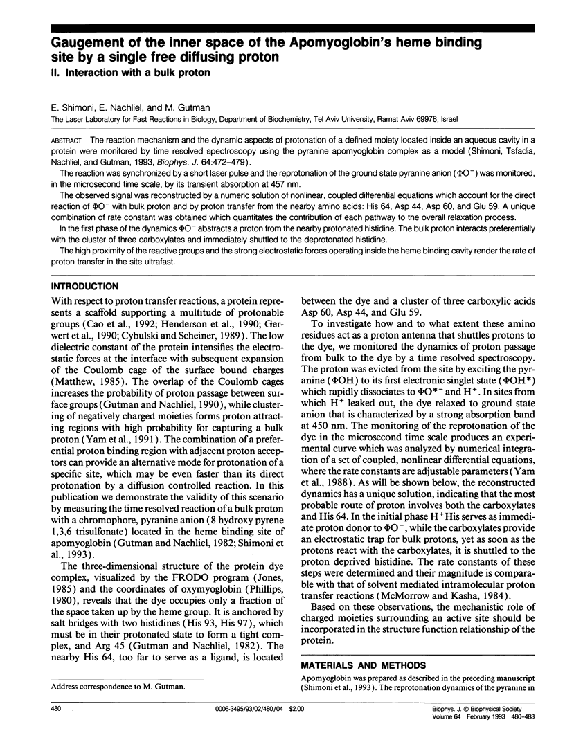
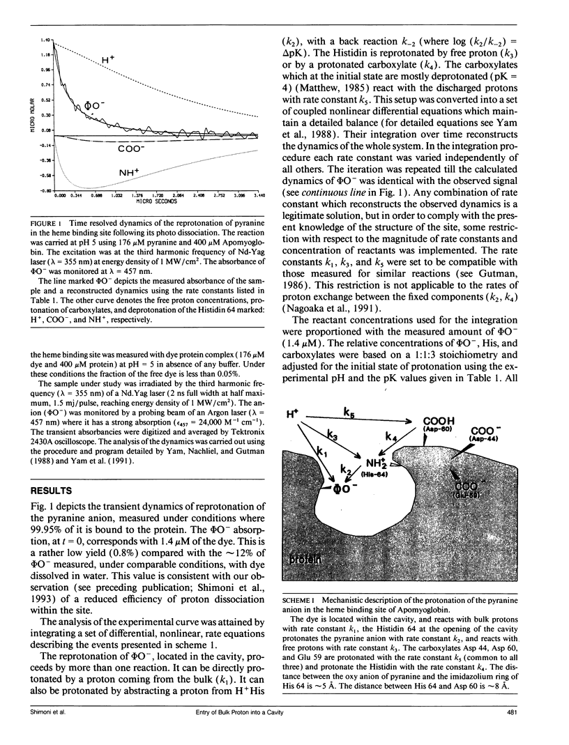
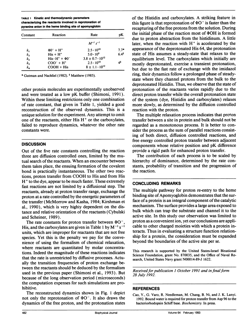
The reaction mechanism and the dynamic aspects of protonation of a defined moiety located inside an aqueous cavity in a protein were monitored by time resolved spectroscopy using the pyranine apomyoglobin complex as a model (Shimoni, Tsfadia, Nachliel, and Gutman, 1993, Biophys. J. 64:472-479). The reaction was synchronized by a short laser pulse and the reprotonation of the ground state pyranine anion (phi O-) was monitored, in the microsecond time scale, by its transient absorption at 457 nm. The observed signal was reconstructed by a numeric solution of nonlinear, coupled differential equations which account for the direct reaction of phi O- with bulk proton and by proton transfer from the nearby amino acids: His 64, Asp 44, Asp 60, and Glu 59. A unique combination of rate constant was obtained which quantitates the contribution of each pathway to the overall relaxation process. In the first phase of the dynamics phi O- abstracts a proton from the nearby protonated histidine. The bulk proton interacts preferentially with the cluster of three carboxylates and immediately shuttled to the deprotonated histidine. The high proximity of the reactive groups and the strong electrostatic forces operating inside the heme binding cavity render the rate of proton transfer in the site ultrafast.
Full text
PDF


Selected References
These references are in PubMed. This may not be the complete list of references from this article.
- Gerwert K., Souvignier G., Hess B. Simultaneous monitoring of light-induced changes in protein side-group protonation, chromophore isomerization, and backbone motion of bacteriorhodopsin by time-resolved Fourier-transform infrared spectroscopy. Proc Natl Acad Sci U S A. 1990 Dec 15;87(24):9774–9778. doi: 10.1073/pnas.87.24.9774. [DOI] [PMC free article] [PubMed] [Google Scholar]
- Gutman M. Application of the laser-induced proton pulse for measuring the protonation rate constants of specific sites on proteins and membranes. Methods Enzymol. 1986;127:522–538. doi: 10.1016/0076-6879(86)27042-1. [DOI] [PubMed] [Google Scholar]
- Gutman M., Nachliel E., Huppert D. Direct measurement of proton transfer as a probing reaction for the microenvironment of the apomyoglobin heme-binding site. Eur J Biochem. 1982 Jun 15;125(1):175–181. doi: 10.1111/j.1432-1033.1982.tb06665.x. [DOI] [PubMed] [Google Scholar]
- Henderson R., Baldwin J. M., Ceska T. A., Zemlin F., Beckmann E., Downing K. H. Model for the structure of bacteriorhodopsin based on high-resolution electron cryo-microscopy. J Mol Biol. 1990 Jun 20;213(4):899–929. doi: 10.1016/S0022-2836(05)80271-2. [DOI] [PubMed] [Google Scholar]
- Johnson K. A., Olson J. S., Phillips G. N., Jr Structure of myoglobin-ethyl isocyanide. Histidine as a swinging door for ligand entry. J Mol Biol. 1989 May 20;207(2):459–463. doi: 10.1016/0022-2836(89)90269-6. [DOI] [PubMed] [Google Scholar]
- Matthew J. B. Electrostatic effects in proteins. Annu Rev Biophys Biophys Chem. 1985;14:387–417. doi: 10.1146/annurev.bb.14.060185.002131. [DOI] [PubMed] [Google Scholar]
- Phillips S. E. Structure and refinement of oxymyoglobin at 1.6 A resolution. J Mol Biol. 1980 Oct 5;142(4):531–554. doi: 10.1016/0022-2836(80)90262-4. [DOI] [PubMed] [Google Scholar]
- Shimoni E., Tsfadia Y., Nachliel E., Gutman M. Gaugement of the inner space of the apomyoglobin's heme binding site by a single free diffusing proton. I. Proton in the cavity. Biophys J. 1993 Feb;64(2):472–479. doi: 10.1016/S0006-3495(93)81389-4. [DOI] [PMC free article] [PubMed] [Google Scholar]
- Yam R., Nachliel E., Kiryati S., Gutman M., Huppert D. Proton transfer dynamics in the nonhomogeneous electric field of a protein. Biophys J. 1991 Jan;59(1):4–11. doi: 10.1016/S0006-3495(91)82192-0. [DOI] [PMC free article] [PubMed] [Google Scholar]


