Abstract
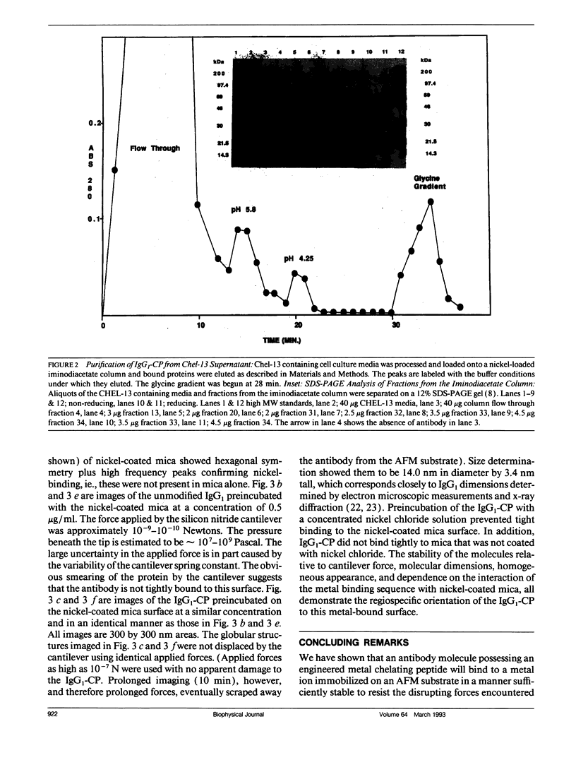
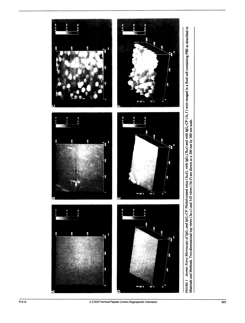
An antibody (IgG1) was designed for oriented adherence to a metal-containing surface. This was achieved by adding a metal-chelating peptide, (CP = His-Trp-His-His-His-Pro), to the COOH-terminus of the heavy chain through genetic engineering. Electroporation of the engineered heavy chain gene into cells expressing the complimentary light chain yielded colonies secreting an intact antibody containing the metal-chelating peptide (IgG1-CP) which had high affinity for a nickel-loaded iminodiacetate column. Purified IgG1-CP was bound to nickel-treated mica and imaged by atomic force microscopy (AFM). Antibody lacking the COOH-terminal metal binding peptide failed to produce discernible AFM images. The AFM images of individual IgG1-CP molecules and their calculated dimensions demonstrated that regiospecific binding and uniform orientation of the antibody was imparted by the peptide. The ability to stably orient macromolecules in their native state to a surface may be used advantageously to visualize them.
Full text
PDF
Images in this article
Selected References
These references are in PubMed. This may not be the complete list of references from this article.
- Andreason G. L., Evans G. A. Introduction and expression of DNA molecules in eukaryotic cells by electroporation. Biotechniques. 1988 Jul-Aug;6(7):650–660. [PubMed] [Google Scholar]
- Beidler C. B., Ludwig J. R., Cardenas J., Phelps J., Papworth C. G., Melcher E., Sierzega M., Myers L. J., Unger B. W., Fisher M. Cloning and high level expression of a chimeric antibody with specificity for human carcinoembryonic antigen. J Immunol. 1988 Dec 1;141(11):4053–4060. [PubMed] [Google Scholar]
- Chu G., Hayakawa H., Berg P. Electroporation for the efficient transfection of mammalian cells with DNA. Nucleic Acids Res. 1987 Feb 11;15(3):1311–1326. doi: 10.1093/nar/15.3.1311. [DOI] [PMC free article] [PubMed] [Google Scholar]
- Deisenhofer J. Crystallographic refinement and atomic models of a human Fc fragment and its complex with fragment B of protein A from Staphylococcus aureus at 2.9- and 2.8-A resolution. Biochemistry. 1981 Apr 28;20(9):2361–2370. [PubMed] [Google Scholar]
- Drake B., Prater C. B., Weisenhorn A. L., Gould S. A., Albrecht T. R., Quate C. F., Cannell D. S., Hansma H. G., Hansma P. K. Imaging crystals, polymers, and processes in water with the atomic force microscope. Science. 1989 Mar 24;243(4898):1586–1589. doi: 10.1126/science.2928794. [DOI] [PubMed] [Google Scholar]
- Hansma H. G., Vesenka J., Siegerist C., Kelderman G., Morrett H., Sinsheimer R. L., Elings V., Bustamante C., Hansma P. K. Reproducible imaging and dissection of plasmid DNA under liquid with the atomic force microscope. Science. 1992 May 22;256(5060):1180–1184. doi: 10.1126/science.256.5060.1180. [DOI] [PubMed] [Google Scholar]
- Hansma P. K., Elings V. B., Marti O., Bracker C. E. Scanning tunneling microscopy and atomic force microscopy: application to biology and technology. Science. 1988 Oct 14;242(4876):209–216. doi: 10.1126/science.3051380. [DOI] [PubMed] [Google Scholar]
- Heuser J. Protocol for 3-D visualization of molecules on mica via the quick-freeze, deep-etch technique. J Electron Microsc Tech. 1989 Nov;13(3):244–263. doi: 10.1002/jemt.1060130310. [DOI] [PubMed] [Google Scholar]
- Hutchens T. W., Yip T. T., Porath J. Protein interaction with immobilized ligands: quantitative analyses of equilibrium partition data and comparison with analytical chromatographic approaches using immobilized metal affinity adsorbents. Anal Biochem. 1988 Apr;170(1):168–182. doi: 10.1016/0003-2697(88)90105-4. [DOI] [PubMed] [Google Scholar]
- Ill C. R., Brehm T., Lydersen B. K., Hernandez R., Burnett K. G. Species specificity of iron delivery in hybridomas. In Vitro Cell Dev Biol. 1988 May;24(5):413–419. doi: 10.1007/BF02628492. [DOI] [PubMed] [Google Scholar]
- Jue R. A., Doolittle R. F. Determination of the relative positions of amino acids by partial specific cleavages of end-labeled proteins. Biochemistry. 1985 Jan 1;24(1):162–170. doi: 10.1021/bi00322a023. [DOI] [PubMed] [Google Scholar]
- Loetscher P., Mottlau L., Hochuli E. Immobilization of monoclonal antibodies for affinity chromatography using a chelating peptide. J Chromatogr. 1992 Mar 20;595(1-2):113–119. doi: 10.1016/0021-9673(92)85151-i. [DOI] [PubMed] [Google Scholar]
- Sarma V. R., Silverton E. W., Davies D. R., Terry W. D. The three-dimensional structure at 6 A resolution of a human gamma Gl immunoglobulin molecule. J Biol Chem. 1971 Jun 10;246(11):3753–3759. [PubMed] [Google Scholar]
- Smith M. C., Furman T. C., Ingolia T. D., Pidgeon C. Chelating peptide-immobilized metal ion affinity chromatography. A new concept in affinity chromatography for recombinant proteins. J Biol Chem. 1988 May 25;263(15):7211–7215. [PubMed] [Google Scholar]
- Toneguzzo F., Hayday A. C., Keating A. Electric field-mediated DNA transfer: transient and stable gene expression in human and mouse lymphoid cells. Mol Cell Biol. 1986 Feb;6(2):703–706. doi: 10.1128/mcb.6.2.703. [DOI] [PMC free article] [PubMed] [Google Scholar]
- Weisenhorn A. L., Drake B., Prater C. B., Gould S. A., Hansma P. K., Ohnesorge F., Egger M., Heyn S. P., Gaub H. E. Immobilized proteins in buffer imaged at molecular resolution by atomic force microscopy. Biophys J. 1990 Nov;58(5):1251–1258. doi: 10.1016/S0006-3495(90)82465-6. [DOI] [PMC free article] [PubMed] [Google Scholar]


