Abstract
Helicobacter pylori can transform, in vivo as well as in vitro, from dividing spiral-shaped forms into nonculturable coccoids, with intermediate forms called U forms. The importance of nonculturable coccoid forms of H. pylori in disease transmission and antibiotic treatment failures is unclear. Metabolic activities of actively growing as well as nonculturable H. pylori were investigated by comparing the concentrations of cellular ATP and total RNA, gene expression, presence of cytoplasmic polyphosphate granules and iron inclusions, and cellular morphology during extended broth culture and nutritional cold starvation. In addition, the effect of exposing broth-cultured or cold-starved cells to a nutrient-rich or acidic environment on the metabolic activities was investigated. ATP was detectable up to 14 days and for at least 25 days after transformation from the spiral form to the coccoid form or U form in broth-cultured and cold-starved cells, respectively. mRNAs of VacA, a 26-kDa protein, and urease A were detected by using reverse transcription-PCR in cells cultured for 2 months in broth or cold starved for at least 28 months. The ATP concentration was not affected during exposure to fresh or acidified broth, while 4- to 12-h exposures of nonculturable cells to lysed human erythrocytes increased cellular ATP 12- to 150-fold. Incubation of nonculturable cold-starved cells with an erythrocyte lysate increased total RNA expression and ureA mRNA transcription as measured by quantitative real-time reverse transcription-PCR. Furthermore, the number of structurally intact starved coccoids containing polyphosphate granules increased almost fourfold (P = 0.0022) under the same conditions. In conclusion, a specific environmental stimulus can induce ATP, polyphosphate, and RNA metabolism in nonculturable H. pylori, indicating viability of such morphological forms.
Helicobacter pylori, a gram-negative spiral-shaped bacterium, colonizes primarily the mucosal layer of the gastric epithelium and is recognized as the main cause of gastritis and peptic ulcer disease in humans (27, 33). During infection, a majority of the H. pylori cells are present as actively dividing spiral-shaped forms (45). A morphological switch from the spiral form, possibly through a so-called U form, into a nondividing coccoid form occurs under various environmental conditions, such as aerobiosis (8), temperature changes (37), extended incubation (40), and antibiotic treatment (4, 6). A high density of coccoid forms of H. pylori has been observed in the human stomach (10), often in close association with damaged mucous cells (19).
Formation of nonculturable coccoid forms was also described for Campylobacter jejuni, some marine Vibrio species, and some other gram-negative bacteria in response to temperature changes and nutrient starvation (3, 21, 34). Coccoid forms of some of these organisms have been shown to revert into dividing organisms upon improvement of the environmental conditions (21, 34).
The coccoid form of H. pylori is not culturable in vitro, and it has been reported that this form represents the morphological manifestation of cellular degeneration and cell death (26). However, studies of nonculturable H. pylori indicate viability of such forms (9, 15, 37, 46). In a study by West et al. (46), H. pylori was able to survive in water under certain conditions, retaining the ability to take up tritium-labeled thymidine (37). Oxidative metabolism has been observed in coccoid forms of H. pylori (9), and respiration was measured in cells starved for as long as 8 months (15). Moreover, coccoid forms of H. pylori were shown to adhere to eucaryotic cells and induce cellular changes similar to those induced by spiral forms, including tyrosine phosphorylation of specific proteins (36). Finally, BALB/cA mice orally infected with the coccoid form of H. pylori developed gastric inflammation with the same severity as animals infected with the spiral form (44).
Both an oral-oral route and a fecal-oral route seem to be involved in the transmission of infection (24, 28, 35). However,H. pylori has not been cultured from environmental specimens, from the oral cavity, or to a significant extent from feces, suggesting a role of nonculturable viable forms in disease transmission. Therefore, to understand the epidemiology of H. pylori infection, it is important to investigate the role of nonculturable cells as potential survival forms in extragastric environments. This study compared concentrations of cellular ATP, gene expression, total RNA, morphology, and cytoplasmic granules of polyphosphate (poly-P) and iron of culturable and nonculturable H. pylori cells maintained under different conditions. The metabolic response to nutrient stimulus or acid stress was analyzed to measure potential viability in suspensions of nonculturable H. pylori devoid of detectable spiral forms.
MATERIALS AND METHODS
Bacterial strain and culture conditions.
H. pylori strain CCUG 17874 was used throughout this study. The strain was cultured on GAB-Camp agar (39) without antibiotics for 2 days at 37°C in a smicroaerobic atmosphere (5% O2, 10% CO2, and 85% N2). Single colonies were inoculated into flasks containing 50 ml of a gonococcal broth (GB) (pH 7.6), originally described for culturing of Neisseria gonorrhoeae (38), supplemented with 5% horse serum (Gibco, Paisley, Scotland). Cultures were incubated for 30 h at 37°C on a rotary shaker (200 rpm) in anaerobic jars with a microaerobic atmosphere generated by Anaerocult C envelopes (Merck, Darmstadt, Germany). An inoculum of 1%, corresponding to approximately 108 CFU of H. pylori per ml, was suspended in 50 ml of fresh GB and incubated as described above. The microaerobic atmosphere was regenerated every 24 h. Cultures were performed in triplicate. The CFU number in each culture was monitored regularly. Aliquots (10 ml) were centrifuged at 4,000 × g for 10 min, resuspended in 1 ml of phosphate-buffered saline (PBS) (pH 7.2), diluted, and seeded on agar. Single colonies were counted after 5 days of culture.
In addition, at different time points during stationary-phase culture, 5 ml was washed once in PBS and resuspended in either spent broth, fresh broth, or fresh broth containing 0.2% (vol/vol) lysed human erythrocytes, prepared as previously described (1). After microaerobic incubation at 37°C, the cellular ATP, gene expression, and total RNA were analyzed as described below.
Cold starvation.
H. pylori cells were cold starved as described by Mizoguchi et al. (29), with the exception that GB was used for the initial culture. Aliquots (5 ml) of aging cold-starved H. pylori were harvested, centrifuged, and resuspended in (i) fresh broth, (ii) fresh broth adjusted to pH 5, (iii) fresh broth adjusted to pH 5 containing 5 mM urea, or (iv) fresh broth containing 0.2% (vol/vol) lysed human erythrocytes. Cellular ATP, total RNA, gene expression, bacterial morphology, and poly-P granule content were subsequently determined.
TEM.
Fixation, staining, and embedding of H. pylori cells for transmission electron microscopy (TEM) were as previously described with few modifications (2). Approximately 109 H. pylori cells were washed once with 5 ml of minimal essential medium (Gibco), resuspended in 5 ml of equal volumes of minimal essential medium and 0.1 M sodium cacodylate buffer (pH 7.4) containing 3% (vol/vol) glutaraldehyde (2), and fixed overnight at 4°C. After centrifugation at 3,000 × g, cells were resuspended in glutaraldehyde-cacodylate buffer. Dehydration and embedding in Epon 100 (Merck) were performed according to a standard protocol (2). For each time point, eight fields of bacteria from two separate grids were examined.
EFTEM.
The presence of iron in crystalline inclusions and of phosphate in poly-P granules was determined by energy-filtering TEM (EFTEM) as previously described (12). Samples were prepared as described above for TEM, with the exception that postfixation in osmium and uranyl acetate was excluded. Thin sections (70 nm) were examined at 120 kV in a Philips electron microscope (CM120 BioTWIN) equipped with a Gatan biofilter (GIF) including a cooled charge-coupled device camera (Gatan MSC 791). The elemental compositions of the crystalline inclusions and poly-P granules were analyzed and visualized using electrons with a filter energy of 59 eV for iron and 152 eV for phosphate.
ATP determination.
The intracellular ATP concentration of H. pylori was determined using a quantitative reaction based on bioluminescence as previously described (41). The samples were either directly analyzed for ATP or frozen at −20°C until assayed. Each culture supernatant was treated similarly to compare the extracellular and intracellular ATP contents (17). Light emission was measured with a 1251 luminometer (Bio-Orbit, Turku, Finland).
RNA isolation.
At regular intervals total RNA was prepared from approximately 5 × 10⁸ cells of broth-cultured or cold-starved H. pylori using an RNeasy minikit (Qiagen, Hilden, Germany) according to the instructions of the manufacturer, with the exception that DNase was also added during the initial cell lysis step. RNA concentrations were determined in a Gene Quant spectrophotometer (Pharmacia Biotech, Uppsala, Sweden) and stored at − 80°C. Aliquots of RNA preparations were treated with 50 μg of RNase A (Roche Molecular Biochemicals) per ml for 60 min at 37°C.
RT-PCR.
Reverse transcription (RT) and PCR assays were carried out in a single step using a Titan one-tube RT-PCR kit (Roche Molecular Biochemicals) according to sthe manufacturer’s instructions with a 0.2 μM concentration of each deoxynucleoside triphosphate (dNTP) and a 0.5 μM concentration of each primer. Primers specific for ureA (11), cagA (43), and the gene (tsaA) for a 26-kDa protein of H. pylori (16) were used. Primers for vacA were constructed and selected based on computer-assisted analysis of published sequences (GenBank/EMBL data bank accession numbers U05676, U5677, and U0714500). The forward primer, Vac1 (5′-GGCACACTGGATTTGTGGCA-3′), and the reverse primer, Vac2 (5′-CGCTCGCTTGATTGGACAGA-3′), amplified a 372-bp product of H. pylori strain CCUG 17874. cDNA was synthesized for 40 min at 50°C, and after incubation at 94°C for 4 min, the mixtures were subjected to 35 cycles of denaturation at 94°C, annealing at 47°C (ureA), 55°C (cagA), 49°C (vacA), or 62°C (26-kDa protein gene); and extension at 70°C. The initial 10 PCR cycles were carried out with 30-s intervals at each indicated temperature. PCR cycles 11 to 20 lasted for 45 s, and cycles 21 to 35 were for 1 min. After a final extension of 7 min, the PCR products were detected by 1.5% (wt/vol) agarose gel electrophoresis.
Real-time RT-PCR.
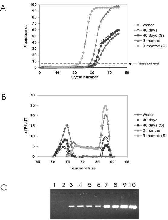
The ability of H. pylori to express urease A during extended culture, cold starvation, and nutrient stimulation was studied quantitatively by real-time RT-PCR with ureA primers as described above. The RT-PCR was done in two steps using rTth DNA polymerase (Applied Biosystems) in both steps. The RT reaction mixture volume was 10 μl, comprising 1× reverse transcriptase buffer (Applied Biosystems), 1 mM MnCl2, a 0.2 mM concentration of each dNTP, 0.5 μM reverse primer, 4 mg of bovine serum albumin per ml, and 1 U of rTth DNA polymerase. Two microliters of total RNA was added to the RT mixture. The incubation conditions for the RT were 70°C for 30 s, 47°C for 30 s, and 70°C for 15 min. PCR amplification of the synthesized cDNA was monitored on-line using the LightCycler instrument (Roche Molecular Biochemicals). PCR was carried out in 20-μl volumes containing 1× chelating buffer (Applied Biosystems), 2.5 mM MgCl2, 0.2 mM dNTPs, a 30,000-fold diluted stock solution of SYBR Green I (Roche Molecular Biochemicals), and 1.25 U of rTth DNA polymerase. Each reaction mixture was loaded into a glass capillary tube, and 2 μl of the cDNA was added. After denaturation at 94°C for 30 s, the samples were subjected to 45 cycles of denaturation (95°C for 0 s), annealing (47°C for 5 s), and extension (72°C for 15 s). The temperature transition rate was set to 20°C/s, and fluorescence was monitored at the end of each extension. The specificity of the amplification was determined by melting-curve analysis (a linear temperature increase from 60 to 95°C at a rate of 0.2°C/s with continuous signal acquisition) and gel electrophoresis. For each sample, a log-linear line was fitted automatically by selecting three points above the threshold band that represented a log-linear increase in fluorescence. The intersection of the extended line with the threshold band was used to determine the fractional cycle number of the crossing point (Cp), calculated automatically by the LightCycler software.
Statistical analysis.
The chi-square test was used to determine the poly-P granule content of nutritionally stimulated H. pylori cells versus controls. A P value of 0.05 was considered significant.
RESULTS
H. pylori growth in broth culture and during cold starvation.
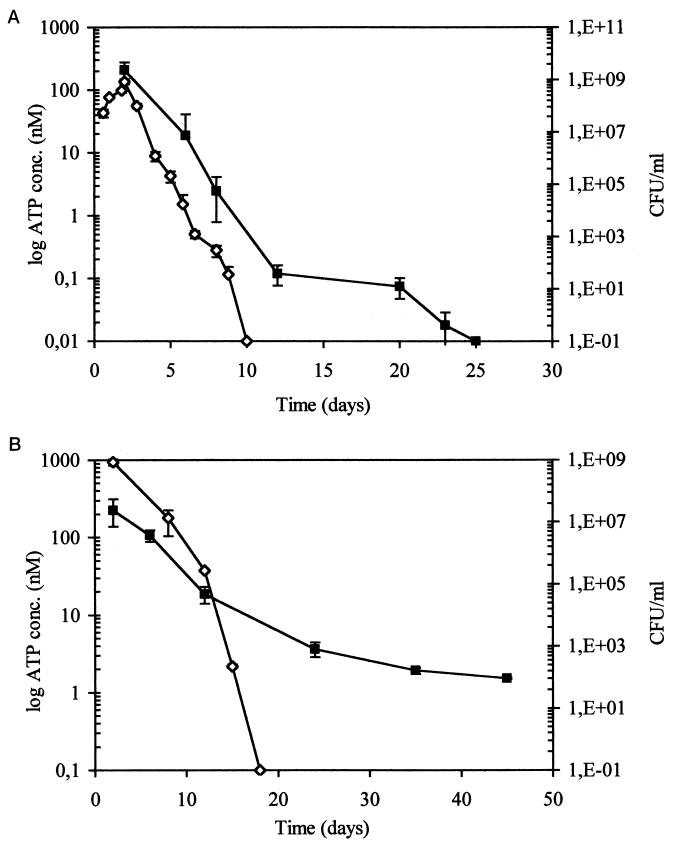
During broth cultures the highest CFU counts were obtained after 48 h. A decline of the CFU count was recorded at day 4, and no colonies (<0.1 CFU/ml) were formed after 9 days of culture (Fig. 1A). For cold starvation experiments, 3-day broth cultures of late-exponential-phase H. pylori were suspended in PBS and maintained at 8°C. In contrast to broth-cultured cells, cold-starved bacteria were culturable for an extended period of time. Eighteen days after suspension of H. pylori in PBS at 8°C, the cells were nonculturable (<0.1 CFU/ml) after seeding onto GAB-Camp agar (Fig. 1B).
FIG. 1.
Viable counts and intracellular ATP content of H. pylori CCUG 17874 during extended culture in GB (A) or during cold starvation in PBS (B). Viable counts (◊) were estimated by dilution plating (CFU per milliliter), and cellular ATP (▪) was determined by bioluminescence. Error bars represent the standard deviations of the means of triplicate samples.
Electron microscopy studies.
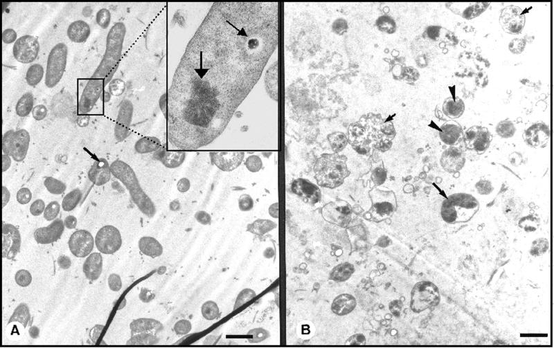
The majority of the spiral forms were 2 to 4 μm long and 0.5 to 0.8 μm wide (Fig. 2A) and were found in broth cultures up to day 8 (Table 1). U forms, with the cytoplasmic body inside a distended outer membrane (Fig. 2B), were observed in small numbers in all broth culture samples; however, these forms were abundant during cold starvation (Table 1). Coccoid forms of H. pylori were subdivided ultrastructurally into an intact (IC) or a degenerative (DC) form, depending on the size and density of the cytoplasm (Fig. 2B). The cytoplasmic body of the IC forms was 1 to 2 μm in diameter, and that of the DC forms was 3 to 4 μm in diameter (Fig. 2B). The frequencies of the IC forms were similar in 22-day broth cultures and after 45 days of cold starvation, being approximately 50% of the total cell count (Table 1). In contrast, DC forms were present in very small numbers during cold starvation (Table 1), whereas these constituted about 50% of the cells in 22-day broth cultures. Sheathed flagella, often in small bundles, were found in all preparations of H. pylori. Broth-cultured and cold-starved forms contained distinct dense cytoplasmic granules (poly-P) and crystalline inclusions, and through analysis by EFTEM at 152 and 59 eV, distinct signals corresponding to phosphate and iron, respectively, were detected (data not shown). Some poly-P granules were electron dense, while others showed sublimation characteristics, i.e., loss of the granule content during sectioning (Fig. 2A). The distributions of poly-P and iron inclusions in the different morphological forms of H. pylori are shown in Table 1.
FIG. 2.
(A) TEM of thin sections of H. pylori cultured in broth for 2 days. Most of these cells were spiral and contained poly-P granules showing sublimation characteristics (arrow). The inset (magnified) shows part of a cell with an intact poly-P granule (small arrow) and crystalline iron (large arrow). (B) TEM of thin sections of H. pylori cultured in broth for 16 days. Three different morphological types are illustrated: a U form (large arrow), IC forms (arrowheads), and DC forms (small arrows). Bars, 1μm.
TABLE 1.
Cell morphology and cytoplasmic content of iron inclusions and poly-P granules in H. pylori during extended culture in GB (2 to 22 days) and during cold starvation in PBS (30 days to 28 months) determined by TEM
| Time | Spirals
|
U forms
|
IC forms
|
DC forms
|
||||||||
|---|---|---|---|---|---|---|---|---|---|---|---|---|
| % of total | % Containing:
|
% of total | % Containing:
|
% of total | % Containing:
|
% of total | % Containing:
|
|||||
| Fea | Pb | Fe | P | Fe | P | Fe | P | |||||
| 2 days | 83 | 13 | 18 | 0.5 | 16 | 2 | 11 | 0.5 | ||||
| 5 days | 72 | 1 | 22 | 2 | 20 | 21 | 8 | 8 | 5 | 8 | ||
| 8 days | 6 | 42 | 8 | 79 | 49 | 0.5 | 15 | 29 | 3 | |||
| 22 days | 4 | 33 | 42 | 54 | 54 | 45 | ||||||
| 30 days | 37.5 | 21 | 7 | 60 | 38 | 4.5 | 2.5 | |||||
| 45 days | 48.5 | 20 | 22 | 49.5 | 35 | 27 | 1 | 1 | 1 | |||
| 3 mo | 10 | 34 | 19 | 71.5 | 39 | 16 | 18.5 | 20 | 6 | |||
| 28 mo | 4.5 | 10 | 86.5 | 4 | 11 | 9 | ||||||
Iron-containing inclusions.
Poly-P-containing granules.
Cytoplasmic ATP content.
Trichloroacetic acid-treated cells were frozen for up to 2 weeks before the ATP assay without significant loss of ATP content. Interestingly, a freeze-thaw step was crucial to obtain optimal release of intracellular ATP. Organisms of 2-day broth cultures, predominately spiral forms, contained on average 200 nM ATP. ATP successively decreased to day 12, when the content leveled out at 0.1 nM on average. Cells were then nonculturable, being predominately coccoids with occasional U forms. At day 20 the ATP content was further decreased, and at day 25 ATP was not detectable (Fig. 1A). In comparison, cold-starved H. pylori generally retained higher concentrations of cellular ATP, with ATP concentrations decreasing less rapidly than during broth culture and being detectable during a longer period of time (Fig. 1B). Thirty-day nonculturable cold-starved cells contained on average 1.9 nM intracellular ATP, and 45-day cells contained 1.5 nM (Fig. 1B). Hence, nonculturable preparations of H. pylori, kept at low temperature and nutritionally starved, contained more than 10-fold-higher concentrations of ATP than nonculturable H. pylori in extended broth cultures. The ATP contents of both broth culture and cold starvation supernatants were negligible (10- to 100-fold less) compared with the intracellular ATP in all samples tested. ATP leakage was detected almost exclusively in the supernatant of cells during the first week of broth culture, while extracellular ATP was not detected in supernatants of cultures containing a majority of coccoid forms (data not shown).
Total RNA content.
Total RNA was purified from 5 × 10⁸ cells of either exponential-phase broth cultures, extended broth cultures, or cold-starved cells. Exponential-phase, 2-day broth cultures, comprising >80% spiral-shaped organisms, contained on average 123 μg of total RNA per ml. Extended broth cultures, with >95% of the organisms being of coccoid morphology and a few percent being U forms, contained 21μg/ml after 10 days, 8.2 μg/ml after 20 days, 5.4 μg/ml after 40 days, and 4.3 μg/ml after 60 days of culture. Cold-starved cells contained 105 μg of RNA per ml after 3 months and 5.7 μg/ml after 28 months.
Gene expression.
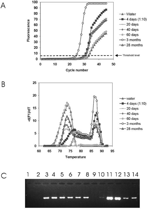
All RNA preparations were negative with conventional PCR if the RT reaction was omitted or if the preparations were pretreated with RNase. Conventional RT-PCR was performed on broth-cultured H. pylori after 4, 20, 30, 40, and 60 days and after 1, 3, and 28 months of cold starvation. Expression of the vacA and ureA genes, as well as the gene for a 26-kDa protein (tsaA), was detected at all time points tested for both broth-cultured and cold-starved cells, demonstrating that these genes are expressed in nonculturable H. pylori. The cagA gene was expressed after 40 days but not after 60 days of broth culture and for up to 3 months of cold starvation. Expression of the ureA gene, detected by real-time RT-PCR, was performed on broth-cultured H. pylori after 4, 20, 40, and 60 days and after 3, and 28 months of cold starvation (Fig. 3). The Cp value, i.e., the cycle number where amplification is detected, is related to the target concentration. A low Cp value indicates an early detection of amplification, corresponding to a high concentration of target cDNA, and vice versa. Clear differences in the levels of ureA gene expression were noted (Fig. 3A). The Cp values were approximately 30 for broth-cultured H. pylori after 4, 20, and 40 days; 32 for broth-cultured H. pylori after 60 days; and 21 and 30 for cold-starved H. pylori after 3 and 28 months, respectively. The specificity of the amplification was verified by melting-curve analysis and gel electrophoresis (Fig. 3B and C).
FIG. 3.
Real-time quantitative PCR analysis of urease A gene expression of 4-, 20-, 40-, and 60-day broth-cultured and 3- and 28-month cold-starved H. pylori. (A and B) Real-time amplification curve (A) and melting-curve (B) analysis of amplified PCR products, using SYBR Green I and the LightCycler. (C) Agarose gel electrophoresis. Lanes 1 and 2, water control; lanes 3 and 4, 4-day cells; lanes 5 and 6, 20-day cells; lanes 7 and 8, 40-day cells; lanes 9 and 10, 60-day cells; lanes 11 and 12, 3-month cells; lanes 13 and 14, 28-month cells. The experiments were done in duplicate.
Response to acid stress and nutrient stimuli. (i) Broth culture.
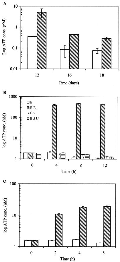
Twelve-day cultures contained mainly coccoid forms and a few U-forms. After cells of 12-, 16-, and 18-day cultures were suspended in fresh broth containing lysed erythrocytes and reincubated for 12 h, the cellular ATP content increased 16-, 4-, and 2-fold in the 12-, 16-, and 18-day cultures, respectively, compared with the cellular ATP content of control cells kept in spent broth (Fig. 4A). Total RNA concentrations did not vary for 40-day cells incubated in fresh medium with or without erythrocyte lysate. In 4- to 60-day cultures RT-PCR products were amplified using primers for ureA,vacA, cagA, and the gene encoding a 26-kDa protein. Nutritionally (erythrocyte lysate) stimulated cells from these cultures were also positive in RT-PCR using ureA primers. With real-time RT-PCR using ureA primers, no effect of erythrocyte lysate was detected on gene expression of 40-day cells (Fig. 5A).
FIG. 4.
Effect of human erythrocyte lysate (0.2%, vol/vol) on cellular ATP content of H. pylori. (A) H. pylori was broth cultured for 12, 16, and 18 days, harvested, and transferred to either fresh broth (□) or fresh broth containing erythrocyte lysate (▧). Cellular ATP was determined after subculture at 37°C for 12 h. (B) Intracellular ATP content of 30-day suspensions of cold-starved nonculturable H. pylori exposed to various nutrient and pH conditions. Cellular ATP was determined prior to exposure of the cells to (i) GB (B); (ii) broth containing lysed human erythrocytes (BE); (iii) broth adjusted to pH 5 (B5); or (iv) broth adjusted to pH 5 and containing 5 mM urea (B5U). Cells were analyzed after 4, 8, and 12 h of exposure to these conditions. (C) Intracellular ATP content of 45-day cold-starved nonculturable H. pylori exposed to fresh broth with (▧) or without (□) the erythrocyte lysate. Cellular ATP was determined at 2, 4, and 8 h of stimulation. Duplicate experiments were performed twice. Means and standard Sdeviations are indicated.
FIG. 5.
Comparison of urease A gene expression by real time RT-PCR of broth-cultured (40 days) and cold-starved (3 months) H. pylori prior to and after erythrocyte lysate stimulation (S). (A and B) Real-time detection (A) and melting-curve analysis (B) of amplified PCR products, using SYBR Green I and the LightCycler. (C) Agarose gel electrophoresis. Lanes 1 and 2, water control; lanes 3 and 4, 40-day cells; lanes 5 and 6, stimulated 40-day cells; lanes 7 and 8, 3-month cells; lanes 9 and 10, stimulated 3-month cells. The experiments were done in duplicate.
(ii) Cold starvation.
The intracellular ATP content of cold-starved cells was monitored during exposure to different environmental conditions at 30 and 45 days postinoculation in PBS at 8°C. Cellular ATP was measured prior to incubation in broth with erythrocyte lysate. A low increase of cellular ATP was recorded for 30-day cells stimulated with fresh broth. A decline of the cellular ATP was detected in cells exposed to fresh broth adjusted to pH 5 or fresh broth adjusted to pH 5 containing 5 mM urea (Fig. 4B). Stimulation of 30-day cells with broth containing lysed erythrocytes increased cellular ATP 150-fold within 4 h at 37°C (Fig. 4B), whereas ATP in 45-day cells increased 7- and 12-fold after 2 and 4 h, respectively (Fig. 4C). Exposure of 45-day cells to fresh broth adjusted to pH 5 with and without urea reduced the cellular ATP content in comparison with nonstimulated cells (data not shown).
The cell morphology and iron and poly-P contents of cells incubated for 12 h in fresh broth compared with nontreated (spent) organisms showed similar ultrastructural patterns (data not shown). However, after 12 h of incubation of 30-day cells with broth and erythrocyte lysate, the relative number of cells containing poly-P (46 of 274, or 16.8%) increased 3.7-fold (P = 0.0022) compared with cells incubated in fresh broth (8 of 160, or 5%). Three-month cells contained 105 μg of total RNA per ml. Similar cells incubated with erythrocyte lysate for 4 h contained on average 188μg of RNA per ml. All RNA preparations of cold-starved bacteria, except those from 28-month cells analyzed with cagA primers, were RT-PCR positive with either of the four primer sets. When 3-month cells were analyzed after 8 h of erythrocyte lysate stimulus using real-time RT-PCR, a clear increase in the urease A mRNA level was found compared with nonstimulated cells. The Cp value of stimulated cells was reduced to 22, compared with 29 for spent cells (Fig. 5A). The specificity of the amplification was verified by melting-curve analysis and gel electrophoresis (Fig. 5B and C).
DISCUSSION
Spiral forms of H. pylori switch to coccoid forms through intermediate C or U forms (20, 23, 32, 40). Our present findings support this classification and the classification of coccoids into IC and DC forms, according to ultrastructural characteristics observed by TEM studies. The DC forms were enlarged and contained less nuclear material with diffuse characteristics. In contrast, coccoid forms with an intact outer membrane had a nuclear composition similar to that of spiral forms and showed no signs of cellular degeneration.
In this study, the culturability of H. pylori was highly correlated with the presence of spiral forms as observed by TEM. These results were not in accordance with a study by Kusters et al. (26) reporting that culturability was lost when approximately 50% of the H. pylori cells were still in the spiral form. This may be related to the use of optimized agar culture conditions. Andersen et al. (1) used a medium containing an erythrocyte lysate to culture H. pylori, with a morphological switch occurring at 18 h postinoculation, after 72 h of culture spherical cells devoid of urease activity regained this activity, and spiral-shaped cells appeared after transfer to fresh medium. However, it is difficult to know if a 72-h culture is completely devoid of spiral forms.
Several bacteria that transform into coccoids, such as H. pylori, Salmonella, and Vibrio cholerae, have homologues to poly-P kinase, which converts poly-P and ADP to ATP (25). Poly-P may be an important energy and phosphorus source also for cells during starvation, essential for endogenous metabolism, as proposed by Bode et al. (5). In this study, the presence of poly-P in all forms of H. pylori except DC forms correlated well with culturability and the presence of ultrastructurally intact cells. In addition, poly-P was abundant in nonculturable H. pylori with an ability to synthesize cellular ATP and mRNA in response to a nutrient stimulus. In Escherichia coli, poly-P accumulates as a result of cellular responses to deficiencies in an amino acid or nitrogen, and mutants of E. coli lacking poly-P kinase die after only a few days in stationary phase (25).
Iron is an essential growth factor for H. pylori, and the ability of tissue-invading pathogens to cause disease is generally related to iron-scavenging capacity (13). Sequence analysis indicates that H. pylori has such a capacity (42). In this study we showed, based on EFTEM and ultrastructural similarities, that crystalline inclusions in both spiral and coccoid forms of H. pylori contain iron. H. pylori has been found to bind human lactoferrin (13), but the lactoferrin binding capacity of the coccoid form was significantly higher than that of the spiral form (22). Intracellular crystal structures, associated with rapid growth, were described for Bacteroides nodosus (18). Frazier et al. (14) demonstrated such iron inclusions in H. pylori, and by expressing the H. pylori ferritin in E. coli, the growth rate significantly increased.
In agreement with Sörberg et al. (40), we found high levels of cellular ATP (≧10 nM) in 2- to 5-day broth cultures, while 8-day cells of nonculturable coccoid and U forms contained approximately 1 nM. After day 12 of broth culture, cellular ATP declined (to ∼0.1 nM), and it was detectable up to day 23; however, cold-starved cells retained higher cellular levels of ATP over a prolonged period of time. Nonculturable structurally intact cells in such cultures may be biologically active although in the process of a slow cellular degeneration.
Bacterial mRNA usually has a half-life of 2 to 3 min and is rapidly degraded by enzymes in processes that regulate gene expression (7). Here, a clear loss of total RNA was observed after conversion to coccoid forms, which corresponds well to the findings of Narikawa et al. (31). However, in contrast to a complete degradation of rRNA observed in E. coli, it was shown by Monstein et al. (30) that coccoid H. pylori cells cleave rRNA in a specific pattern. Surprisingly, we found expression of mRNAs for three genes, i.e., vacA,ureA, and tsaA, in more than 2-year-old nonculturable cold-starved cell suspensions of H. pylori, comprising a majority of IC forms and few DC and U forms. Narikawa et al. (31) found mRNAs for urease A and a 26-kDa protein in bismuth- or bile salt-induced coccoids for an additional 5 days after negative cultures and suggested that this reflects residual life during cellular degradation.
In conclusion, spiral forms of H. pylori convert to coccoid forms through intermediate U forms. We found that nonculturable H. pylori cells rapidly produce ATP and synthesize ureA mRNA in responses to a human erythrocyte lysate stimulus. Moreover, in coccoid forms different inclusions containing iron and poly-P were accumulated, possibly representing energy stores during nutrient deprivation, suggesting that some coccoid forms of H. pylori could represent a survival mechanism in the human stomach or oral cavity, as well as outside the human host. Further studies are needed to establish the possible pathophysiological role of nonculturable H. pylori.
Acknowledgments
The expert technical assistance of Maria Stollenwerk and Elisabeth Brakti is greatly appreciated.
This study was supported by a grant from the Swedish Medical Research Council (16x04723), a Lund University Hospital ALF grant (to T. W), and grants from the Swedish Agricultural Research Council (50.0497), the Board for Technical Development, and the Danish National Research Foundation.
REFERENCES
- 1.Andersen, A. P., D. A. Elliott, M. Lawson, P. Barland, V. B. Hatcher, and E. G. Puszkin. 1997. Growth and morphological transformations of Helicobacter pylori in broth media. J. Clin. Microbiol. 35:2918–2922. [DOI] [PMC free article] [PubMed] [Google Scholar]
- 2.Andersen, L. P., J. Blom, and H. Nielsen. 1993. Survival and ultrastructural changes of Helicobacter pylori after phagocytosis by human polymorphonuclear leukocytes and monocytes. APMIS 101:61–72. [PubMed] [Google Scholar]
- 3.Barer, M. R., L. T. Gribbon, C. R. Harwood, and C. E. Nwoguh. 1993. The viable but non-culturable hypothesis and medical bacteriology. Rev. Med. Microbiol. 4:183–191. [Google Scholar]
- 4.Bell, G. D., K. U. Powell, S. M. Burridge, G. Harrison, B. Rameh, J. Weil, P. W. Gant, P. H. Jones, and J. E. Trowell. 1993. Reinfection or recrudescence after apparently successful eradication of Helicobacter pylori infection: implications for treatment of patients with duodenal ulcer disease. Q. J. Med. 86:375–382. [PubMed] [Google Scholar]
- 5.Bode, G., F. Mauch, H. Ditschuneit, and P. Malfertheiner. 1993. Identification of structures containing polyphosphate in Helicobacter pylori. J. Gen. Microbiol. 139:3029–3033. [DOI] [PubMed] [Google Scholar]
- 6.Bode, G., F. Mauch, and P. Malfertheiner. 1993. The coccoid forms of Helicobacter pylori. Criteria for their viability. Epidemiol. Infect. 111:483–490. [DOI] [PMC free article] [PubMed] [Google Scholar]
- 7.Brawerman, G. 1987. Determinants of messenger RNA stability. Cell 48:5–6. [DOI] [PubMed] [Google Scholar]
- 8.Catrenich, C. E., and K. M. Makin. 1991. Characterization of the morphological conversion of Helicobacter pylori from bacillary to coccoid forms. Scand. J. Gastroenterol. 26(Suppl. 181):58–64. [PubMed] [Google Scholar]
- 9.Cellini, L., I. Robuffo, E. Di Campli, S. Di Bartolemeo, T. Taraborelli, and B. Dainelli. 1998. Recovery of Helicobacter pylori ATCC 43504 from a viable but not culturable state: regrowth or resuscitation? APMIS 106:571–579. [PubMed] [Google Scholar]
- 10.Chan, W. Y., P. K. Hui, K. M. Leung, J. Chow, F. Kwok, and C. S. Ng. 1994. Coccoid forms of Helicobacter pylori in the human stomach. Am. J. Clin. Pathol. 10:503–507. [DOI] [PubMed] [Google Scholar]
- 11.Clayton, C. L., H. Kleanthous, P. J. Coates, D. D. Morgan, and S. Tabaqchali. 1992. Sensitive detection of Helicobacter pylori by using polymerase chain reaction. J. Clin. Microbiol. 30:192–200. [DOI] [PMC free article] [PubMed] [Google Scholar]
- 12.De Bruijn, W. C., C. W. Sorber, E. S. Gelsema, A. L. Beckers, and J. F. Jongkind. 1993. Energy-filtering transmission electron microscopy of biological specimens. Scanning Microsc. 7:693–709. [PubMed] [Google Scholar]
- 13.Dhaenens, L., F. Szczebara, and M. O. Husson. 1997. Identification, characterization, and immunogenicity of the lactoferrin-binding protein from Helicobacter pylori. Infect. Immun. 65:514–518. [DOI] [PMC free article] [PubMed] [Google Scholar]
- 14.Frazier, B. A., J. D. Pfeifer, D. G. Russell, P. Falk, A. N. Olsen, M. Hammar, T. U. Westblom, and S. J. Normark. 1993. Paracrystalline inclusions of a novel ferritin containing nonheme iron, produced by the human gastric pathogen Helicobacter pylori: evidence for a third class of ferritins. J. Bacteriol. 175:966–972. [DOI] [PMC free article] [PubMed] [Google Scholar]
- 15.Gribbon, L. T., and M. R. Barer. 1995. Oxidative metabolism in nonculturable Helicobacter pylori and Vibrio vulnificus studied by substrate-enhanced tetrazolium reduction and digital image processing. Appl. Environ. Microbiol. 61:3379–3384. [DOI] [PMC free article] [PubMed] [Google Scholar]
- 16.Hammar, M., T. Tyszkiewicz, T. Wadström and P. W. O’Toole. 1991. Rapid detection of Helicobacter pylori in gastric biopsy material by polymerase chain reaction. J. Clin. Microbiol. 30:54–58. [DOI] [PMC free article] [PubMed] [Google Scholar]
- 17.Hazeleger, W. C., J. D. Janse, P. M. F. J. Koenraad, R. R. Beumer, F. M. Rombauts, and T. Abee. 1995. Temperature-dependent membrane fatty acid and cell physiology changes in coccoid forms of Campylobacter jejuni. Appl. Environ. Microbiol. 61:2713–2719. [DOI] [PMC free article] [PubMed] [Google Scholar]
- 18.Hine, P. M. 1988. Intracellular crystal formation in Bacteroides nodusus. J. Ultrastruct. Mol. Struct. Res. 100:263–268. [DOI] [PubMed] [Google Scholar]
- 19.Janas, B., E. Czkwianianc, L. Bak-Romaniszyn, H. Bartel, D. Tosik, and I. Planeta-Malecka. 1995. Electron microscopic study of association between coccoid forms of Helicobacter pylori and gastric epithelial cells. Am. J. Gastroenterol. 10:1829–1833. [PubMed] [Google Scholar]
- 20.Jones, D. M., and A. Curry. 1990. The genesis of coccal forms of Helicobacter pylori, p.29–37.In P. Malfertheiner and H. Ditschuneit (ed.),Helicobacter pylori. Gastritis and peptic ulcer . Springer-Verlag, Berlin, Germany.
- 21.Jones, D. M., E. M. Sutcliffe, and A. Curry. 1991. Recovery of viable but non-culturable Campylobacter jejuni. J. Gen. Microbiol. 137:2477–2482. [DOI] [PubMed] [Google Scholar]
- 22.Khin, M. M., M. Rignér, P. Aleljung, T. Wadström, and B. Ho. 1996. Binding of human plasminogen and lactoferrin by Helicobacter pylori coccoid forms. J. Med. Microbiol. 45:433–439. [DOI] [PubMed] [Google Scholar]
- 23.Kitsos, C. M., and C. T. K.-H. Stadtländer. 1998. Helicobacter pylori in liquid culture: evaluation of growth rates and ultrastructure. Curr. Microbiol. 37:88–93. [DOI] [PubMed] [Google Scholar]
- 24.Klein, P. H., D. Y. Graham, A. Gaillour, A. R. Opekun, and E. O’Brian Smith. 1991. Water source as a risk factor for Helicobacter pylori infection in Peruvian children. Lancet 337:1503–1506. [DOI] [PubMed] [Google Scholar]
- 25.Kornberg, A., N. N. Rao., and D. Ault-Riche. 1999. Inorganic polyphosphate: a molecule of many functions. Annu. Rev. Biochem. 68:89–125. [DOI] [PubMed] [Google Scholar]
- 26.Kusters, J. G., M. M. Gerrits, J. A. G. Van Strijp, and C. M. J. E. Vandenbroucke-Grauls. 1997. Coccoid forms of Helicobacter pylori are the morphologic manifestation of cell death. Infect. Immun. 65:3672–3679. [DOI] [PMC free article] [PubMed] [Google Scholar]
- 27.Marshall, B. J. 1994. Helicobacter pylori. Am. J. Gastroenterol. 89(Suppl. 8):116–128. [PubMed] [Google Scholar]
- 28.Mendall, M. A., and T. C. Northfield. 1995. Transmission of Helicobacter pylori infection. Gut 37:1–3. [DOI] [PMC free article] [PubMed] [Google Scholar]
- 29.Mizoguchi, H., T. Fujioka, K. Kishi, A. Nishizono, R. Kodama, and M. Nasu. 1998. Diversity in protein synthesis and viability of Helicobacter pylori coccoid forms in response to various stimuli. Infect. Immun. 66:5555–5560. [DOI] [PMC free article] [PubMed] [Google Scholar]
- 30.Monstein, H.-J., A. Tiveljung, and J. Jonasson. 1998. Non-random fragmentation of ribosomal RNA in Helicobacter pylori during conversion to the coccoid form. FEMS Immunol. Med. Microbiol. 22:217–224. [DOI] [PubMed] [Google Scholar]
- 31.Narikawa, S., S. Kawai, H. Aoshima, O. Kawamata, R. Kawaguchi, K. Hikiji, M. Kato, S. Iino, and Y. Mizushima. 1997. Comparison of the nucleic acids of helical and coccal forms of Helicobacter pylori. Clin. Diagn. Lab. Immunol. 4:285–290. [DOI] [PMC free article] [PubMed] [Google Scholar]
- 32.Nilius, M., A. Ströhle, G. Bode, and P. Malfertheiner. 1993. Coccoid like forms (CLF) of Helicobacter pylori. Enzyme activity and antigenicity. Zbl. Bakteriol. 280:259–272. [DOI] [PubMed] [Google Scholar]
- 33.Nomura, A., G. N. Stemmermann, P. H. Chyou, G. I. Perez-Perez, and M. J. Blaser. 1994.Helicobacter pylori and the risk for duodenal and gastric ulceration. Ann. Intern. Med. 120:977–981. [DOI] [PubMed] [Google Scholar]
- 34.Oliver, J. D. 1995. The viable but non-culturable state in the human pathogen Vibrio vulnificus. FEMS Microbiol. Lett. 133:203–208. [DOI] [PubMed] [Google Scholar]
- 35.Sarker, S. A., D. Mahalanabis, P. Hildebrand, M. M. Rahaman, P. K. Bardhan, G. Fuchs, C. Beglinger, and K. Gyr. 1997. Helicobacter pylori: prevalence, transmission, and serum pepsinogen II concentrations in children of a poor periurban community in Bangladesh. Clin. Infect. Dis. 25:990–995. [DOI] [PubMed] [Google Scholar]
- 36.Segal, E. D., S. Falkow, and L. S. Tompkins. 1996. Helicobacter pylori attachment to gastric cells induces cytoskeletal rearrangements and tyrosine phosphorylation of host cell proteins. Proc. Natl. Acad. Sci. USA 93:1259–1264. [DOI] [PMC free article] [PubMed] [Google Scholar]
- 37.Shamamat, M., U. Mai, C. Pazsko-Kolva, M. Kessel, and R. R. Colwell. 1993. Use of autoradiography to assess viability of Helicobacter pylori in water. Appl. Environ. Microbiol. 59:1231–1235. [DOI] [PMC free article] [PubMed] [Google Scholar]
- 38.Soltesz, V. L., and P.-A. Mårdh. 1980. Serum-free liquid medium for Neisseria gonorrhoeae. Curr. Microbiol. 4:45–49. [Google Scholar]
- 39.Soltesz, V., B. Zeeberg, and T. Wadström. 1992. Optimal survival of Helicobacter pylori under various transport conditions. J. Clin. Microbiol. 30:1453–1456. [DOI] [PMC free article] [PubMed] [Google Scholar]
- 40.Sörberg, M., M. Nilsson, H. Hanberger, and L. E. Nilsson. 1996. Morphologic conversion of Helicobacter pylori from bacillary to coccoid form. Eur. J. Clin. Microbiol. Infect. Dis. 15:216–219. [DOI] [PubMed] [Google Scholar]
- 41.Stollenwerk, M., C. Fallgren, F. Lundberg, J. O. Tegenfeldt, L. Montelius, and Å. Ljungh. 1998. Quantitation of bacterial adhesion to polymer surfaces by bioluminiscence. Zbl. Bakteriol. 287:7–18. [DOI] [PubMed] [Google Scholar]
- 42.Tomb, J.-F., O. White, A. R. Kerlavage, R. A. Clayton, G. G. Sutton, R. D. Fleischmann, K. A. Ketchum, H. P. Klenk, S. Gill, B. A. Dougherty, K. Nelson, J. Quackenbush, L. Zhou, E. F. Kirkness, S. Peterson, B. Loftus, D. Richardson, R. Dodson, H. G. Khalak, A. Glodek, K. McKenney, L. M. Fitzegerald, N. Lee, M. D. Adams, E. K. Hickey, D. E. Berg, J. D. Gocayne, T. R. Utterback, J. D. Peterson, J. M. Kelley, M. D. Cotton, J. M. Weidman, C. Fujii, C. Bowman, L. Watthey, E. Wallin, W. S. Hayes, M. Borodovsky, P. D. Karp, H. O. Smith, C. M. Fraser, and J. C. Venter. 1997. The complete genome sequence of the gastric pathogen Helicobacter pylori. Nature 338:539–547. [DOI] [PubMed] [Google Scholar]
- 43.Tummuru, M. K. R., T. L. Cover, and M. J. Blaser. 1993. Cloning and expression of a high-molecular-weight major antigen of Helicobacter pylori: evidence of linkage to cytotoxin production. Infect. Immun. 61:1799–1809. [DOI] [PMC free article] [PubMed] [Google Scholar]
- 44.Wang, X., E. Sturegård, R. Rupar, H.-O. Nilsson, P. Aleljung, B. Carlén, R. Willén, and T. Wadström. 1997. Infection of BALB/cA mice by spiral and coccoid forms of Helicobacter pylori. J. Med. Microbiol. 46:657–663. [DOI] [PubMed] [Google Scholar]
- 45.Warren, J. R., and B. J. Marshall. 1983. Unidentified curved bacilli on gastric epithelium in active chronic gastritis. Lancet i:1273–1275. [PubMed]
- 46.West, A. P., M. R. Millar, and D. S. Tompkins. 1992. Effect of physical environment on survival of Helicobacter pylori. J. Clin. Pathol. 45:228–231. [DOI] [PMC free article] [PubMed] [Google Scholar]


