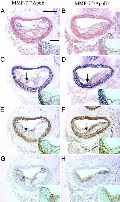
Fig. 2.
Effect of knocking out MMP-7 on apoE knockout mouse atherosclerosis. Histological appearance of representative sections of brachiocephalic arteries from apoE single knockout control mice (A, C, E, and G) and MMP-7/apoE double knockout mice (B, D, F and H). Sections are stained with hematoxylin and eosin (A and B) or elastin van Gieson (C and D) or immunostained for α-smooth muscle actin (smooth muscle cells; E and F) or Mac-2 (macrophages; G and H). Arrows indicate buried fibrous layers rich in smooth muscle cells and invested with elastin. (Scale bars: 200 μm; Inset, 50 μm.)

