Abstract
The interaction between two Fusarium mycotoxins, zearalenone (ZEN) and its derivative ¯α-zearalenol (¯α-ZOL), with two food-grade strains of Lactobacillus was investigated. The mycotoxins (2 μg ml−1) were incubated with either Lactobacillus rhamnosus strain GG or L. rhamnosus strain LC705. A considerable proportion (38 to 46%) of both toxins was recovered from the bacterial pellet, and no degradation products of ZEN and ¯α-ZOL were detected in the high-performance liquid chromatograms of the supernatant of the culturing media and the methanol extract of the pellet. Both heat-treated and acid-treated bacteria were capable of removing the toxins, indicating that binding, not metabolism, is the mechanism by which the toxins are removed from the media. Binding of ZEN or ¯α-ZOL by lyophilized L. rhamnosus GG and L. rhamnosus LC705 was a rapid reaction: approximately 55% of the toxins were bound instantly after mixing with the bacteria. Binding was dependent on the bacterial concentration, and coincubation of ZEN with ¯α-ZOL significantly affected the percentage of the toxin bound, indicating that these toxins may share the same binding site on the bacterial surface. These results can be exploited in developing a new approach for detoxification of mycotoxins from foods and feeds.
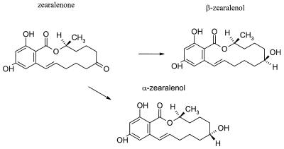
Zearalenone (ZEN) (Fig. 1) is a nonsteroidal estrogenic mycotoxin produced by several species of Fusarium (F. graminareum, F. crookwellemse, F. culmorum, and F. semitectum) which primarily colonize maize and also colonize, to a lesser extent, barley, oats, wheat, and sorghum (12, 17, 20, 30). The level of ZEN in human food can be as high as 289 μg/g (15, 31). Several derivatives of ZEN, including ¯α-zearalenol (¯α-ZOL) and β-zearalenol (β-ZOL) as well as monohydroxylated, dihydroxylated, and formylated ZEN, have been isolated from cultures of Fusarium (27).
FIG. 1.
ZEN and its naturally occurring derivatives, ¯α-ZOL and β-ZOL.
ZEN has predominantly estrogenic properties that are manifested in female swine, cattle, and sheep as reproductive problems (13). Concentrations of 1 to 5 μg of ZEN/g of feed are sufficient to produce clinical symptoms in swine (29). The alpha-reduction of the keto group increases the estrogenic activity of ZEN. ¯α-ZOL has about 10 to 20 times the activity of ZEN and some 100 times that of β-ZOL (24).
ZEN and its metabolites act as growth stimulants, and their occurrence in food has been related to the early onset of puberty in children from Puerto Rico (28). The cooccurrence of ZEN and trichothecenes in contaminated corn has been correlated with the incidence of human esophageal cancer in China (8). The estimated safe human intake of ZEN has been reported to be 0.05 μg/kg of body weight/day (17).
A small number of studies on the degradation and biotransformation of ZEN by various microorganisms have been published. El-Sharkawy et al. (7) investigated the conversion of ZEN by seven genera (23 species) of microorganisms. The metabolites formed included ¯α-ZOL and β-ZOL and another polar metabolite, zearalenone-4-O-sulfate. ZEN was reduced stereoselectively by cultures of Candida tropicalis, Zygosaccharomyces rouxii, and seven Saccharomyces strains to both ¯α-ZOL and β-ZOL (1). When ZEN was incubated with rumen and pig microflora in vitro, it was also reduced to ¯α-ZOL and β-ZOL (14, 16).
Except for some yeast strains and some beneficial rumen microbes, none of the microorganisms tested can be used by either the food and or feed industry for the purpose of ZEN detoxification. In addition, the products of the metabolism of ZEN by the tested microorganisms were more toxic or as toxic as the parent compound. We have found that specific strains of bacteria of both food and intestinal origin, and with a good safety record in the human diet, effectively bind aflatoxin (3) and trichothecenes (2) in vitro. In the present study we have investigated the ability of two food-grade Lactobacillus strains, which are efficient in binding aflatoxins and trichothecenes, to remove ZEN and its main derivative, ¯α-ZOL, from liquid media under variable experimental conditions.
MATERIALS AND METHODS
Bacterial strains.
The bacteria used were Lactobacillus rhamnosus strain GG and L. rhamnosus strain LC705. The strains were obtained from Valio Ltd. (Helsinki, Finland) as a freeze-dried powder. These strains were selected based on their common use by the food industry and on available information regarding their effects on aflatoxins and trichothecenes.
Bacterial counts were determined by flow cytometry with the Coulter Electronics EPICS Elite ESP cytometer equipped with an air-cooled 488-nm argon-ion laser at 15 mV. Total bacterial counts were enumerated by using the fluorescent emission from SYTO9 (LIVE/DEAD BacLight bacterial viability kit, catalog no. L-7012; Molecular Probes, Eugene, Oreg.) at a concentration of 3.34 μM per 106 to 107 bacteria. A 525-nm-pore-size band-pass filter was used to collect the emission for both strains, and Fluoresbrite beads (2.0-μm diameter; Polysciences, Inc., Warrington, Pa.) were used as an internal calibration.
Interaction of ZEN and ¯α-ZOL with bacteria.
ZEN and ¯α-ZOL (Sigma, St. Louis, Mo.) were dissolved in methanol, and their concentrations were determined spectrophotometrically at 236 nm (ɛ236 = 26,030 M−1 cm−1). Aqueous ZEN and ¯α-ZOL solutions were prepared by evaporating the methanol with nitrogen, adding 50 μl of methanol, and then adding phosphate-buffered saline (PBS) (pH 7.3, 0.01 M) to achieve the desired volume.
Bacterial cultures of L. rhamnosus GG and L. rhamnosus LC705 were obtained by incubating 0.1 g of lyophilized bacteria (containing approximately 1010 bacteria) in 10 ml of deMan-Rogosa-Sharpe (MRS) broth (Oxoid, Basingstoke, Hampshire, United Kingdom) under aerobic conditions at 37°C for 24 h. An aliquot of the bacterial culture was transferred into a 15-ml tissue culture tube containing 9 ml of MRS broth and 2 μg of either ZEN or ¯α-ZOL ml−1. The tubes were incubated at 37°C. At 24, 48, and 72 h, 2 ml of the culture was collected and centrifuged at 3,000 × g for 10 min (<10°C). The supernatant was collected and analyzed for the toxins by high-performance liquid chromatography (HPLC). The bacterial pellet was suspended in methanol and centrifuged (3,000 × g for 10 min at <10°C), and the supernatant was analyzed for toxins.
After the initial experiments with cultured bacteria, lyophilized bacteria were used to examine the ability of bacteria to remove ZEN and ¯α-ZOL. The bacteria were first washed once with 3 ml of PBS and then either incubated in 3 ml of PBS for 1 h (viable bacteria), boiled in 3 ml of PBS for 1 h (heat-treated bacteria), or incubated in 3 ml of 2 M HCl for 1 h and washed twice with 3 ml of PBS (acid-treated bacteria). After these treatments, the bacterial samples were centrifuged (3,000 × g for 10 min at <10°C) and the supernatant was removed. The bacterial pellet was suspended in 1.5 ml of PBS containing 4 μg of either ZEN or ¯α-ZOL. The mixture was incubated at 37°C for 30 min and centrifuged at 3,000 × g, and the supernatant was analyzed for toxins by HPLC. All assays were performed in triplicate, and both positive controls (PBS substituted for bacteria) and negative controls (PBS substituted for ZEN and ¯α-ZOL) were included.
The removal of ZEN and ¯α-ZOL from the media was tested at different conditions (incubation time, 0, 4, 24, 48, or 72 h; incubation temperature, 4, 25, or 37°C; bacterial concentration, 1 × 10⁸ to 5 × 10¹⁰ viable cells/ml).
Determination of ZEN and ¯α-ZOL by HPLC.
Reverse-phase HPLC (model LC-10ADvp solvent delivery system and model SIL-10Advp autoinjector; Shimadzu, Kyoto, Japan) was used to quantify the ZEN and ¯α-ZOL remaining in the supernatant of bacteria incubated with either ZEN or ¯α-ZOL. Both toxins were separated on an Allsphere ODS-2 column (250 by 4.6 mm; particle size, 5 μm; Alltech, Deerfield, Ill.) fitted with a Spherisorb ODS-2 guard column (Alltech) with a mobile phase of water-methanol (35:65 [vol/vol]) at a flow rate of 1 ml/min, detected by fluorescence (fluorescence detector RF-10AXL; Shimadzu) at 280 (excitation) and 440 (emission) nm, and quantified by Class VP 5.0 software (Shimadzu). The assay temperature was 30°C with an injection volume of 10 μl, and the retention times were 12 and 13.7 min for ¯α-ZOL and ZEN, respectively.
The percentage of the toxin removed was calculated by using the following equation: 100 × [1 − (peak area of ZEN or ¯α-ZOL in the supernatant/peak area of ZEN or ¯α-ZOL in the positive control)].
Statistical analysis.
SPSS, version 9.0, for Windows was used for the statistical analysis of the data. Analysis of variance was used to test the differences in toxin binding between the strains and at various conditions. Significant differences in the mean values are reported at P values of <0.05.
RESULTS
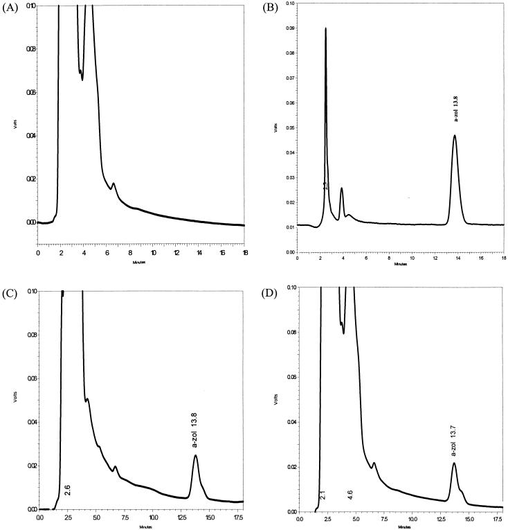
After 72 h of culturing L. rhamnosus GG or L. rhamnosus LC705 with ZEN and ¯α-ZOL, no degradation products were observed on the HPLC chromatograms (Fig. 2), indicating that the strains used in this study were unable to metabolize either ZEN or ¯α-ZOL. ZEN and ¯α-ZOL were fully recovered from the bacterial cells and the culture media, and 38 to 46% of the toxins were found in the bacterial pellet (Table 1).
FIG. 2.
HPLC chromatograms of the supernatant from L. rhamnosus GG culture in MRS broth incubated for 24 h (A), a standard solution of ZEN (2 μg ml−1) (B), the supernatant of L. rhamnosus GG culture with ZEN (2 μg ml−1) (C), and the methanolic extraction of the L. rhamnosus GG cell pellet incubated with ZEN (2 μg ml−1) (D). See the text for HPLC conditions. The retention time for ZEN was 13.7 ± 0.1 min. No other metabolites or degradation products were detected when ZEN was incubated with L. rhamnosus GG.
TABLE 1.
Recovery of ZEN and ¯α-ZOL from the supernatant and cell pellet of the Lactobacillus culturea
| L. rhamnosus strain and incubation period (h) | % Recovery of toxin
|
|||
|---|---|---|---|---|
| ZEN
|
¯α-ZOL
|
|||
| Supernatant | Cell pellet | Supernatant | Cell pellet | |
| GG | ||||
| 24 | 55 | 39 | 68 | 43 |
| 48 | 58 | 46 | 69 | 44 |
| 72 | 63 | 43 | 69 | 44 |
| LC705 | ||||
| 24 | 53 | 38 | 64 | 42 |
| 48 | 50 | 46 | 66 | 44 |
| 72 | 54 | 43 | 66 | 44 |
ZEN and ¯α-ZOL (2 μg ml−1) were added to MRS broth containing either L. rhamnosus GG or L. rhamnosus LC705. The supernatant and pellet fractions were analyzed by HPLC after 24, 48, and 72 h.
Heat treatment and acid treatment significantly enhanced the ability of the bacteria to remove both ZEN and ¯α-ZOL (Table 2).
TABLE 2.
Removal of ZEN and ¯α-ZOL (2.67 μg ml−1) by viable, heat-treated, or acid-treated lyophilized L. rhamnosus GG and L. rhamnosus LC705 (1010 bacteria/ml)
| Toxin | % Removal of toxin from L. rhamnosus straina:
|
|||||
|---|---|---|---|---|---|---|
| GG
|
LC705
|
|||||
| Viable | Heat treated | Acid treated | Viable | Heat treated | Acid treated | |
| ZEN | 52 ± 3 | 70 ± 0 | 69 ± 2 | 47 ± 4 | 76 ± 3 | 76 ± 0 |
| ¯α-ZOL | 50 ± 0 | 68 ± 0 | 68 ± 1 | 43 ± 1 | 75 ± 3 | 76 ± 1 |
Results are averages ± standard deviations (each measurement was done in triplicate and repeated on three consecutive days). There was no significant difference (P < 0.05) between values for the removal of ZEN or ¯α-ZOL by heat- or acid-treated cells of the same strain.
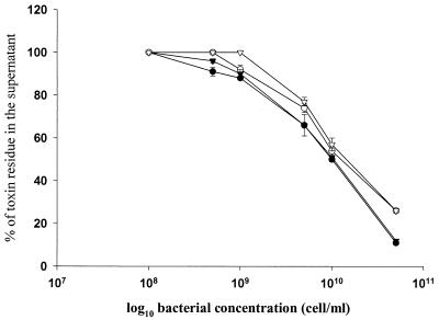
The removal of both toxins by L. rhamnosus GG and L. rhamnosus LC705 from the liquid media was dependent on the concentration of the bacteria in the incubation medium (Fig. 3). A minimum of 109 bacterial cells/ml was required for significant removal of ZEN and ¯α-ZOL.
FIG. 3.
Effect of bacterial (L. rhamnosus GG and L. rhamnosus LC705) concentrations on the removal of ZEN and ¯α-ZOL from liquid media. Error bars represent standard deviations. •, L. rhamnosus GG plus ZEN; ○, L. rhamnosus LC705 plus ZEN; ▾, L. rhamnosus GG plus ¯α-ZOL; ▿, L. rhamnosus LC705 plus ¯α-ZOL.
The removal of ZEN and ¯α-ZOL by both L. rhamnosus GG and L. rhamnosus LC705 was not dependent on the incubation temperature, since both toxins were removed by both strains at all incubation temperatures used. The percentage of ZEN removed was 58, 60, and 56 at 4, 25, and 37°C, respectively. Similar results were also obtained for ¯α-ZOL.
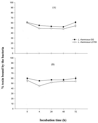
Binding of ZEN and ¯α-ZOL was a rapid reaction since approximately 60% of ZEN and ¯α-ZOL was removed from the liquid media within the 10-min centrifugation after mixing with either L. rhamnosus GG or L. rhamnosus LC705 (Fig. 4). Interestingly, both strains released some ZEN and ¯α-ZOL back into the liquid media during the first 4 h of incubation (0 versus 4 h, P < 0.05). However, the ability of the bacteria to remove ZEN and ¯α-ZOL from liquid media increased again when the incubation was continued. Incubation of a mixture of ZEN and ¯α-ZOL (1:1) significantly decreased the percentage of toxins removed by both strains (Table 3).
FIG. 4.
Effect of incubation time on the removal of ZEN (A) and ¯α-ZOL (B) by L. rhamnosus GG and L. rhamnosus LC705. Error bars represent standard deviations.
TABLE 3.
Removal of ZEN and ¯α-ZOL by L. rhamnosus GG and L. rhamnosus LC705 from a mixture of toxinsb
| L. rhamnosus strain | % Removal of toxin
|
|||
|---|---|---|---|---|
| ZENa
|
¯α-ZOLa
|
|||
| Alone | With ¯α-ZOL | Alone | With ZEN | |
| IGG | 51 ± 1 | 30 ± 3 | 51 ± 0.5 | 45 ± 1 |
| LC705 | 46 ± 3 | 37 ± 1 | 46 ± 0.5 | 39 ± 2 |
The percentage of toxin removed differed significantly (P < 0.05) when the toxin was incubated alone or coincubated with the other toxin.
ZEN and/or ¯α-ZOL (2.67 μg ml−1) was incubated with L. rhamnosus GG or L. rhamnosus LC705 (1010 bacteria), and the residue in the supernatant was measured by HPLC. Results are the averages ± standard deviations of triplicate samples.
DISCUSSION
The present study demonstrates that selected food-grade strains of lactic acid bacteria have the ability to remove ZEN and its derivative ¯α-ZOL, known food and feed contaminants, from the liquid media. The same strains have been previously shown to bind aflatoxins (3, 25) and trichothecenes (2).
To distinguish between true biodegradation, abiotic degradation, and sequestration (19) of the mycotoxins, the fate of ZEN and its derivative ¯α-ZOL was examined in growing cultures of the lactobacilli. The observation that all of the added ZEN and ¯α-ZOL was recovered from the bacterial cells and the culture media indicates that ZEN and ¯α-ZOL were chemically stable and potentially bioavailable under the incubation conditions. However, no degradation products of either ZEN or ¯α-ZOL were observed on the HPLC chromatogram after 72 h of incubation with either L. rhamnosus GG or L. rhamnosus LC705, indicating that these strains were unable to metabolize either ZEN or ¯α-ZOL. Megharaj et al. (21) reported that after 18 days of incubation nearly 90% of the ZEN added to the medium containing Pseudomonas fluorescens was associated with the bacterial cells or dissolved in the culture media.
Recovery of nearly 50% of ZEN and ¯α-ZOL from the bacterial cells by methanol extraction indicates that these toxins were associated with the bacterial surface and not absorbed by the bacteria. In the present study, the percentage of the added ZEN or ¯α-ZOL associated with the bacterial cells was slightly lower than that reported by Megharaj et al. (21) but similar to the binding of aflatoxin B1 by the same strains (9, 22). In some other studies, transformation of ZEN by microorganisms to more-potent estrogenic zearalenols has been observed (1, 7, 14, 16). This indicates that biodegradation of ZEN by bacteria is not a suitable approach to detoxify ZEN and its derivatives. Consequently, we decided to investigate in more detail the conditions under which these strains remove ZEN and ¯α-ZOL from liquid media.
Treatment of the bacteria by heat and acid significantly enhanced the ability of the bacteria to remove both ZEN and ¯α-ZOL from liquid media. Cell wall polysaccharide and peptidoglycan (26) are the two main elements responsible for the binding of mutagens to Streptococcus and Lactobacillus (10, 11, 32). Both of these components are expected to be affected by heating and acids. Heating may cause protein denaturation or the formation of Maillard reaction products between polysaccharides and peptides and proteins, whereas under acidic conditions, the glycosidic linkages in polysaccharides break down releasing monomers that may then be further fragmented into aldehydes. Acids may also break the amide linkages in peptides and proteins, producing peptides and the component amino acids. The peptidoglycan structure of the cell wall is usually quite thick in these organisms (9), but its thickness may be reduced and/or its pore size may be increased via heat and acid treatments. This perturbation of the bacterial cell wall may allow both ZEN and ¯α-ZOL to bind to cell wall and plasma membrane constituents that are not available when the bacterial cell is intact. The debris observed by flow cytometry in acid-treated bacterial samples may have resulted from peptidoglycan breakdown (unpublished observation). The effective removal of ZEN and ¯α-ZOL by all nonviable bacteria suggests that binding, rather than metabolism, is involved. This is in agreement with previous findings on aflatoxins and trichothecenes (4, 23, 25).
The concentration of bacteria in the incubation medium needed for significant removal of ZEN and ¯α-ZOL by L. rhamnosus GG or L. rhamnosus LC705 was similar to that reported for aflatoxin binding by the same lactobacilli (3, 18).
Upon mixing ZEN and ¯α-ZOL with either L. rhamnosus GG or L. rhamnosus LC705, significant amounts of both toxins were removed from the incubation medium, indicating that the removal was a rapid reaction. When incubation was continued, the toxins were first released to the liquid media and then rebound by the bacteria. Similar fluctuations in binding have been reported for the removal of aflatoxin B1 from liquid media by these same strains (3).
The ability of both Lactobacillus strains to remove ZEN and ¯α-ZOL similarly to aflatoxin B1 and trichothecenes led us to believe that these bacteria have many binding sites for different toxins. To investigate this, we incubated ZEN with or without ¯α-ZOL and ¯α-ZOL with or without ZEN together with L. rhamnosus GG or L. rhamnosus LC705. Incubation of a mixture of toxins (1:1) significantly decreased the percentage of toxins removed by both strains. The data indicate that the toxins may share the same binding sites, with L. rhamnosus GG having a higher affinity to ¯α-ZOL than to ZEN.
This study clearly shows that both L. rhamnosus GG and L. rhamnosus LC705 significantly reduce the levels of ZEN and its main derivative ¯α-ZOL) in liquid media. Both L. rhamnosus GG and L. rhamnosus LC705 are probiotic strains and are currently used by the food industry in different dairy products. These strains can bind aflatoxin B1 in the chicken duodenum (5) and influence the absorption of aflatoxins in humans (6). Similar studies are planned for ZEN and ¯α-ZOL to investigate whether the binding ability is functional under physiological conditions and if such binding will reduce the absorption of these toxins from the intestine and hence reduce the estrogenic effects of these toxins. These studies, together with the ability of these strains to bind aflatoxins (3) and trichothecenes (2), offer a potential approach to reduce the intestinal absorption of mycotoxins from the human diet and animal feeds.
Acknowledgments
This work was supported by a grant to H.M. from the Academy of Finland.
We thank Valio Ltd., Helsinki, Finland, for providing the strains used in this study.
REFERENCES
- 1.Böswald, C., G. Engelhardt, H. Vogel, and P. R. Wallnöfer. 1995. Metabolism of the Fusarium mycotoxins zearalenone and deoxynivalenol by yeast strains of technological relevance. Nat. Toxins 3:138-144. [DOI] [PubMed] [Google Scholar]
- 2.El-Nezami, H. S., A. Chrevatidis, S. Auriola, S. Salminen, and H. Mykkänen. Removal of common Fusarium toxins in vitro by strains of Lactobacillus and Propionibacterium. Food Addit. Contam., in press. [DOI] [PubMed]
- 3.El-Nezami, H. S., P. E. Kankaanpää, S. Salminen, and J. T. Ahokas. 1998. Ability of dairy strains of lactic acid bacteria to bind food carcinogens. Food Chem. Toxicol. 36:321-326. [DOI] [PubMed] [Google Scholar]
- 4.El-Nezami, H. S., P. E. Kankaanpää, S. Salminen, and J. T. Ahokas. 1998. Physico-chemical alterations enhance the ability of dairy strains of lactic acid bacteria to remove aflatoxins from contaminated media. J. Food Prot. 61:466-468. [DOI] [PubMed] [Google Scholar]
- 5.El-Nezami, H. S., H. Mykkänen, P. Kankaanpää, S. Salminen, and J. T. Ahokas. 2000. Ability of Lactobacillus and Propionibacterium strains to remove aflatoxin B1, from the chicken duodenum. J. Food. Prot. 63:549-552. [DOI] [PubMed] [Google Scholar]
- 6.El-Nezami, H. S., H. Mykkänen, P. Kankaanpää, T. Suomalainen, J. T. Ahokas, and S. Salminen. 2000. The ability of a mixture of Lactobacillus and Propionibacterium examining to influence the faecal recovery of aflatoxins in healthy Egyptian volunteers: a pilot clinical study. Biosci. Microflora 19:41-45. [Google Scholar]
- 7.El-Sharkawy, S., M. Selim, M. Afifi, and F. Halaweish. 1991. Microbial transformation of zearalenone to a zearalenone sulfate. Appl. Environ. Microbiol. 57:549-552. [DOI] [PMC free article] [PubMed] [Google Scholar]
- 8.Gao, H.-P., and T. Yoshizawa. 1997. Further study on Fusarium mycotoxins in corn and wheat from a high-risk area for human esophageal cancer in China. Mycotoxins 45:51-55. [Google Scholar]
- 9.Haskard C., H. El-Nezami, P. Kankaanpää, S. Salminen, and J. T. Ahokas. 2001. Surface binding of aflatoxin B1 by lactic acid bacteria. Appl. Environ. Microbiol. 67:3086-3091. [DOI] [PMC free article] [PubMed] [Google Scholar]
- 10.Hosono, A., T. Tanabe, and H. Otani. 1990. Binding properties of lactic acid bacteria isolates from kefir milk with mutagenic amino acid pyrolyzates. Milchwissenschaft 45:647-651. [Google Scholar]
- 11.Hosono, A., A. Yoshimura, and H. Otani. 1988. Desmutagenic property of cell walls of Streptococcus faecalis on the mutagenicities induced by amino acid pyrolysates. Milchwissenschaft 43:168-170. [Google Scholar]
- 12.International Agency for Research on Cancer. 1993. Some naturally occurring substances: food items and constituents, heterocyclic aromatic amines and mycotoxins, p. 397-444. In IARC monographs on the evaluation of carcinogenic risks to humans, vol. 56. International Agency for Research on Cancer, Lyon, France.
- 13.Jemmali, M. 1987. Trade and economic implications of mycotoxins: needs for greater uniformity, MYC 87/6, p. 164. In Joint FAO/W. H. O./UNEP Second International Conference on Mycotoxins. FAO, Bangkok, Thailand.
- 14.Kiessling, K.-H., H. Pettersson, K. Sandholm, and M. Olsen. 1984. Metabolism of aflatoxin, ochratoxin, zearalenone, and three trichothecenes by intact rumen fluid, rumen protozoa, and rumen bacteria. Appl. Environ. Microbiol. 47:1070-1073. [DOI] [PMC free article] [PubMed] [Google Scholar]
- 15.Kim, J.-C., H.-J. Kang, D.-H. Lee, Y.-W. Lee, and T. Yoshizawa. 1993. Natural occurrence of Fusarium mycotoxins (trichothecenes and zearalenone) in barley and corn in Korea. Appl. Environ. Microbiol. 59:3798-3802. [DOI] [PMC free article] [PubMed] [Google Scholar]
- 16.Kollarczik, B., M. Gareis, and M. Hanelt. 1994. In vitro transformation of the Fusarium mycotoxins deoxynivalenol and zearalenone by the normal gut microflora of pigs. Nat. Toxins 2:105-110. [DOI] [PubMed] [Google Scholar]
- 17.Kuiper-Godman, T., P. M. Scott, and H. Watanabe. 1987. Risk assessment of the mycotoxin zearalenone. Regul. Toxicol. Pharmacol. 7:253-306. [DOI] [PubMed] [Google Scholar]
- 18.Line, J. E., and R. E. Brackett. 1995. Factors affecting aflatoxin B1 removal by Flavobacterium aurantiacum. J. Food Prot. 58:91-94. [DOI] [PubMed] [Google Scholar]
- 19.Madsen, E. L. 1991. Determining in situ bioremediation, facts and challenges. Environ. Sci. Technol. 25:1663-1673. [Google Scholar]
- 20.Marasas, W., P. Nelson, and T. Toussoun. 1984. Toxigenic Fusarium species. Identity and mycotoxicology. Pennsylvania State University Press, University Park, Pa.
- 21.Megharaj, M., I. Garthwaite, and J. H. Thiele. 1997. Total biodegradation of the oestrogenic mycotoxin zearalenone by a bacterial culture. Lett. Appl. Microbiol. 24:329-333. [DOI] [PubMed] [Google Scholar]
- 22.Oatley, J. T., M. D. Rarick, G. E. Ji, and J. E. Linz. 2000. Binding of aflatoxin B1 to bifidobacteria in vitro. J. Food Prot. 63:1133-1136. [DOI] [PubMed] [Google Scholar]
- 23.Peltonen, K., H. El-Nezami, S. Salminen, and J. Ahokas. 2000. Binding of aflatoxin B1 by probiotic bacteria. J. Sci. Food Agric. 80:1-4. [Google Scholar]
- 24.Pfohl-Leszkowicz, A., L. Chekir-Ghedira, and H. Bacha. 1995. Genotoxicity of zearalenone, an estrogenic mycotoxin: DNA adduct formation in female mouse tissues. Carcinogenesis 16:2315-2320. [DOI] [PubMed] [Google Scholar]
- 25.Pieridis, M., H. El-Nezami, K. Peltonen, S. Salminen, and J. Ahokas. 2000. Ability of dairy strains of lactic acid bacteria to bind aflatoxin M1 in a food model. J. Food Prot. 63:645-650. [DOI] [PubMed] [Google Scholar]
- 26.Rajendran, R., and Y. Ohta. 1998. Binding of heterocyclic amines by lactic acid bacteria from miso, a fermented Japanese food. Can. J. Microbiol. 44:109-115. [DOI] [PubMed] [Google Scholar]
- 27.Richardson, K. E., W. M. Hagler, C. A. Haney, P. B. Hamilton. 1985. Zearalenone and trichothecene production in soyabeans by toxigenic Fusarium. J. Food Prot. 48:240-243. [DOI] [PubMed] [Google Scholar]
- 28.Schoental, R. 1983. Precocious sexual development in Puerto Rico and oestrogenic mycotoxins (zearalenone). Lancet i:537. [DOI] [PubMed]
- 29.Scott, P. M., H. L. Trenholm, and M. D. Sutton (ed.). 1985. Mycotoxins: a Canadian perspective. National Research Council of Canada, Ottawa, Canada.
- 30.Thrane, U. 1989. Fusarium species and their specific profiles of secondary metabolites, p. 199-225. In J. Chelkowski (ed.), Fusarium mycotoxins. Taxonomy and pathogenicity. Elsevier, Amsterdam, The Netherlands.
- 31.Yuwai, K. E., K. L. Rao, K. Singh, T. Tanaka, and Y. Ueno. 1994. Occurrence of nivalenol, deoxynivalenol and zearalenone in imported cereals in Papua New Guinea. Nat. Toxins 2:19-21. [DOI] [PubMed] [Google Scholar]
- 32.Zhang, X. B., and Y. Ohta. 1993. Antimutagenicity of cell fractions of microorganisms on potent mutagenic pyrolysates. Mutat. Res. 298:247-253. [DOI] [PubMed] [Google Scholar]


