

Abstract
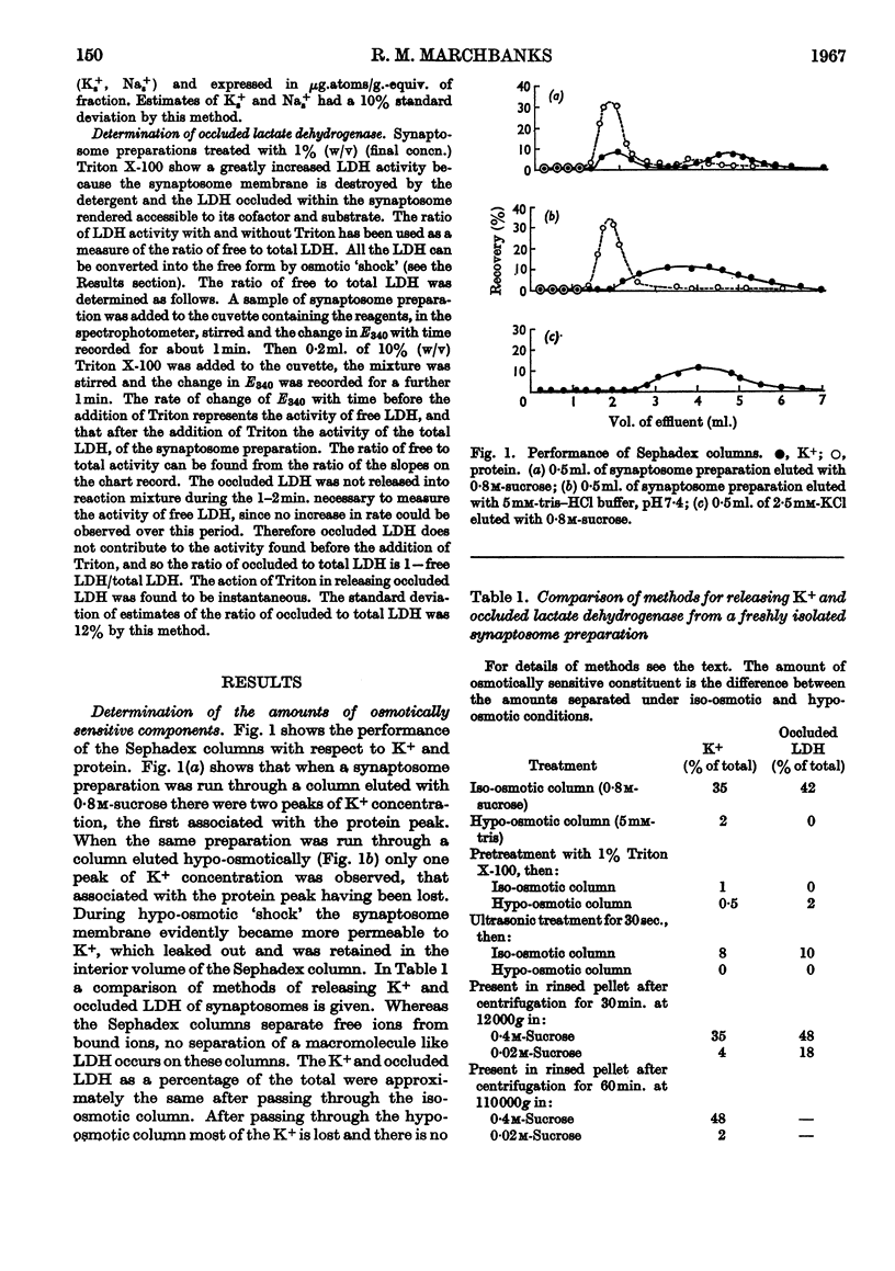
1. Synaptosomes are pinched-off nerve terminals whose components can be liberated by osmotic `shock'. A synaptosome preparation run through a Sephadex column that was eluted with an iso-osmotic solution retained its small ions, whereas when the column was eluted hypo-osmotically the small ions were lost. In this way the osmotically sensitive Na+ and K+ of synaptosomes were measured. Measurements of the lactate dehydrogenase occluded within the synaptosome were also made. The release of osmotically sensitive Na+ and K+ and occluded lactate dehydrogenase had similar characteristics with respect to the degree of osmotic `shock' necessary and the action of lytic agents. 2. The distribution of osmotically sensitive Na+, K+ and occluded lactate dehydrogenase in the subfractions of a crude mitochondrial preparation was examined. The synaptosome fraction was the richest source of these constituents. 3. On standing at 5° in iso-osmotic solution Na+ and K+ were lost from synaptosomes, whereas the amount of occluded lactate dehydrogenase remained stable, suggesting that the synaptosome membrane retained its integrity but that Na+ and K+ diffused through it out of the osmotically sensitive compartment. 4. The uptake of Na+ and K+ into the osmotically sensitive compartment was examined. At 5° the rates of uptake of Na+ and K+ were found to be equal to the rates of loss of these ions when correction to a uniform concentration gradient had been made. K+ travelled across the membrane slightly faster than Na+, the rate of K+ movement being about 1·0μμequiv.cm.−2sec.−1 under a concentration gradient of 0·1m. Active transport is not thought to contribute to the ion movements under the conditions used. 5. The amount of K+ taken up into the osmotically sensitive compartment as a function of the external concentration was examined. Since the uncharged molecule d-[14C]galactose distributes across the synaptosome membrane similarly to K+ there is not thought to be a synaptosomal trans-membrane potential. The volume of the osmotically sensitive compartment was measured by this method and found to agree with estimates of the synaptosomal volume made from morphological studies. In media of low ionic strength synaptosomes exhibit a Donnan effect. 6. It is concluded from these studies that the osmotically sensitive compartment represents the inner volume of the synaptosome, which is completely separated from the outside environment by a diffusion barrier having many of the general properties of a biological membrane.
Full text
PDF


Selected References
These references are in PubMed. This may not be the complete list of references from this article.
- Clementi F., Whittaker V. P., Sheridan M. N. The yield of synaptosomes from the cerebral cortex of guinea pigs estimated by a polystyrene bead "tagging" procedure. Z Zellforsch Mikrosk Anat. 1966;72(1):126–138. doi: 10.1007/BF00336902. [DOI] [PubMed] [Google Scholar]
- DE ROBERTIS E., PELLEGRINO DE IRALDI A., RODRIGUEZ DE LORES GARNAIZ G., SALGANICOFF L. Cholinergic and non-cholinergic nerve endings in rat brain. I. Isolation and subcellular distribution of acetylcholine and acetylcholinesterase. J Neurochem. 1962 Jan-Feb;9:23–35. doi: 10.1111/j.1471-4159.1962.tb07489.x. [DOI] [PubMed] [Google Scholar]
- GUILLET M., FOWLER R. C. Experimental variations in sonic fragility of red cells. J Gen Physiol. 1954 May 20;37(5):621–630. doi: 10.1085/jgp.37.5.621. [DOI] [PMC free article] [PubMed] [Google Scholar]
- Hosie R. J. The localization of adenosine triphosphatases in morphologically characterized subcellular fractions of guinea-pig brain. Biochem J. 1965 Aug;96(2):404–412. doi: 10.1042/bj0960404. [DOI] [PMC free article] [PubMed] [Google Scholar]
- JOHNSON M. K. The intracellular distribution of glycolytic and other enzymes in rat-brain homogenates and mitochondrial preparations. Biochem J. 1960 Dec;77:610–618. doi: 10.1042/bj0770610. [DOI] [PMC free article] [PubMed] [Google Scholar]
- Keynes R. D., Ritchie J. M. The movements of labelled ions in mammalian non-myelinated nerve fibres. J Physiol. 1965 Jul;179(2):333–367. doi: 10.1113/jphysiol.1965.sp007666. [DOI] [PMC free article] [PubMed] [Google Scholar]
- LOWRY O. H., ROSEBROUGH N. J., FARR A. L., RANDALL R. J. Protein measurement with the Folin phenol reagent. J Biol Chem. 1951 Nov;193(1):265–275. [PubMed] [Google Scholar]
- PETHICA B. A., SCHULMAN J. H. The physical chemistry of haemolysis by surface-active agents. Biochem J. 1953 Jan;53(2):177–185. doi: 10.1042/bj0530177. [DOI] [PMC free article] [PubMed] [Google Scholar]
- USSING H. H. Some aspects of the application of tracers in permeability studies. Adv Enzymol Relat Subj Biochem. 1952;13:21–65. doi: 10.1002/9780470122587.ch2. [DOI] [PubMed] [Google Scholar]
- Whittaker V. P., Michaelson I. A., Kirkland R. J. The separation of synaptic vesicles from nerve-ending particles ('synaptosomes'). Biochem J. 1964 Feb;90(2):293–303. doi: 10.1042/bj0900293. [DOI] [PMC free article] [PubMed] [Google Scholar]