

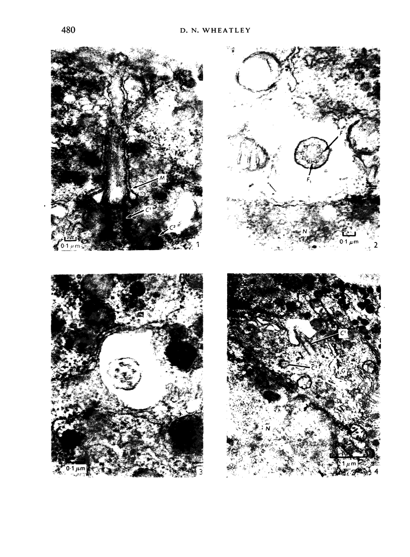
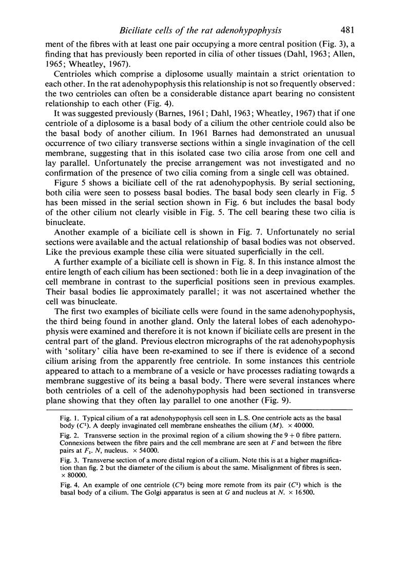
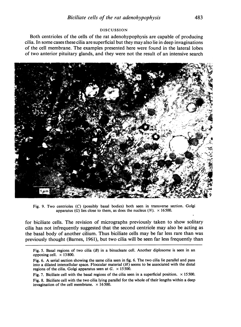
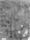
Full text
PDF
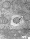
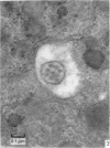
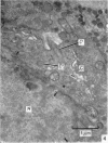
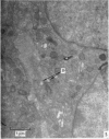
Images in this article
Selected References
These references are in PubMed. This may not be the complete list of references from this article.
- Allen R. A. Isolated cilia in inner retinal neurons and in retinal pigment epithelium. J Ultrastruct Res. 1965 Jun;12(5):730–747. doi: 10.1016/s0022-5320(65)80058-2. [DOI] [PubMed] [Google Scholar]
- BARNES B. G. Ciliated secretory cells in the pars distalis of the mouse hypophysis. J Ultrastruct Res. 1961 Oct;5:453–467. doi: 10.1016/s0022-5320(61)80019-1. [DOI] [PubMed] [Google Scholar]
- COUPLAND R. E. (ELECTRON MICROSCOPIC OBSERVATIONS ON THE STRUCTURE OF THE RAT ADRENAL MEDULLA. I. THE ULTRASTRUCTURE AND ORGANIZATION OF CHROMAFFIN CELLS IN THE NORMAL ADRENAL MEDULLA.) J Anat. 1965 Apr;99:231–254. [PMC free article] [PubMed] [Google Scholar]
- DAHL H. A. Fine structure of cilia in rat cerebral cortex. Z Zellforsch Mikrosk Anat. 1963;60:369–386. doi: 10.1007/BF00336612. [DOI] [PubMed] [Google Scholar]
- GRILLO M. A., PALAY S. L. Ciliated Schwann cells in the autonomic nervous system of the adult rat. J Cell Biol. 1963 Feb;16:430–436. doi: 10.1083/jcb.16.2.430. [DOI] [PMC free article] [PubMed] [Google Scholar]
- KARNOVSKY M. J. Simple methods for "staining with lead" at high pH in electron microscopy. J Biophys Biochem Cytol. 1961 Dec;11:729–732. doi: 10.1083/jcb.11.3.729. [DOI] [PMC free article] [PubMed] [Google Scholar]
- Kagayama M. The follicular cell in the pars distalis of the dog pituitary gland: an electron microscope study. Endocrinology. 1965 Dec;77(6):1053–1060. doi: 10.1210/endo-77-6-1053. [DOI] [PubMed] [Google Scholar]
- LUFT J. H. Improvements in epoxy resin embedding methods. J Biophys Biochem Cytol. 1961 Feb;9:409–414. doi: 10.1083/jcb.9.2.409. [DOI] [PMC free article] [PubMed] [Google Scholar]
- MUNGER B. L. A light and electron microscopic study of cellular differentiation in the pancreatic islets of the mouse. Am J Anat. 1958 Sep;103(2):275–311. doi: 10.1002/aja.1001030207. [DOI] [PubMed] [Google Scholar]
- MUNGER B. L., ROTH S. I. The cytology of the normal parathyroid glands of man and Virginia deer; a light and electron microscopic study with morphologic evidence of secretory activity. J Cell Biol. 1963 Feb;16:379–400. doi: 10.1083/jcb.16.2.379. [DOI] [PMC free article] [PubMed] [Google Scholar]
- PALADE G. E. A study of fixation for electron microscopy. J Exp Med. 1952 Mar;95(3):285–298. doi: 10.1084/jem.95.3.285. [DOI] [PMC free article] [PubMed] [Google Scholar]
- SALAZAR H. THE PARS DISTALIS OF THE FEMALE RABBIT HYPOPHYSIS: AN ELECTRON MICROSCOPIC STUDY. Anat Rec. 1963 Dec;147:469–497. doi: 10.1002/ar.1091470404. [DOI] [PubMed] [Google Scholar]
- VEGGE T. Ultrastructure of normal human trabecular endothelium. Acta Ophthalmol (Copenh) 1963;41:193–199. doi: 10.1111/j.1755-3768.1963.tb03540.x. [DOI] [PubMed] [Google Scholar]
- WILSON R. B., McWHORTER C. A. Isolated flagella in human skin. Election microscopic observations. Lab Invest. 1963 Feb;12:242–249. [PubMed] [Google Scholar]
- Wheatley D. N. Cilia and centrioles of the rat adrenal cortex. J Anat. 1967 Apr;101(Pt 2):223–237. [PMC free article] [PubMed] [Google Scholar]
- ZEIGEL R. F. On the occurrence of cilia in several cell types of the chick pancreas. J Ultrastruct Res. 1962 Oct;7:286–292. doi: 10.1016/s0022-5320(62)90024-2. [DOI] [PubMed] [Google Scholar]