

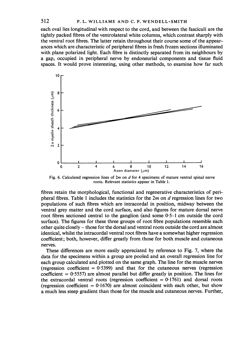
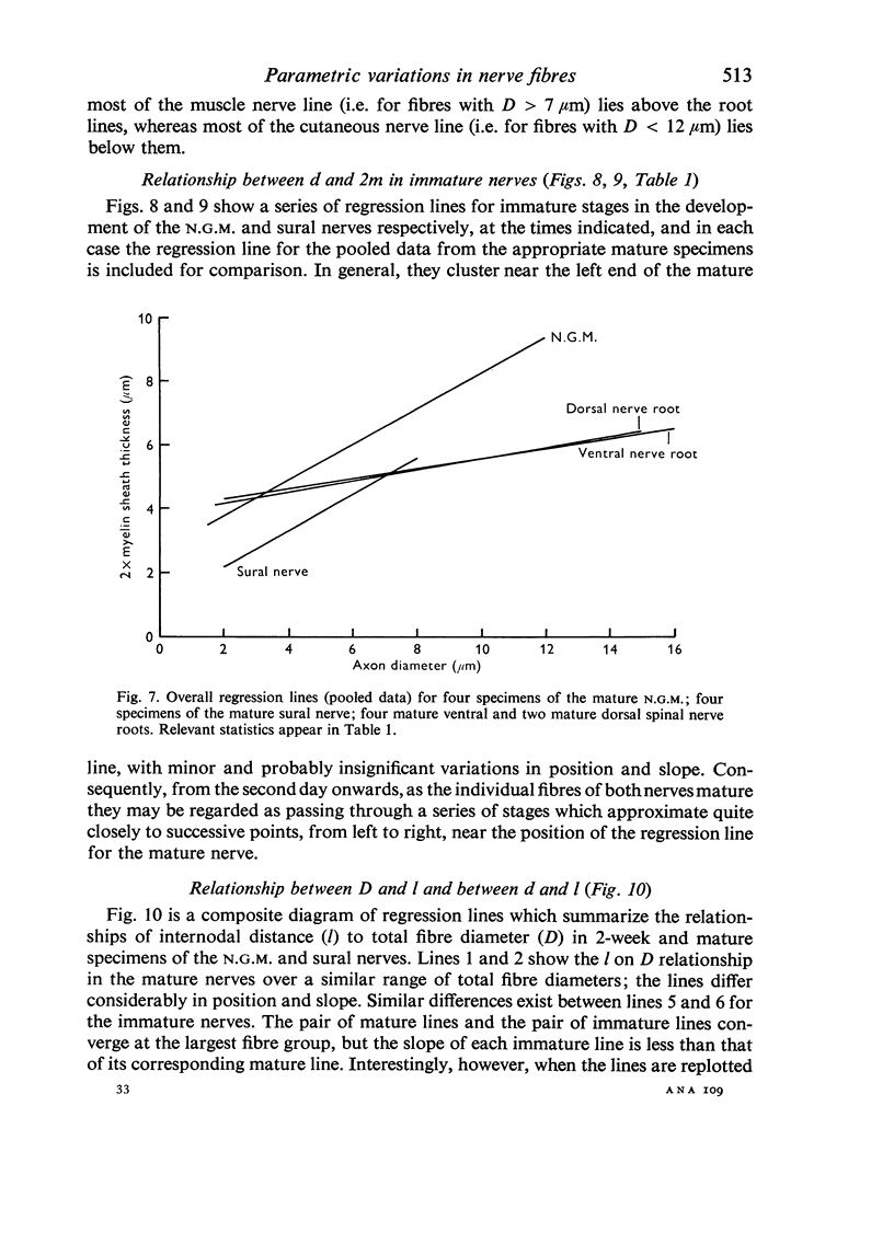
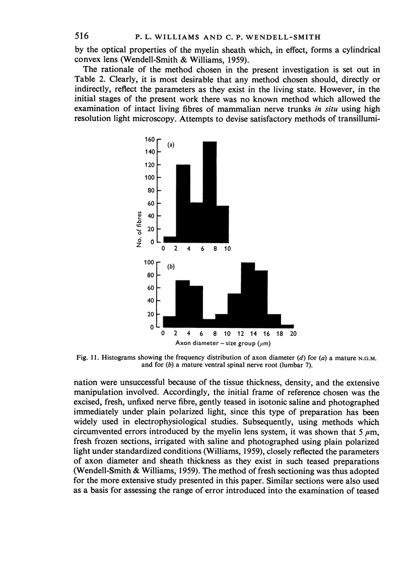
Full text
PDF


Selected References
These references are in PubMed. This may not be the complete list of references from this article.
- CRAGG B. G., THOMAS P. K. The relationships between conduction velocity and the diameter and internodal length of peripheral nerve fibres. J Physiol. 1957 May 23;136(3):606–614. doi: 10.1113/jphysiol.1957.sp005785. [DOI] [PMC free article] [PubMed] [Google Scholar]
- Donovan A. The nerve fibre composition of the cat optic nerve. J Anat. 1967 Jan;101(Pt 1):1–11. [PMC free article] [PubMed] [Google Scholar]
- Dyck P. J. Histologic measurements and fine structure of biopsied sural nerve: normal, and in peroneal muscular atrophy, hypertrophic neuropathy, and congenital sensory neuropathy. Mayo Clin Proc. 1966 Nov;41(11):742–774. [PubMed] [Google Scholar]
- ESMOND W. G., SMITH A. The structure of adult peripheral myelinated nerve fibers as revealed by phase microscopy. Exp Cell Res. 1958 Apr;14(2):430–433. doi: 10.1016/0014-4827(58)90202-7. [DOI] [PubMed] [Google Scholar]
- EVANS D. H. L., VIZOSO A. D. Observations on the mode of growth of motor nerve fibers in rabbits during post-natal development. J Comp Neurol. 1951 Dec;95(3):429–461. doi: 10.1002/cne.900950304. [DOI] [PubMed] [Google Scholar]
- FERNAND V. S. V., YOUNG J. Z. The sizes of the nerve fibres of muscle nerves. Proc R Soc Lond B Biol Sci. 1951 Dec 31;139(894):38–58. doi: 10.1098/rspb.1951.0045. [DOI] [PubMed] [Google Scholar]
- FINEAN J. B. X-ray diffraction studies on the myelin sheath in peripheral and central nerve fibres. Exp Cell Res. 1958;14(Suppl 5):18–32. [PubMed] [Google Scholar]
- Friede R. L., Samorajski T. Axon caliber related to neurofilaments and microtubules in sciatic nerve fibers of rats and mice. Anat Rec. 1970 Aug;167(4):379–387. doi: 10.1002/ar.1091670402. [DOI] [PubMed] [Google Scholar]
- Friede R. L., Samorajski T. Myelin formation in the sciatic nerve of the rat. A quantitative electron microscopic, histochemical and radioautographic study. J Neuropathol Exp Neurol. 1968 Oct;27(4):546–570. [PubMed] [Google Scholar]
- Friede R. L., Samorajski T. Relation between the number of myelin lamellae and axon circumference in fibers of vagus and sciatic nerves of mice. J Comp Neurol. 1967 Jul;130(3):223–231. doi: 10.1002/cne.901300304. [DOI] [PubMed] [Google Scholar]
- HESS A., YOUNG J. Z. The nodes of Ranvier. Proc R Soc Lond B Biol Sci. 1952 Nov 20;140(900):301–320. doi: 10.1098/rspb.1952.0063. [DOI] [PubMed] [Google Scholar]
- HODLER J., STAMPFLI R., TASAKI I. Role of potential wave spreading along myelinated nerve fiber in exictation and conduction. Am J Physiol. 1952 Aug;170(2):375–389. doi: 10.1152/ajplegacy.1952.170.2.375. [DOI] [PubMed] [Google Scholar]
- Hall S. M., Williams P. L. The distribution of electron-dense tracers in peripheral nerve fibres. J Cell Sci. 1971 Mar;8(2):541–555. doi: 10.1242/jcs.8.2.541. [DOI] [PubMed] [Google Scholar]
- Huxley A. F., Stämpfli R. Evidence for saltatory conduction in peripheral myelinated nerve fibres. J Physiol. 1949 May 15;108(3):315–339. [PMC free article] [PubMed] [Google Scholar]
- LANDON D. N., WILLIAMS P. L. ULTRASTRUCTURE OF THE NODE OF RANVIER. Nature. 1963 Aug 10;199:575–577. doi: 10.1038/199575a0. [DOI] [PubMed] [Google Scholar]
- Landon D. N., Langley O. K. The local chemical environment of nodes of Ranvier: a study of cation binding. J Anat. 1971 Apr;108(Pt 3):419–432. [PMC free article] [PubMed] [Google Scholar]
- Ochoa J. The sural nerve of the human foetus: electron microscope observations and counts of axons. J Anat. 1971 Feb;108(Pt 2):231–245. [PMC free article] [PubMed] [Google Scholar]
- QUILLIAM T. A. Some characteristics of myelinated fibre populations. J Anat. 1956 Apr;90(2):172–187. [PMC free article] [PubMed] [Google Scholar]
- SCHWARZACHER H. G. Markscheidendicke und Achsenzylinderdurchmesser in peripheren menschlichen Nerven. Acta Anat (Basel) 1954;21(1):26–46. [PubMed] [Google Scholar]
- STAMPFLI R. Saltatory conduction in nerve. Physiol Rev. 1954 Jan;34(1):101–112. doi: 10.1152/physrev.1954.34.1.101. [DOI] [PubMed] [Google Scholar]
- SUNDERLAND S., ROCHE A. F. Axon-myelin relationships in peripheral nerve fibres. Acta Anat (Basel) 1958;33(1-2):1–37. doi: 10.1159/000141338. [DOI] [PubMed] [Google Scholar]
- Sanders F. K., Whitteridge D. Conduction velocity and myelin thickness in regenerating nerve fibres. J Physiol. 1946 Sep 18;105(2):152–174. [PMC free article] [PubMed] [Google Scholar]
- TASAKI I. New measurements of the capacity and the resistance of the myelin sheath and the nodal membrane of the isolated frog nerve fiber. Am J Physiol. 1955 Jun;181(3):639–650. doi: 10.1152/ajplegacy.1955.181.3.639. [DOI] [PubMed] [Google Scholar]
- THOMAS P. K., YOUNG J. Z. Internode lengths in the nerves of fishes. J Anat. 1949 Oct;83(4):336-50, pl. [PMC free article] [PubMed] [Google Scholar]
- TIEGS O. W. Innervation of voluntary muscle. Physiol Rev. 1953 Jan;33(1):90–144. doi: 10.1152/physrev.1953.33.1.90. [DOI] [PubMed] [Google Scholar]
- Vizoso A. D., Young J. Z. Internode length and fibre diameter in developing and regenerating nerves. J Anat. 1948 Apr;82(Pt 1-2):110–134.1. [PMC free article] [PubMed] [Google Scholar]
- WENDELL-SMITH C. P., WILLIAMS P. L. Some structural characteristics of myelinated nerve fibres. Nature. 1958 Dec 6;182(4649):1608–1609. doi: 10.1038/1821608b0. [DOI] [PubMed] [Google Scholar]
- WILLIAMS P. L., LANDON D. N. Paranodal apparatus of peripheral myelinated nerve fibres of mammals. Nature. 1963 May 18;198:670–673. doi: 10.1038/198670a0. [DOI] [PubMed] [Google Scholar]
- Williams P. L., Hall S. M. In vivo observations on mature myelinated nerve fibres of the mouse. J Anat. 1970 Jul;107(Pt 1):31–38. [PMC free article] [PubMed] [Google Scholar]
- Williams P. L., Hall S. M. Prolonged in vivo observations of normal peripheral nerve fibres and their acute reactions to crush and deliberate trauma. J Anat. 1971 Apr;108(Pt 3):397–408. [PMC free article] [PubMed] [Google Scholar]