Abstract
Gatifloxacin is a new 8-methoxy fluoroquinolone with enhanced activity against gram-positive cocci. We used the neutropenic murine thigh infection model to characterize the time course of antimicrobial activity of gatifloxacin and determine which pharmacokinetic (PK)-pharmacodynamic (PD) parameter best correlated with efficacy. The thighs of mice were infected with 106.5 to 107.4 CFU of strains of Staphylococcus aureus, Streptococcus pneumoniae, or Escherichia coli, and the mice were then treated for 24 h with 0.29 to 600 mg of gatifloxacin per kg of body weight per day, with the dose fractionated for dosing every 3, 6, 12, and 24 h. Levels in serum were measured by microbiologic assay. In vivo postantibiotic effects (PAEs) were calculated from serial values of the log10 numbers of CFU per thigh 2 to 4 h after the administration of doses of 8 and 32 mg/kg. Nonlinear regression analysis was used to determine which PK-PD parameter best correlated with the numbers of CFU per thigh at 24 h. Pharmacokinetic studies revealed peak/dose values of 0.23 to 0.32, area under the concentration-time curve (AUC)/dose values of 0.47 to 0.62, and half-lives of 0.6 to 1.1 h. Gatifloxacin produced in vivo PAEs of 0.2 to 3.1 h for S. pneumoniae and 0.4 to 2.3 h for S. aureus. The 24-h AUC/MIC was the PK-PD parameter that best correlated with efficacy (R2 = 90 to 94% for the three organisms, whereas R2 = 70 to 81% for peak level/MIC and R2 = 48 to 73% for the time that the concentration in serum was greater than the MIC). There was some reduced activity when dosing every 24 h was used due to the short half-life of gatifloxacin in mice. In subsequent studies we used the neutropenic and nonneutropenic murine thigh and lung infection models to determine if the magnitude of the AUC/MIC needed for the efficacy of gatifloxacin varied among pathogens (including resistant strains) and infection sites. The mice were infected with 106.5 to 107.4 CFU of four isolates of S. aureus (one methicillin resistant) per thigh, nine isolates of S. pneumoniae (two penicillin intermediate, four penicillin resistant, and two ciprofloxacin resistant) per thigh, four isolates of the family Enterobacteriaceae per thigh, a single isolate of Pseudomonas aeruginosa per thigh, and 108.3 CFU of Klebsiella pneumoniae per lung. The mice were then treated for 24 h with 0.29 to 600 mg of gatifloxacin per kg every 6 or 12 h. A sigmoid dose-response model was used to estimate the dose (in milligrams per kilogram per 24 h) required to achieve a net bacteriostatic effect over 24 h. MICs ranged from 0.015 to 8 μg/ml. The 24-h AUC/MICs for each static dose (1.7 to 592) varied from 16 to 72. Mean ± standard deviation 24-h AUC/MICs for isolates of the family Enterobacteriaceae, S. pneumoniae, and S. aureus were 41 ± 21, 52 ± 20, and 36 ± 9, respectively. Methicillin, penicillin, or ciprofloxacin resistance did not alter the magnitude of the AUC/MIC required for efficacy. The 24-h AUC/MICs required to achieve bacteriostatic effects against K. pneumoniae were quite similar in the thigh and lung (70 versus 56 in neutropenic mice and 32 versus 43 in nonneutropenic mice, respectively). The magnitude of the 24-h AUC/MIC of gatifloxacin required for efficacy against multiple pathogens varied only fourfold and was not significantly altered by drug resistance or site of infection.
Gatifloxacin is a new 8-methoxy fluoroquinolone with broad-spectrum antimicrobial activity that has recently been approved for the treatment of infections of the respiratory and genitourinary tracts. Similar to several other new fluoroquinolones, gatifloxacin has enhanced potency against gram-positive cocci, including multiple-drug-resistant Streptococcus pneumoniae isolates (14).
The goals of our experiments were to (i) characterize the in vivo time course of the antimicrobial activity of gatifloxacin and (ii) determine the pharmacokinetic (PK) or pharmacodynamic (PD) parameter and parameter magnitude predictive of efficacy.
(Part of this work was presented at the 39th Interscience Conference on Antimicrobial Agents and Chemotherapy, San Francisco, Calif., September 1999.)
MATERIALS AND METHODS
Bacteria, media, and antibiotic.
Nine strains of S. pneumoniae (three penicillin susceptible, two penicillin intermediate, four penicillin resistant, and two ciprofloxacin resistant), four strains of Staphylococcus aureus (three methicillin susceptible and one methicillin resistant), four strains of members of the family Enterobacteriaceae (one Escherichia coli strain, two Enterobacter cloacae strains, and one Klebsiella pneumoniae strain), and a single isolate of Pseudomonas aeruginosa were used for these experiments. All organisms except S. pneumoniae were grown, subcultured, and quantified in Mueller-Hinton broth (Difco Laboratories, Detroit, Mich.) and Mueller-Hinton agar (Difco Laboratories). Sheep blood agar plates (Remel, Milwaukee, Wis.) were used for S. pneumoniae. The lower limit of organism quantification in these studies was 100 CFU/thigh and 100 CFU/lung. Gatifloxacin was supplied by Bristol-Myers Squibb, Princeton, N.J.
In vitro susceptibility studies.
The MICs and minimal bactericidal concentrations (MBCs) of gatifloxacin, penicillin, methicillin, and ciprofloxacin for the various isolates were determined by standard microdilution methods of the National Committee for Clinical Laboratory Standards.
Murine infection model.
The animals used for the murine infection model were maintained in accordance with the criteria of the American Association for Accreditation of Laboratory Animal Care. All studies with animals were approved by the Animal Research Committee of the William S. Middleton Memorial Veteran Affairs Hospital.
Thigh model.
Six-week-old, specific-pathogen-free, female ICR/Swiss mice (weight, 23 to 27 g; Harlan Sprague-Dawley, Madison, Wis.) were used for all studies. For most studies, mice were rendered neutropenic (neutrophil counts, <100/mm3) by injecting cyclophosphamide (Mead Johnson Pharmaceuticals, Evansville, Ind.) intraperitoneally 4 days (150 mg/kg of body weight) and 1 day (100 mg/kg) before experimental infection. Previous studies have shown that this regimen produces neutropenia in this model for 5 days (1). Two experiments used nonneutropenic animals (models of K. pneumoniae infection of the lung and thigh). Broth cultures of freshly plated bacteria were grown overnight to the logarithmic phase to an absorbance of 0.3 at 580 nm (Spectronic 88; Bausch & Lomb, Inc., Rochester, N.Y.). After dilution 1:10 in fresh Mueller-Hinton broth, the bacterial counts of the inoculum ranged from 106 to 107 CFU/ml. Thigh infections with each of the isolates were produced by injection of 0.1 ml of inoculum into the thighs of halothane-anesthetized mice 2 h before therapy with gatifloxacin.
Lung model.
Stationary-phase broth cultures of K. pneumoniae ATCC 43816 were obtained by overnight incubation. Cultures were centrifuged at 10,000 × g for 20 min and washed twice in 0.9% saline before being resuspended in saline. Diffuse pneumonia in mice was induced by a 45-min exposure to an aerosol of 108 CFU/ml produced with a Collison nebulizer set at a rate of 4 to 5 liters/min (10). Antimicrobial therapy was initiated 14 h after the infection procedure.
Drug pharmacokinetics.
Studies of PK in serum after the administration of a single dose were performed with thigh-infected mice given subcutaneous doses (0.2 ml/dose) of gatifloxacin (18.75 and 75 mg/kg). For each of the doses examined, four groups of three mice each were sampled by retroorbital puncture at 15- to 60-min intervals over 5 h. The total volume collected from individual animals was less than 10% of the total blood volume. The samples were then centrifuged at 10,000 × g for 5 min, and the serum was removed. Serum gatifloxacin concentrations were determined by standard microbiologic assay with S. aureus ATCC 6538p as the test organism and antibiotic medium 1 as the agar diffusion medium. The lower limit of detection of this assay was 0.5 μg/ml. The intraday variation was less than 7%. All studies were performed on the same day. Pharmacokinetic constants, including the elimination half-life, area under the concentration-time curve (AUC), and peak level, were calculated by use of a noncompartmental model. The level of protein binding in the serum of neutropenic infected mice was determined by ultrafiltration methods.
Treatment protocols. (i) In vivo PAE.
Two hours after infection with either S. pneumoniae ATCC 10813 or S. aureus ATCC 6538p, neutropenic mice were treated with single subcutaneous doses of gatifloxacin (8 or 32 mg/kg). Groups of two treated and two untreated control mice were killed at sampling intervals ranging from 2 to 6 h. The control groups were sampled six times over 12 h. The treated groups were sampled seven times over 24 h. The thighs were removed at each time point and were immediately processed for determination of the numbers of CFUs (13). The times following administration of the two doses that the levels of gatifloxacin in serum remained above the MIC for the organisms (T>MIC) were calculated from our pharmacokinetic studies. The postantibiotic effect (PAE) was calculated by subtracting the time that it took for the organisms to increase 1 log in the thighs of saline-treated animals from the time that it took the organisms to grow the same amount in treated animals after levels in serum fell below the MIC for the infecting organism (6).
(ii) PK and PD parameter determination.
Neutropenic mice were infected with penicillin-susceptible S. pneumoniae strain ATCC 10813, methicillin-susceptible S. aureus strain ATCC 6538p, or E. coli ATCC 25922. Treatment with gatifloxacin was initiated 2 h after infection. Groups of two mice were treated for 24 h with 20 to 24 different gatifloxacin dosing regimens by use of fourfold increasing total doses divided into one, two, four, or eight doses. The total doses of gatifloxacin covered more than a 2,000-fold range (0.29 to 600 mg/kg/24 h). Drug doses were administered subcutaneously in 0.2-ml volumes. The mice were killed after 24 h of therapy, and the thighs were removed and processed for determination of the numbers of CFU. Untreated control mice were killed just before treatment and after 24 h.
(iii) PK and PD parameter magnitude studies.
Dosing studies similar to those described above were performed with four to six fourfold increasing gatifloxacin doses, administered every 6 or 12 h to thigh-infected neutropenic animals. Nine strains of S. pneumoniae (two penicillin-intermediate, four penicillin-resistant, and two ciprofloxacin-resistant pneumococci), four strains of S. aureus (three methicillin-susceptible strains and one-methicillin resistant strain), four strains of the family Enterobacteriaceae (one E. coli strain, two E. cloacae strains, and one K. pneumoniae strain), and a single isolate of P. aeruginosa were used to infect the animals. We also used a model of K. pneumoniae pneumonia in both neutropenic and nonneutropenic animals to determine the impact of the infection site and the presence of neutrophils on the magnitude of the PK or PD parameter necessary for efficacy. The gatifloxacin MICs for the organisms studied varied more than 500-fold. The total daily dose of gatifloxacin used in these studies varied from 0.29 to 1,200 mg/kg.
Data analysis.
The results of these studies were analyzed by use of the sigmoid dose-effect model. The model is derived from the Hill equation, E = (Emax × DN)/(ED50N + DN), where E is the effect or, in this case, the log change in the number of CFU per thigh or lung between treated mice and untreated control mice after the 24-h period of study; Emax is the maximum effect; D is the total dose administered over 24 h; ED50 is the dose required to achieve 50% of Emax; and N is the slope of the dose-effect curve. The indices Emax, ED50, and N were calculated by nonlinear least-squares regression. The correlations between efficacy and each of the three PK or PD parameters (T>MIC, AUC/MIC, peak level/MIC) studied were determined by nonlinear least-squares multivariate regression (Sigma Stat; Jandel Scientific Software, San Rafael, Calif.). The coefficient of determination (R2) was used to estimate the variance that could be due to regression for each of the PK or PD parameters.
We used the 24-h static dose as well as the doses necessary to achieve both 1 and 2 log10 reductions in colony counts compared to the numbers at the start of therapy to allow a more meaningful comparison of the potency of gatifloxacin against a variety of organisms. The magnitude of the PK or PD parameter associated with each endpoint dose was calculated from the following equation: log10 D = {log10[E/(Emax − E)]/N} + log ED50, where E is equal to control growth for D equal to static dose, E is equal to control growth + 1 log10 for D that results in 1 log10 killing, and E is equal to control + 2 log10s for D that results in 2 log10 killing. The significance of differences among the various dosing endpoints was determined by using analysis of variance on ranks.
RESULTS
In vitro susceptibility testing.
The MICs and MBCs of gatifloxacin, penicillin, methicillin, and ciprofloxacin for the 18 strains studied are shown in Table 1. Gatifloxacin MICs varied more than 500-fold (range, 0.015 to 8.0 μg/ml).
TABLE 1.
In vitro susceptibilities of the strains tested to gatifloxacin, penicillin, methicillin, and ciprofloxacin
| Organism | MIC/MBC (μg/ml)
|
|||
|---|---|---|---|---|
| Gati- floxacin | Peni- cillin | Methi- cillin | Cipro- floxacin | |
| S. pneumoniae CDC 145 | 0.25/0.25 | 4.0/4.0 | ||
| S. pneumoniae CDC 146 | 0.25/0.25 | 4.0/4.0 | ||
| S. pneumoniae CDC 673 | 0.25/0.25 | 8.0/8/0 | ||
| S. pneumoniae CDC 1020 | 0.50/0.50 | 2.0/2.0 | ||
| S. pneumoniae ATCC 10813 | 0.25/0.25 | 0.008/0.008 | ||
| S. pneumoniae ATCC 49619 | 0.25/0.25 | 0.50/0.50 | ||
| S. pneumoniae CDC 1396 | 0.50/1.0 | 0.50/0.50 | ||
| S. pneumoniae MNO-418 | 8.0/8.0 | 8.0/8.0 | ||
| S. pneumoniae T6-2668 | 2.0/2.0 | 4.0/4.0 | ||
| S. aureus ATCC 25923 | 0.12/0.12 | 0.25/0.25 | ||
| S. aureus ATCC 33591 | 0.12/0.12 | >8.0/>8.0 | ||
| S. aureus ATCC 29213 | 0.12/0.12 | 0.50/1.0 | ||
| S. aureus ATCC 6538p | 0.06/0.06 | 0.06/0.06 | ||
| E. coli ATCC 25922 | 0.015/0.015 | 0.008/0.008 | ||
| K. pneumoniae ATCC 43816 | 0.06/0.06 | 0.06/0.06 | ||
| E. cloacae L2249 | 0.12/0.12 | <0.03/<0.03 | ||
| E. cloacae L4567 | 0.12/0.12 | <0.03/<0.03 | ||
| P. aeruginosa ATCC 27853 | 2.0/4.0 | 0.50/0.50 | ||
Pharmacokinetics.
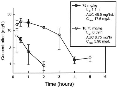
The time course of serum gatifloxacin levels in infected neutropenic mice following the administration of subcutaneous doses of 18.75 and 75 mg/kg are shown in Fig. 1. For the two doses studied, gatifloxacin kinetics were nonlinear, with the elimination half-life increasing nearly twofold with the higher dose. The elimination half-life was 0.59 h with the 18.75-mg/kg dose and 1.1 h with the 75-mg/kg dose. The AUC/dose and peak level/dose values for the escalating single doses were 0.47 to 0.62 and 0.23 to 0.32, respectively. The level of gatifloxacin binding in mouse serum was 23% at drug concentrations of 10 and 100 μg/ml. This is similar to the low degree of binding reported for gatifloxacin binding in human serum. Because of the similar and low degree of binding, total drug levels are considered in PK calculations throughout the report.
FIG. 1.
Serum gatifloxacin concentrations after administration of single doses of 18.75 and 75 mg/kg to neutropenic infected mice. Each symbol represents the mean ± standard deviation of the levels in the sera of three mice. t1/2, serum elimination half-live (in hours); Cmax, peak level in serum.
In vivo time-kill assays and PAEs.
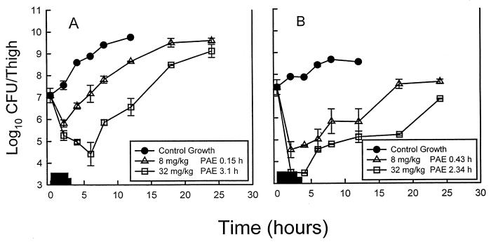
At the start of therapy, the mice were infected with 107.8 CFU of S. pneumoniae per ml and 106.4 CFU of S. aureus per ml. The growth of organisms in the thighs of saline-treated mice increased 2.30 ± 0.13 and 1.70 ± 0.10 log10 CFU/thigh, respectively, over 12 h. The growth of 1 log10 CFU/thigh in saline-treated animals occurred in 3.0 and 3.6 h in S. pneumoniae- and S. aureus-infected animals, respectively. On the basis of determination of the PK of gatifloxacin in serum, serum gatifloxacin levels following the administration of single doses of 8.0 and 32 mg/kg remained above the MIC for S. pneumoniae ATCC 10813 (MIC, 0.25 mg/liter) for 2.3 and 4.0 h, respectively. The values of T>MIC for these doses for S. aureus ATCC 6538p (MIC, 0.06 mg/liter) were 2.9 and 4.7 h, respectively. These gatifloxacin doses produced reductions in colony counts of S. pneumoniae of 1.27 ± 0.16 and 1.82 ± 0.22 log10 CFU/thigh, respectively, at 2 h. These doses also produced significant concentration-dependent killing of S. aureus of 2.86 ± 0.37 and 3.90 ± 0.12 log10 CFU/thigh, respectively, at 2 h. The growth curves of both S. pneumoniae and S. aureus for the control groups and treated mice are shown in Fig. 2. Against S. pneumoniae escalating doses produced PAEs of 0.15 and 3.10 h for the two doses, respectively. Similarly, the study with S. aureus demonstrated PAEs of 0.43 and 2.34 h for the two doses, respectively. No detectable drug carryover was observed in either treatment group.
FIG. 2.
In vivo PAEs of gatifloxacin after administration of single doses of 8 and 32 mg/kg against S. pneumoniae ATCC 10813 (A) and S. aureus ATCC 6538p (B). Each symbol represents the mean ± standard deviation for two mice. The widths of the bars represent the duration of time that levels in serum exceeded the MIC for the infecting pathogen.
PK and PD parameter determination.
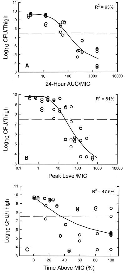
At the start of therapy the mice were infected with 7.49 ± 0.05, 6.29 ± 0.02, and 7.37 ± 0.01 log10 CFU of S. pneumoniae ATCC 10813, S. aureus ATCC 6538p, and E. coli ATCC 25922 per thigh, respectively. The organisms grew 2.87 ± 0.13, 1.39 ± 0.26, and 2.23 ± 0.11 log10 CFU/thigh, respectively, after 24 h in untreated control mice. Escalating doses of gatifloxacin again resulted in the concentration-dependent killing of each of the strains. The highest doses studied reduced the organism burden from 3.21 ± 0.12 to 5.40 ± 0.01 log10 CFU/thigh. The relationships between the microbiologic effect and each of the PD parameters, percent T>MIC, 24-h AUC/MIC, and peak level/MIC, for S. pneumoniae ATCC 10813 are shown in Fig. 3. The strongest relationship was seen when the results were correlated with the 24-h AUC/MIC ratio, with an R2 value of 93%. The correlation with the peak level/MIC was stronger than that with the percent T>MIC, with R2 values of 81 and 48%, respectively. Analysis of studies with S. aureus and E. coli similarly established the strength of the correlation of the 24-h AUC/MIC with efficacy (R2 for 24-h AUC/MIC = 90 to 94%, R2 for peak/MIC = 70 to 72%, and R2 for T>MIC = 71 to 73%).
FIG. 3.
Relationships of the gatifloxacin 24-h AUC/MIC (A), peak level/MIC (B), and T>MIC (C) for S. pneumoniae ATCC 10813 with the log10 numbers of CFU per thigh after 24 h of therapy. Each symbol represents the mean for two thighs per mouse. The horizontal dashed lines represent the organism burden at the start of therapy.
Twenty-four-hour static dose determination.
The doses necessary to achieve a static effect, determined from the multiple-dosing-regimen studies, are shown in Table 2. The bacteriostatic doses for the regimens administered every 3, 6, and 12 h were nearly identical. However, administration once daily was somewhat less effective. The amount of gatifloxacin necessary to achieve a static effect was from 1.3- to 4.5-fold higher with the 24-h regimen than with the shorter dosing intervals. It is likely that this reduction in activity with the 24-h regimen was due to the relatively short elimination half-life of gatifloxacin in this model.
TABLE 2.
Relationship between gatifloxacin dosing interval and bacteriostatic efficacy
| Organism | 24-h static dose (mg/kg) with dosing everya:
|
|||
|---|---|---|---|---|
| 24 h | 12 h | 6 h | 3 h | |
| S. pneumoniae ATCC 10813 | 103 ± 65 | 78.0 ± 9.40 | 67.0 ± 4.07 | 68.0 ± 47.20 |
| S. aureus ATCC 25923 | 30.2 ± 7.79 | 11.0 ± 4.45 | 13.2 ± 1.57 | 13.0 ± 0.74 |
| E. coli 25922 | 4.82 ± 0.25 | 1.75 ± 0.35 | 1.07 ± 0.06 | 1.28 ± 0.15 |
The values are means ± standard errors.
The growth curves of the nine pneumococcal strains, four staphylococcal strains, four strains of the family Enterobacteriaceae, and the P. aeruginosa strain in the thighs of control animals were relatively similar. At the start of therapy the mice were infected with between 6.50 ± 0.13 and 7.40 ± 0.05 log10 CFU/thigh. The organisms grew 1.59 ± 0.06 to 2.80 ± 0.01 log10 CFU/thigh (mean, 2.28 ± 0.30 log10 CFU/thigh) in untreated control mice. The growth curve for K. pneumoniae in the lungs of untreated mice was similar to that observed for the organism in the thighs: 2.04 ± 0.09 and 2.27 ± 0.07 log10 CFU/lung, respectively. Organism growth dynamics in both the thighs and the lungs of untreated animals were not affected by the presence or the absence of neutrophils. The maximal reduction in S. pneumoniae counts in gatifloxacin-treated mice ranged from 3.20 ± 0.05 to 4.95 ± 0.11 log10 CFU/thigh (mean, 4.30 ± 0.65 log10 CFU/thigh). A similar degree of killing was observed in those animals infected with S. aureus (mean, 4.39 ± 0.66 log10 CFU/thigh). Significant organism killing was also observed in studies with gram-negative bacilli (mean, 4.23 ± 0.73 log10 CFU/thigh).
Table 3 shows the 24-h AUC/MIC ratios necessary to achieve a static effect and 1 log10 and 2 log10 reductions in CFU over 24 h. Against the 18 organisms studied, the static doses covered a 350-fold range (1.7 to 592 mg/kg/day). However, the 24-h AUC/MIC ratios corresponding to these static doses varied only fourfold (16 to 70). The 24-h AUC/MIC ratios associated with a static effect were relatively similar for all of the organisms studied (mean 24-h AUC/MIC ratios, 52 for S. pneumoniae, 36 for S. aureus, and 41 for members of the family Enterobacteriaceae). Penicillin, methicillin, and ciprofloxacin resistance did not alter the magnitude of the 24-h AUC/MIC ratio necessary for efficacy. The magnitudes of the AUC/MIC ratio required to achieve a static effect against K. pneumoniae in both the lung and thigh infection models were also similar: 70 and 56, respectively. Neutrophils had only a modest effect on the magnitude of the 24-h AUC/MIC necessary for efficacy. The magnitudes of the parameter needed to achieve a static effect were 1.3- to 2.2-fold lower in the nonneutropenic infection model.
TABLE 3.
Relationship between gatifloxacin MIC and AUC/MIC necessary to achieve a static effect and 1 log10 and 2 log10 killing
| Organism | MIC (μg/ml) | 24-h AUC/MIC ratio required for:
|
||
|---|---|---|---|---|
| Static effect | 1 log10 kill | 2 log10 killing | ||
| S. pneumoniae CDC 145 | 0.25 | 37.0 | 45.8 | 56.1 |
| S. pneumoniae CDC 146 | 0.25 | 54.8 | 66.7 | 81.4 |
| S. pneumoniae CDC 673 | 0.25 | 53.6 | 64.4 | 78.4 |
| S. pneumoniae CDC 1020 | 0.50 | 57.6 | 81.1 | 114 |
| S. pneumoniae ATCC 10813 | 0.25 | 72.4 | 107.7 | 160.0 |
| S. pneumoniae ATCC 49619 | 0.25 | 40.0 | 45.2 | 50.9 |
| S. pneumoniae CDC 1396 | 0.50 | 18.4 | 24.9 | 33.8 |
| S. pneumoniae MNO-418 | 8.0 | 23.1 | 33.4 | 45.3 |
| S. pneumoniae T6-2668 | 2.0 | 51.3 | 80.5 | 128 |
| Mean ± SD | 52.0 ± 20.3 | 61.1 ± 26.4 | 83.1 ± 42.6 | |
| S. aureus ATCC 25923 | 0.12 | 27.2 | 47.8 | 76.5 |
| S. aureus ATCC 33591 | 0.12 | 45.6 | 74.2 | 113 |
| S. aureus ATCC 29213 | 0.12 | 29.7 | 54.5 | 90.9 |
| S. aureus ATCC 6538p | 0.06 | 43.2 | 131 | 507 |
| Mean ± SD | 36.4 ± 9.3 | 76.9 ± 37.8 | 197 ± 207 | |
| E. coli ATCC 25922 | 0.015 | 54.1 | 86.5 | 133 |
| K. pneumoniae ATCC 43816 (thigh model, neutropenic mice) | 0.06 | 69.7 | 129 | 235 |
| K. pneumoniae ATCC 43816 (thigh model, nonneutropenic mice) | 0.06 | 32.2 | 62.2 | 165a |
| K. pneumoniae ATCC 43816 (lung model, neutropenic mice) | 0.06 | 55.9 | 96.1 | 153 |
| K. pneumoniae ATCC 43816 (lung model, nonneutropenic mice) | 0.06 | 43.2 | 63.2 | 91a |
| E. cloacae L2249 | 0.12 | 16.4 | 32.8 | 64.8 |
| E. cloacae L4567 | 0.12 | 27.3 | 57.4 | 132 |
| P. aeruginosa ATCC 27853 | 2.0 | 23.6 | 31.2 | 41.0 |
| Mean ± SD | 41.2 ± 21.5 | 72.2 ± 38.6 | 126 ± 68.8 | |
Datum not included in calculation of the mean.
The dose-response curves for the various organisms were similar; thus, the 24-h AUC/MIC ratios associated with other microbiologic endpoints such as 1 or 2 log10 killing were also similar for the organisms. The 24-h AUC/MIC ratios necessary to achieve a 1 log10 reduction in organism counts were 1.2- to 2.1-fold larger than the ratios necessary to achieve a static effect. The magnitude of the parameter required to produce a 2 log10 killing varied more than the other endpoints for the organisms tested (mean, 119 ± 108) and was 1.8- to 5.5-fold greater than the AUC/MIC required to achieve a bacteriostatic effect.
DISCUSSION
Antimicrobial PD studies characterize the time course of the antibiotic effect (5). The time course of antimicrobial activity can be determined by two characteristics: (i) the effect of drug concentrations on the extent of organism killing and (ii) the presence or absence of antimicrobial effects after levels in serum have fallen below the MIC.
A variety of in vitro and in vivo studies have demonstrated that the fluoroquinolones exhibit concentration-dependent killing and produce prolonged PAEs against susceptible gram-positive and gram-negative pathogens (3, 4, 11, 14, 16; J. E. Leggett, S. Ebert, B. Fantin, and W. A. Craig, Program Abstr. 29th Intersci. Conf. Antimicrob. Agents Chemother., abstr. 313, p. 153, 1989; Y. Wantanabe, S. Ebert, and W. Craig, Program Abstr. 32nd Intersci. Conf. Agents Chemother., abstr. 42, 1992). The efficacies of antibiotics characterized by this pattern of activity are best correlated with one of the concentration-dependent PK-PD parameters. Several animal infection models that use multiple-dosing regimens have identified the AUC/MIC ratio as the principal PK-PD parameter predictive of efficacy, while the peak level/MIC ratio has been suggested to be important for prevention of the selection of resistant mutants during therapy (3, 4, 7, 16; Leggett et al., 29th ICAAC; Watanabe et al., 32nd ICAAC).
The current studies characterized the in vivo PD activities of the new fluoroquinolone gatifloxacin. Penicillin resistance in S. pneumoniae and methicillin resistance in S. aureus had no impact upon the in vitro and in vivo potencies of gatifloxacin. Elevated gatifloxacin MICs were also demonstrated for the two pneumococcal organisms resistant to ciprofloxacin. The fluoroquinolone resistance mechanisms of these organisms have not yet been determined. Similar to studies with other fluoroquinolones, the antimicrobial activities of gatifloxacin were enhanced by escalating drug concentrations (6, 8, 11, 14, 16). The in vivo PAEs, however, were only modest in these studies. Given these PD characteristics, one would predict that either the AUC/MIC or the peak level/MIC would be the PK-PD parameter that most strongly correlates with the efficacy of gatifloxacin. Similar to previous reports, our data from multiple-dosing-regimen studies confirmed that the 24-h AUC/MIC is the PK or PD parameter that best predicts the efficacies of this new fluoroquinolone against both gram-positive and gram-negative pathogens (4).
Leggett et al. (10) have shown that the 24-h cumulative doses of a fluoroquinolone, an aminoglycoside, and a β-lactam required to produce a net bacteriostatic effect are very similar to the doses required to protect 50% of the neutropenic animals infected with a strain of K. pneumoniae from death during a longer course of therapy. In those studies, an AUC/MIC of 35 was an excellent predictor of the 24-h static dose and the 50% protective dose of ciprofloxacin. On the other hand, an AUC/MIC value of 100 was necessary to achieve 100% survival in this model. This magnitude of the AUC/MIC is similar to the value of 125 shown in a retrospective analysis to be associated with successful outcomes for critically ill patients treated with ciprofloxacin, further suggesting that antimicrobial PD in humans may be predicted from studies with animal models (9). In a prospective multivariate analysis, Preston et al. (15) found that both peak level/MIC and AUC/MIC were important predictors of successful levofloxacin therapy in pulmonary, soft tissue, and urinary tract infections. When the peak/MIC and AUC/MIC ratios reached 12.2 and 100, respectively, rates of clinical and microbiologic cures approached 100%. A 24-h AUC/MIC of 35 is equivalent to a constant concentration of 1.5 times the in vitro MIC for 24 h. In other words, a value of 35 means that the in vivo MIC is very similar to the in vitro MIC. The value of this parameter is independent of the dosing interval, the fluoroquinolone used, the animal species, and the site of infection (2, 5, 7, 9; Leggett et al., 29th ICAAC).
The magnitude of the 24-h AUC/MIC required for efficacy in these studies is similar to those observed with other compounds in the fluoroquinolone class (4). The magnitude of this parameter was not affected by drug resistance or site of infection. The presence of neutrophils reduced the static dose by 23 to 54% in the thigh and lung infection models, respectively. Prior studies with fluoroquinolones in a pneumococcal infection model suggested a more prominent contribution of neutrophils, with static dose reductions approaching sevenfold in the nonneutropenic model (D. R. Andes, M. L. van Ogtrop, and W. A. Craig, Program Abstr. Infect. Dis. Soc. Am. Annu. Meet., abstr. 73, p. 52, 1999).
The pharmacokinetics of a 400-mg oral or intravenous dose of gatifloxacin in humans would produce a 24-h AUC of 35 mg · h/liter (12, 17). On the basis of a PD goal of an AUC/MIC of 35, this model would predict that gatifloxacin would be successful in the treatment of organisms for which MICs are as high as 1.0 μg/ml. If one considers the gatifloxacin PK in relation to the MIC at which 90% of S. pneumoniae isolates are inhibited (0.25 μg/ml), current dosing regimens would achieve a 24-h AUC/MIC ratio of 140 on the basis of total drug levels (AUC/MIC for free drug, 112). These PD studies support the current once-daily gatifloxacin dosing regimen used for empirical therapy of community-acquired respiratory tract infections.
REFERENCES
- 1.Andes, D., and W. A. Craig. 1998. In vivo activities of amoxicillin and amoxicillin-clavulanate against Streptococcus pneumoniae: application to breakpoint determinations. Antimicrob. Agents Chemother. 42:2375-2379. [DOI] [PMC free article] [PubMed] [Google Scholar]
- 2.Andes, D. R., and W. A. Craig. 1998. Pharmacodynamics of fluoroquinolones in experimental models of endocarditis. Clin. Infect. Dis. 27:47-50. [DOI] [PubMed] [Google Scholar]
- 3.Blaser, J., B. B. Stone, M. C. Groner, and S. H. Zinner. 1987. Comparative study with enoxacin and netilmicin in a pharmacodynamic model to determine importance of ratio of antibiotic peak concentration to MIC for bacterial activity and emergence of resistance. Antimicrob. Agents Chemother. 31:1054-1060. [DOI] [PMC free article] [PubMed] [Google Scholar]
- 4.Craig, W., and A. Dalhoff. 1998. Pharmacodynamics of fluoroquinolones in experimental animals, p. 208-232. In J. Kuhlman, A. Dalhoff, and H. J. Zeiller (ed.), Handbook of experimental pharmacology, vol. 127. Quinolone antibacterials. Springer-Verlag, Berlin, Germany. [Google Scholar]
- 5.Craig, W. A. 1998. Pharmacokinetics and pharmacodynamics of antibiotics in mice and men. Clin. Infect. Dis. 26:1-12. [DOI] [PubMed] [Google Scholar]
- 6.Craig, W. A., and S. Gudmundsson. 1996. Postantibiotic effect, p. 296-329. In V. Lorian (ed.), Antibiotics in laboratory medicine, 4th ed. The Williams & Wilkins Co., Baltimore, Md.
- 7.Drusano, G. L., D. Johnson, M. Rosen, and M. C. Standiford. 1993. Pharmacodynamics of a fluoroquinolone antimicrobial agent in a neutropenic rat model of Pseudomonas sepsis. Antimicrob. Agents Chemother. 37:483-490. [DOI] [PMC free article] [PubMed] [Google Scholar]
- 8.Dudley, M. N. 1991. Pharmacodynamics and pharmacokinetics of antibiotics with special reference to the fluoroquinolones. Am. J. Med. 91(Suppl. 6A):45-50. [DOI] [PubMed] [Google Scholar]
- 9.Forrest, A., D. E. Nix, C. H. Ballow, T. F. Goss, M.C. Birmingham, and J. J. Schentag. 1993. Pharmacodynamics of intravenous ciprofloxacin in seriously ill patients. Antimicrob. Agents Chemother. 37:1073-1081. [DOI] [PMC free article] [PubMed] [Google Scholar]
- 10.Leggett, J. E., B. Fantin, S. Ebert, K. Totsuka, B. Vogelman, W. Calamae, H. Mattie, and W. A. Craig. 1989. Comparative antibiotic dose-effect relationships at several dosing intervals in murine pneumonitis and thigh-infection models. J. Infect. Dis. 159:281-292. [DOI] [PubMed] [Google Scholar]
- 11.Lister, P. D., and C. C. Sanders. 1999. Pharmacodynamics of trovafloxacin, ofloxacin, and ciprofloxacin against Streptococcus pneumoniae in an in-vitro pharmacokinetic model. Antimicrob. Agents Chemother. 43:1118-1123. [DOI] [PMC free article] [PubMed] [Google Scholar]
- 12.Nakashima, M., T. Uematsu, K. Kosuge, H. Kusajima, T. Ooie, Y. Masuda, R. Ishida, and H. Uchida. 1995. Single- and multiple-dose pharmacokinetics of AM-1155, a new 6-fluoro-8-methoxy quinolone in humans. Antimicrob. Agents Chemother. 39:2635-2640. [DOI] [PMC free article] [PubMed] [Google Scholar]
- 13.Odland, B. A., R. N. Jones, J. Verhoef, A. Fluit, M. L. Beach, et al. 1999. Antimicrobial activity of gatifloxacin (AM-1155, CG5501), and four other fluoroquinolones tested against 2,284 recent clinical strains of Streptococcus pneumoniae from Europe, Latin America, Canada, and the United States. Diagn. Microbiol. Infect. Dis. 34:315-320. [DOI] [PubMed] [Google Scholar]
- 14.Pankuch, G. A., M. R. Jacobs, and P. C. Appelbaum. 1999. Postantibiotic effects of gatifloxacin against gram-positive and -negative organisms. Antimicrob. Agents Chemother. 43:2574-2575. [DOI] [PMC free article] [PubMed] [Google Scholar]
- 15.Preston, S. L., G. L. Drusano, A. L. Berman, L. Adam, C. L. Fowler, A. T. Chow, B. Dornseif, V. Reichl, J. Natarajan, and M. Corrado. 1998. Pharmacodynamics of levofloxacin. A new paradigm for early clinical trials. JAMA 279:125-129. [DOI] [PubMed] [Google Scholar]
- 16.Vogelman, B., S. Gudmundsson, J. Leggett, J. Turnidge, S. Ebert, and W. A. Craig. 1988. Correlation of antimicrobial pharmacokinetic parameters with therapeutic efficacy in an animal model. J. Infect. Dis. 158:831-847. [DOI] [PubMed] [Google Scholar]
- 17.Wise, R., J. M. Andrews, J. P. Ashby, and J. Marshall. 1999. A study to determine the pharmacokinetics and inflammatory fluid penetration of gatifloxacin following a single oral dose. J. Antimicrob. Chemother. 44:701-704. [DOI] [PubMed] [Google Scholar]


