Abstract
During the natural mammal-tick infection cycle, the Lyme disease spirochete Borrelia burgdorferi comes into contact with components of the alternative complement pathway. B. burgdorferi, like many other human pathogens, has evolved the immune evasion strategy of binding two host-derived fluid-phase regulators of complement, factor H and factor H-like protein 1 (FHL-1). The borrelial complement regulator-acquiring surface protein 1 (CRASP-1) is a surface-exposed lipoprotein that binds both factor H and FHL-1. Analysis of CRASP-1 expression during the mammal-tick infectious cycle indicated that B. burgdorferi expresses this protein during mammalian infection, supporting the hypothesized role for CRASP-1 in immune evasion. However, CRASP-1 synthesis was repressed in bacteria during colonization of vector ticks. Analysis of cultured bacteria indicated that CRASP-1 is differentially expressed in response to changes in pH. Comparisons of CRASP-1 expression patterns with those of other infection-associated B. burgdorferi proteins, including the OspC, OspA, and Erp proteins, indicated that each protein is regulated through a unique mechanism.
Borrelia burgdorferi (sensu lato), the causative agent of Lyme disease, is transmitted to humans and other warm-blooded hosts through the bite of infected Ixodes spp. ticks. The Ixodes tick life cycle consists of three postembryonic stages: larva, nymph, and adult, each of which takes only a single blood meal. Ticks acquire spirochetes during blood feeding on infected vertebrate hosts, since transovarial transmission of B. burgdorferi is extremely rare (7, 56). Larvae that acquire infections with their blood meal retain the spirochetes in their midgut through the molt to nymphs and can transmit B. burgdorferi to their next host during the nymphs' blood feeding. Completion of the transmission cycle requires that the bacteria interact with and adapt to a wide range of environments in both host and vector tissues. Successful infection of vertebrate hosts also necessitates sophisticated means of evading the vertebrates' immune system. One crucial strategy employed by Lyme disease spirochetes to evade immune defense is their innate resistance to complement-mediated killing (29).
The complement system forms a critical line of defense in the innate immunity arsenal against invading microorganisms. Direct activation of complement results in both opsonization and formation of lytic membrane attack complexes, leading to killing of invading microorganisms. B. burgdorferi utilizes a strategy to resist complement-mediated killing shared by many other human pathogens, including Streptococcus pyogenes (25, 27, 57), Streptococcus pneumoniae (50), Neisseria meningitidis (58), Neisseria gonorrhoeae (59), Echinococcus granulosus (16), Yersinia enterocolitica (12), and Borrelia hermsii (24). This effective complement evasion strategy involves coating the bacterial cell surface with host-derived fluid-phase regulators of the alternative complement pathway, factor H, and/or factor H-like protein 1 (FHL-1). Factor H and FHL-1, alternatively spliced variants of the same gene, are composed of short consensus repeats (SCRs), which are individually folded, repeating protein domains (77, 79). Factor H consists of 20 SCR domains, while FHL-1 contains only the first 7 SCRs of factor H, plus an extension of four hydrophobic amino acids at the C terminus. Both of these plasma proteins act to control the alternative pathway of complement activation at the level of C3b. Factor H and FHL-1 compete with factor B for binding to C3b, accelerate the decay of the C3 convertase, support the dissociation of the C3bBb complex (decay-accelerating activity), and act as cofactors for factor I-mediated degradation of C3b (35, 55, 73, 78).
B. burgdorferi produces specific proteins that bind serum factor H and/or FHL-1, which have been collectively termed “CRASPs” (complement regulator-acquiring surface proteins) (28, 30-32, 45). Intriguingly, different genovars of B. burgdorferi (sensu lato) express different numbers of CRASPs, each of which differs in relative affinity for factor H and FHL-1. Strains of B. burgdorferi (sensu stricto) and Borrelia afzelii, which generally display intermediate serum-resistant and serum-resistant phenotypes, respectively, produce several distinct CRASPs (30-32, 45), while Borellia garinii strains appear to completely lack functional CRASPs (28). The B. burgdorferi CRASPs (BbCRASPs) and B. afzelii CRASPs (BaCRASPs) are divided into three groups according to their ability to bind factor H or FHL-1: proteins that bind both factor H and FHL-1 (BbCRASP-1 and -2 and BaCRASP-1 and -2), proteins that preferentially bind FHL-1 (BaCRASP-3), and proteins that bind only factor H (BbCRASP-3, -4, and -5 and BaCRASP-4 and -5) (31, 34).
Several of the spirochetal genes encoding CRASPs have been identified and found to be completely different, evolutionarily distinct genes. BbCRASP-1 and BaCRASP-1 are both encoded by cspA, located on linear plasmid lp54 (13, 29, 42, 72). The proteins are homologous, and we will hereafter refer to both proteins as simply “CRASP-1.” The gene encoding BbCRASP-2 is unrelated to any other CRASP-encoding genes (P. Kraiczy, unpublished results). CRASPs belonging to the third group (BbCRASP-3, -4, and -5 and BaCRASP-4 and -5) are members of the cp32 prophage-encoded Erp protein family (1, 2, 22, 30, 31, 33, 45-47, 67). The extensive differences between cspA and erp genes and their 5′-noncoding regions suggested to us that each may be regulated through different mechanisms and may possibly be expressed at different stages of the spirochetes' infectious cycle. Those hypotheses were tested by examining production of CRASP-1 during several stages of the mammal-tick cycle and by comparison of those results with data of similar studies on Erp and other virulence-associated proteins (49).
MATERIALS AND METHODS
Bacteria and growth conditions.
Infectious, clonal B. burgdorferi strain B31-MI-16 was utilized in all tick-mouse infection studies (49). Bacteria were grown in Barbour-Stoenner-Kelly II (BSK-II) medium (5) at 34°C to mid-exponential phase (1 × 10⁷ to 5 × 10⁷ bacteria/ml) unless otherwise specified. For pH and temperature effect studies, bacteria were grown to mid-exponential phase at either 23 or 34°C in BSK-II medium supplemented with 25 mM HEPES and buffered to a pH of either 6.5, 7.0, or 8.0 (10). The pH values of the media were again measured following cell harvesting.
Tick rearing and infection.
Four to six-week-old female BALB/c mice were injected subcutaneously with 104 B31 MI-16 suspended in BSK-II medium. To assess spirochetal infectivity in mice, ear punch biopsies obtained from each animal were cultured in BSK-II medium at 34°C. Uninfected, adult Ixodes scapularis ticks were obtained from Jerry Bowman (Oklahoma State University, Stillwater). Equivalent numbers of males and females were allowed to mate and feed on New Zealand White rabbits. Completely engorged females were maintained in a humidified chamber until eggs were laid. After hatching, approximately 200 larvae were fed on each infected mouse. Larval ticks were allowed to feed to repletion and then were held in a humidified chamber until completion of molting. Spirochetal infectivity of the ticks was assessed by indirect-immunofluorescence analysis (IFA) utilizing a B. burgdorferi B31 rabbit polyclonal anti-membrane protein antibody (49) and Alexa Fluor 594-labeled goat anti-rabbit immunoglobulin G secondary antibody (IgG; Molecular Probes, Eugene, Oregon) and was approximately 87%.
For studies of B. burgdorferi acquisition by ticks from mice, feeding larval ticks were removed with fine-tipped forceps at 24, 48, and 72 h after placement on infected mice. After 96 h, remaining ticks were fully engorged and had dropped off the mice. At that time point, some ticks were dissected immediately, while the remaining ticks were kept in the humidified chamber until dissection at times 120, 144, 168, 192, and 264 h after the initiation of tick feeding.
For studies of B. burgdorferi transmission from ticks to mice, 20 infected flat nymphal ticks each were fed on 4- to 6-week-old naive female BALB/c mice, as described previously (49). Feeding nymphal ticks were forcibly removed with forceps after 24, 28, and 72 h after placement on mice. Also, engorged nymphs that had just completed feeding at 96 h as well as nymphs 120, 144, 168, 192, and 264 h postplacement on mice were analyzed.
Temporal analysis of CRASP-1 expression in ticks and tick bite sites.
Tick midguts and salivary glands were dissected separately. Occasionally during forcible removal of a feeding tick, a piece of skin remained attached to the hypostome of the tick. These attached skin samples were carefully dissected away for analysis. Ticks and skin pieces were examined immediately after removal from the mouse. A minimum of three ticks and three skin samples were examined at each time point.
Tick midguts, tick salivary glands, or skin samples were dissected in 10 μl of phosphate-buffered saline (PBS) on glass slides and allowed to air dry overnight. Slides were then fixed and permeabilized in acetone for 15 min and then air dried. PBS containing 0.2% bovine serum albumin (BSA) and 10% goat serum was used to block the slides for 1 h at room temperature. Slides were washed in wash buffer (PBS plus 0.2% BSA) and incubated overnight at 4°C in the CRASP-1-specific monoclonal antibody RH1 recognizing CRASP-1 (29, 71), diluted 1:10 in PBS-0.2% BSA. Slides were then washed and incubated for 1 h at room temperature in a 1:32,000 dilution of rabbit polyclonal antiserum raised against B. burgdorferi total membrane proteins (49). After a series of four wash steps, slides were simultaneously incubated in 1:1,000 dilutions of both Alexa Fluor 488-labeled goat anti-mouse IgG and Alexa Fluor 594-labeled goat antirabbit IgG (Molecular Probes) for 45 min at room temperature. Slides were again washed five times, dried, and mounted with either ProLong Anti-Fade mounting medium (Molecular Probes, Eugene, Oregon) or glycerol for viewing. Slides were analyzed and images were captured using an Axiophot epifluorescence microscope at magnification ×400 and a Spot digital high-resolution camera (Zeiss, Hallbergmoos, Germany). Labeled bacteria within 25 random fields per slide were counted to determine the proportions of CRASP-1-positive bacteria relative to the anti-B. burgdorferi B31 membrane protein antibody-positive bacteria, since this antibody labels all B. burgdorferi present in a given field (49). Slides of dissected tick midguts were incubated with either RH1 or anti-membrane protein antibody alone or both secondary antibodies alone to serve as negative fluorescence controls.
Protein electrophoresis and immunoblot analysis.
Cell lysates were prepared as described previously (49). Briefly, equivalent masses (approximately 3 μg) of each cell lysate was separated by a 12.5% sodium dodecyl sulfate-polyacrylamide gel electrophoresis and transferred to nitrocellulose membranes. Membranes were blocked by incubation for >1 h in 5% (wt/vol) nonfat dried milk in Tris-buffered saline-Tween (TBS-T), washed with TBS-T, and incubated for 1 h with monoclonal antibody RH1 (29, 71). Membranes were washed and incubated with conjugated protein A horseradish peroxidase (Amersham) in TBST-T. Bound antibodies were detected by West-Pico enhanced chemiluminescence (Amersham). To ensure equal loading of protein concentrations, blots were stripped and hybridized with murine anti-FlaB monoclonal antibody H9724 (6), since FlaB is a constitutively expressed flagellar protein of B. burgdorferi.
RESULTS
CRASP-1 expression during colonization of larval ticks.
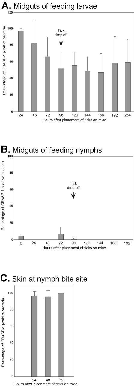
As B. burgdorferi is acquired from infected vertebrates by feeding tick larvae, the bacteria are exposed to the host's blood and are thus susceptible to complement-mediated killing. We therefore examined expression of CRASP-1 during that crucial stage of the bacterial infectious cycle. Double-labeling IFA of tick midgut contents was conducted, using a murine monoclonal antibody specific for CRASP-1 and a rabbit polyclonal antiserum that recognizes all B. burgdorferi cells. Naive larvae were fed upon infected mice and forcibly removed at 24-h intervals. In addition, engorged larvae were examined for several days following completion of blood feeding (>96 h postattachment). Between 100 and 600 B. burgdorferi spirochetes were detected in the midgut of each feeding infected larval tick, with the number of spirochetes increasing as feeding progressed. Twenty-four hours into the infection process, 96% of spirochetes that had migrated into the feeding ticks expressed detectable levels of CRASP-1 (Fig. 1A). During the second and third days of feeding, the proportion of CRASP-1-expressing bacteria began to decline. After 96 h, when the larvae had completed feeding and had naturally detached, the percentage of ingested bacteria expressing detectable levels of CRASP-1 dropped somewhat but remained elevated. This level of expression was maintained for at least 8 days after the completion of feeding. Control analysis lacking one or more labeling reagents for this and all other IFA studies described below indicated that results were specific (data not shown).
FIG. 1.
Temporal analysis of B. burgdorferi CRASP-1 expression during the tick-mammal infectious cycle. Double-labeling IFA was utilized to assess the proportion of spirochetes expressing CRASP-1 at different stages in the infectious cycle. (A) B. burgdorferi-infected mice were infested with naive larval I. scapularis ticks. Ticks were forcibly removed at 24-h intervals until drop-off at 96 h and for several days after the conclusion of feeding. (B) Infected nymphal I. scapularis ticks were allowed to feed on naive mice. Ticks were forcibly removed at 24-h intervals until drop-off at 96 h and for several days after the conclusion of feeding. (C) Mouse skin attached to feeding nymphal tick hypostomes. (A-C) Bars, mean percentages of CRASP-1-positive bacteria counted; error bars, standard deviation of the means.
B. burgdorferi spirochetes found within a feeding tick are a mixed population, consisting of recently acquired bacteria as well as those bacteria that were acquired hours or days earlier and have undergone phenotypic changes to adapt to tick midgut colonization. These data indicate that essentially all B. burgdorferi bacteria acquired by naive ticks from infected mice express CRASP-1 during the acquisition process, consistent with the function of that protein in binding factor H and FHL-1 to protect against killing by complement. For an as-yet-unknown reason, the spirochetes repress synthesis of CRASP-1 upon colonization of the larval midgut.
CRASP-1 expression within unfed nymphs.
During the weeks after completion of a blood meal, engorged larval ticks digest the mammalian blood and molt to the next developmental stage. We examined the level of CRASP-1 expression in unfed nymphs to assess the effect, if any, of the molting process on bacterial CRASP-1 expression. Fifty to 250 spirochetes were detected in the midgut of each unfed nymph. IFA of unfed nymphs revealed that very few of the spirochetes residing in their midguts expressed detectable amounts of CRASP-1 (Fig. 1B). IFA on the same unfed nymphal midgut contents using a rabbit polyclonal antibody that recognizes all B. burgdorferi B31 membrane proteins confirmed tick infection and indicated that each was colonized by large numbers of B. burgdorferi.
CRASP-1 expression within transmitting nymphal ticks.
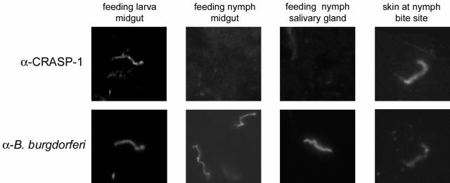
Nymphal ticks readily transmit B. burgdorferi to naive mammalian hosts while taking a blood meal. Theoretically then, the feeding nymphal tick midgut represents a new environment in which the spirochete must be protected from complement-mediated killing. Yet studies have shown that ingested host complement fails to kill spirochetes inside the tick midgut (61), presumably due to tick salivary components that block complement activation (37, 61, 63, 70, 74). For these reasons, we next examined expression levels of CRASP-1 by B. burgdorferi within midguts of transmitting nymphal ticks. Infected nymphs were allowed to feed upon naive mice and forcibly removed at 24-h intervals. Additionally, fully engorged nymphs were examined for several days following natural detachment. Infected ticks harbored a variable number of B. burgdorferi spirochetes in their midguts, ranging between 75 and 500. No detectable CRASP-1 expression was observed on the spirochetes in the midguts of nymphs during the first and second days of feeding (Fig. 1B and 2). By the third day of feeding, only 6% of spirochetes expressed detectable CRASP-1. After tick detachment and for up to 4 days after the completion of feeding, the level of spirochetes expressing CRASP-1 fell below 0.5%. We note that the spirochetes within feeding nymphal midguts are mixed populations, consisting of bacteria that have not yet transmitted to the mammalian host but will do so in the following hours, as well as spirochetes that are destined to remain colonizing the tick. Therefore, IFA of salivary glands from feeding nymphs was conducted to assess CRASP-1 expression by spirochetes further along in the transmission process. Five to ten bacteria were observed in each salivary gland dissection, none of which expressed a detectable level of CRASP-1 (Fig. 2 and data not shown). We conclude that CRASP-1 synthesis is greatly repressed within the midgut of the transmitting nymphal tick and remains poorly expressed by bacteria even after invasion of salivary glands.
FIG. 2.
Representative examples of IFA images of B. burgdorferi during tick and mammalian infection. Infected nymphal I. scapularis ticks were allowed to feed on naive mice. Ticks were forcibly removed at 24-h intervals until drop-off at 96 h and for several days after the conclusion of feeding. Double-labeling IFA was utilized to assess the expression of CRASP-1 by spirochetes within the midguts of the feeding nymphal ticks (24-h pull-off shown), the feeding nymph salivary gland (24-h pull-off shown), and within skin taken from the nymph bite site (72-h sample shown). RH1 antibody labeled only those spirochetes expressing CRASP-1 (top panel), while anti-B. burgdorferi (α-B. burgdorferi; anti-membrane protein) polyclonal antibody labeled all spirochetes in the same field (bottom panel).
CRASP-1 expression in mouse skin at tick bite site.
B. burgdorferi bacteria transmitted into the mammalian host soon lose the protection against host complement afforded by tick saliva, and bacteria must therefore protect themselves in order to disseminate and establish a successful infection. We assessed the role of CRASP-1 at that stage by examining mouse skin that remained attached to the hypostomes of forcibly removed feeding nymphs. This tissue sample consists of the blood pool into which the tick salivated, as well as adjacent skin and other tissues. On average, 5 to 15 bacteria were observed in each skin sample, with at least three skin samples examined for each time point. At all time points examined, nearly every one of the spirochetes within the tick bite site produced CRASP-1 (Fig. 1C and 2). That high level of CRASP-1 expression was observed for all bacteria transmitted to the mouse skin, regardless of the duration of feeding, suggesting that CRASP-1 is extremely important during the early stages of invasion of the mammalian host.
Effects of culture pH on CRASP-1 expression.
We next sought to understand the environmental cues that lead B. burgdorferi to regulate CRASP-1 expression. Since some B. burgdorferi proteins that are preferentially synthesized during infection of vertebrate hosts are also regulated by pH (8-10, 60, 75), we assessed the effect of modulating the pH of culture medium on CRASP-1 expression. B. burgdorferi was cultured for several generations at 34°C or 23°C in medium buffered to a pH of 6.5, 7.0, or 8.0, with no detectable change in the pH of spent growth medium (data not shown). Significantly higher levels of CRASP-1 were produced by bacteria cultivated in medium buffered to a pH of 8.0 than by those grown in medium of pH 7.0 at both temperatures (Fig. 3).
FIG. 3.
Analysis of CRASP-1 protein expression by Borrelia burgdorferi cultivated under different growth conditions. To assess the effect of pH of CRASP-1 expression, bacteria were cultivated at either 23 or 34°C in medium modified to a pH of 6.5, 7.0, or 8.0. Cell lysates were prepared from bacteria grown under each condition, and Western blot analysis using RH1 monoclonal antibody specific for CRASP-1 was conducted. The blots were then stripped and hybridized with murine anti-FlaB monoclonal antibody to ensure equal loading of proteins, since FlaB is a constitutively expressed flagellar protein.
Expression of spirochetal proteins involved in mammalian infection is often regulated by temperature, with greater amounts of protein produced by bacteria cultured at 34°C than by bacteria grown at ambient temperature (9, 11, 26, 60, 65, 66, 68, 75). These temperatures are hypothesized to mimic conditions within feeding ticks and unfed ticks, respectively (65). Temperature also influences expression of borrelial factor H-binding Erp proteins, with greater expression of Erp proteins at 34°C than at 23°C (3, 4, 21, 49, 66, 68). Previous work demonstrated that cultivation of bacteria at 33 or 37°C resulted in greater expression of three different CRASPs (BaCRASP-1, BaCRASP-2, and BaCRASP-5) than did cultivation at 20°C (31). Examination of B. burgdorferi protein expression during continuous cultivation at warmer temperatures is hypothesized to model conditions similar to those experienced by the bacteria during a chronic mammalian infection (64). Expression of the unrelated OspC protein increases upon shift from growth at ambient temperature to 37°C, but its expression declines during continuous cultivation at 37°C (64, 65). In contrast, members of the Erp protein family are continually expressed at high levels throughout multiple passages at 34°C (49). OspC is produced by B. burgdorferi during the initial stages of mammalian infection but repressed shortly thereafter (40, 41, 64), whereas Erp proteins are expressed throughout long-term infection (20, 21, 43, 47-49, 66, 69). To this end, we examined CRASP-1 expression by bacteria grown at 23°C, shifted to 34°C, and then maintained at 34°C for three additional passages. Contrary to the case with OspC, but similar to that with the Erp proteins, high-level expression of CRASP-1 was maintained by the spirochetes (data not shown).
DISCUSSION
This study represents the first temporal analysis of the expression of the B. burgdorferi factor H/FHL-1 binding protein CRASP-1. Consistent with the demonstrated function of CRASP-1 (29-31), it was expressed during points in the infection cycle when the bacteria are exposed to the host complement system. Expression of CRASP-1 increased dramatically upon entrance into the mammalian host. Likewise, all B. burgdorferi acquired by feeding ticks with the blood of infected mice produced high levels of CRASP-1. These data greatly strengthen the hypothesis that CRASP-1 serves as an important virulence factor that protects migrating spirochetes from the complement-mediated killing defense mounted by the host.
Spirochetes expressing CRASP-1 were extremely rare in unfed I. scapularis nymphs. The low level of CRASP-1 expression by B. burgdorferi cultivated at 23°C and at acidic and neutral pH paralleled the low expression of CRASP-1 by bacteria in the midguts of unfed ticks. As nymphal tick feeding progressed, CRASP-1 expression by bacteria in tick midguts increased only marginally. In contrast, essentially all spirochetes detected at tick bite sites expressed CRASP-1. High level expression was detected in mouse skin regardless of duration of tick feeding. Thus, different populations of spirochetes deposited in the mammalian skin all produce CRASP-1. This suggests that B. burgdorferi produces CRASP-1 in response to signals that indicate a need for protection from mammalian host complement. Since bacteria cultivated in medium conditioned to a pH of 8.0 had greater CRASP-1 expression levels than those of cultures grown at a pH of 6.5, the pH of the spirochete's environment appears to play a role in regulation of CRASP-1 expression. Interestingly, tick saliva has a very basic pH of approximately 9.5 to 10 (38, 75). These data suggest that the high pH of the tick saliva serves as a cue that signals B. burgdorferi to increase production of CRASP-1. Since B. burgdorferi produced low levels of CRASP-1 when cultured at lower pH, perhaps the somewhat acidic conditions of the tick midgut (75) signal to keep CRASP-1 repressed in that microenvironment.
A high percentage of spirochetes expressed CRASP-1 during the initial stages of larval acquisition, but those numbers declined during the second day of feeding. This finding indicates both the utility of the CRASP-1 protein for the survival of B. burgdorferi in the mammalian host and a lack of importance when inside the vector. The differential CRASP-1 expression observed for B. burgdorferi within feeding larval and nymphal ticks correlates with the apparent ineffectiveness of complement inside the tick vector. Comparative studies using wild-type and C3-deficient mice demonstrated that the host complement system has no effect on spirochetes inside the I. scapularis vector (61), most likely because complement is inactivated by the tick saliva (37, 61, 63, 70, 74). We hypothesize that the lack of CRASP-1 expression by spirochetes in the midgut of the transmitting nymph is permitted by the lack of a need for protection against complement inside the vector.
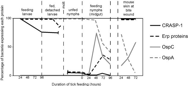
As stated earlier, CRASP-1 is not the only factor H/FHL-1-binding protein in the armament of B. burgdorferi that protects the bacterium from complement-mediated killing. Other outer surface proteins of B. burgdorferi that specifically interact with complement regulator factor H include molecules of the Erp (OspE/F-related proteins) protein family (1, 2, 22, 30, 31, 33, 34, 44, 45, 67). The Erp proteins, encoded by allelic genes on the cp32 plasmids, bind factor H proteins of many different vertebrates, which may allow B. burgdorferi to establish infection in a diverse range of hosts (36, 67). Previous studies demonstrated that B. burgdorferi regulates Erp protein production during the natural tick-mammalian infectious cycle, with high-level expression during mammalian stages of infection but very low levels during tick infection (49). However, unlike the case with CRASP-1, B. burgdorferi newly colonizing tick midguts produces high levels of Erp proteins for a substantial amount of time during feeding and produces Erp proteins when in the midguts and salivary glands of feeding nymphs (21, 49) (Fig. 4). Additionally, erp expression is not influenced by culture pH (3), in contrast to the strong effects of pH we observed in these studies on CRASP-1. These differences suggest that while both the CRASP-1 and Erp proteins bind factor H, they may not be completely redundant. It is possible that the difference in expression pattern for the two classes of proteins reflects differences in their functions. For example, CRASP-1 also binds FHL-1, while Erp proteins do not. Our preliminary results suggest that some Erp proteins may perform additional roles alongside their binding of factor H (unpublished results). Further studies of both the differences and the similarities of the CRASP-1 and Erp proteins will continue to shed light upon their functions in the B. burgdorferi infection processes.
FIG. 4.
Expression profiles of various outer surface proteins of B. burgdorferi (this work and references 14, 49, 51, 64 and 65). Inside the midgut of feeding larval ticks, newly acquired spirochetes do not express OspC. OspA, a spirochetal tick colonization factor, is expressed by all spirochetes within larvae throughout blood feeding. Erp proteins are expressed until a few days after completion of the larval blood meal, whereas CRASP-1 expression declines during the second day of feeding. After the molt, spirochetes continue to express OspA in the unfed nymph, but expression of Erp proteins and CRASP-1 is barely detectable. As the nymph feeds and transmits spirochetes into the mammalian host, expression of the OspC and Erp proteins increases, while OspA expression declines. CRASP-1 expression is not induced in the nymphal midgut. Nearly all transmitted spirochetes within mouse skin at the tick bite wound express detectable levels of the CRASP-1 and Erp proteins. OspC expression by bacteria in the skin does not increase until the second day of tick feeding, just as OspA expression declines.
The expression patterns of two additional, unrelated B. burgdorferi surface proteins, OspA and OspC, have also been well characterized for both ticks and mice (14, 15, 20, 23, 39, 62, 64, 65, 76) (Fig. 4). OspA has been used in a Lyme disease vaccine that works by blocking transmission of B. burgdorferi from an infected tick into a mammalian host (17, 18). B. burgdorferi OspA is abundantly expressed on spirochetes within the arthropod and is essential for pathogen adherence to the vector (52, 64). The I. scapularis TROSPA protein has been implicated in mediating this interaction (53). Conversely, OspC is not expressed in larval ticks acquiring B. burgdorferi from infected mice (64). OspC expression is induced during nymphal tick feeding (49, 64, 65) (Fig. 4), when it apparently functions to assist transmission to the mammalian host (19, 54). The somewhat mirror images of OspC and OspA expression during the infection cycle have given rise to a model of B. burgdorferi gene regulation in which genes can be classified as either group I (ospC-like) or group II (ospA-like) (75). Our data, combined with those of other studies (3, 9, 49, 51), indicate that borrelial gene regulation is far more complex than earlier envisioned. As illustrated in Fig. 4, each of the four proteins discussed has a unique expression pattern during the infection cycle, indicative of four distinct regulatory pathways. Furthermore, studies of additional B. burgdorferi virulence factors are indicating that there are even more components to the complex signaling networks controlling B. burgdorferi gene expression (our unpublished results.)
In summary, we analyzed the expression profile of CRASP-1, the B. burgdorferi surface protein that binds the two central fluid-phase complement regulators of the alternative pathway. We speculate that B. burgdorferi employs differential expression of CRASP-1 and other factor H-binding proteins to circumvent complement-mediated killing by innate defense. Further studies characterizing the role of CRASP-1 from different complement-resistant and -susceptible B. burgdorferi bacteria, as well as cspA mutants, will help elucidate differences in the immune evasion strategies of different genospecies. If proven critical for mammalian infection, CRASP-1 may serve as a novel vaccine candidate for the prevention of Lyme disease.
Acknowledgments
This study was funded by U.S. National Institutes of Health grant RO1-AI44254 to Brian Stevenson and Deutsche Forschungsgemeinschaft grant Br446/11 to Volker Brade. Kate von Lackum, Jennifer C. Miller, Michael E. Woodman, and Sean P. Riley were supported by NIH training program in microbial pathogenesis 5T32AI49795.
We thank Jerry Bowman for providing I. scapularis ticks; Jason Carlyon for providing instruction on salivary gland extraction; Don Cohen for assistance with microscopy; Matthew J. Troese, Kelly Babb, Natalie Mickelson, Sarah Kearns, and Sara Bair for their technical assistance; and all the members of our laboratories for helpful comments on this work and the manuscript.
Editor: D. L. Burns
REFERENCES
- 1.Alitalo, A., T. Meri, H. Lankinen, I. Seppala, P. Lahdenne, S. P. Hefty, D. Akins, and S. Meri. 2002. Complement inhibitor factor H binding to Lyme disease spirochetes is mediated by inducible expression of multiple plasmid-encoded outer surface protein E paralogs. J. Immunol. 169:3847-3853. [DOI] [PubMed] [Google Scholar]
- 2.Alitalo, A., T. Meri, L. Ramo, T. S. Jokiranta, T. Heikkila, I. J. T. Seppala, J. Oksi, M. Viljanen, and S. Meri. 2001. Complement evasion by Borrelia burgdorferi: serum-resistant strains promote C3b inactivation. Infect. Immun. 69:3685-3691. [DOI] [PMC free article] [PubMed] [Google Scholar]
- 3.Babb, K., N. El-Hage, J. C. Miller, J. A. Carroll, and B. Stevenson. 2001. Distinct regulatory pathways control expression of Borrelia burgdorferi infection-associated OspC and Erp surface proteins. Infect. Immun. 69:4146-4153. [DOI] [PMC free article] [PubMed] [Google Scholar]
- 4.Babb, K., J. D. McAlister, J. C. Miller, and B. Stevenson. 2004. Molecular characterization of Borrelia burgdorferi erp promoter/operator elements. J. Bacteriol. 186:2745-2756. [DOI] [PMC free article] [PubMed] [Google Scholar]
- 5.Barbour, A. G. 1984. Isolation and cultivation of Lyme disease spirochetes. Yale J. Biol. Med. 57:521-525. [PMC free article] [PubMed] [Google Scholar]
- 6.Barbour, A. G., S. F. Hayes, R. A. Heiland, M. E. Schrumpf, and S. L. Tessier. 1986. A Borrelia-specific monoclonal antibody binds to a flagellar epitope. Infect. Immun. 52:549-554. [DOI] [PMC free article] [PubMed] [Google Scholar]
- 7.Burgdorfer, W., S. F. Hayes, and D. Corwin. 1989. Pathophysiology of the Lyme disease spirochete, Borrelia burgdorferi, in ixodid ticks. Rev. Infect. Dis. 11 Suppl. 6:S1442-S1450. [DOI] [PubMed] [Google Scholar]
- 8.Carroll, J. A., R. M. Cordova, and C. F. Garon. 2000. Identification of eleven pH-regulated genes in Borrelia burgdorferi localized to linear plasmids. Infect. Immun. 68:6677-6684. [DOI] [PMC free article] [PubMed] [Google Scholar]
- 9.Carroll, J. A., N. El-Hage, J. C. Miller, K. Babb, and B. Stevenson. 2001. Borrelia burgdorferi RevA antigen is a surface-exposed outer membrane protein whose expression is regulated in response to environmental temperature and pH. Infect. Immun. 69:5286-5293. [DOI] [PMC free article] [PubMed] [Google Scholar]
- 10.Carroll, J. A., C. F. Garon, and T. G. Schwan. 1999. Effects of environmental pH on membrane proteins in Borrelia burgdorferi. Infect. Immun. 67:3181-3187. [DOI] [PMC free article] [PubMed] [Google Scholar]
- 11.Cassatt, D. R., N. K. Patel, N. D. Ulbrandt, and M. S. Hanson. 1998. DbpA, but not OspA, is expressed by Borrelia burgdorferi during spirochetemia and is a target for protective antibodies. Infect. Immun. 66:5379-5387. [DOI] [PMC free article] [PubMed] [Google Scholar]
- 12.China, B., M. P. Sory, B. T. N′Guyen, M. De Bruyere, and G. R. Cornelis. 1993. Role of the YadA protein in prevention of opsonization of Yersinia enterocolitica by C3b molecules. Infect. Immun. 61:3129-3136. [DOI] [PMC free article] [PubMed] [Google Scholar]
- 13.Cordes, F. S., P. Roversi, P. Kraiczy, M. M. Simon, V. Brade, O. Jahraus, R. Wallis, C. Skerka, P. F. Zipfel, R. Wallich, and S. M. Lea. 2005. A novel fold for the factor H-binding protein BbCRASP-1 of Borrelia burgdorferi. Nat. Struct. Mol. Biol. 12:276-277. [DOI] [PubMed] [Google Scholar]
- 14.de Silva, A. M., and E. Fikrig. 1997. Arthropod- and host-specific gene expression by Borrelia burgdorferi. J. Clin. Investig. 99:377-379. [DOI] [PMC free article] [PubMed] [Google Scholar]
- 15.de Silva, A. M., S. R. Telford III, L. R. Brunet, S. W. Barthold, and E. Fikrig. 1996. Borrelia burgdorferi OspA is an arthropod-specific transmission-blocking Lyme disease vaccine. J. Exp. Med. 183:271-275. [DOI] [PMC free article] [PubMed] [Google Scholar]
- 16.Diaz, A., A. Ferreira, and R. B. Sim. 1997. Complement evasion by Echinococcus granulosus: sequestration of host factor H in the hydatid cyst wall. J. Immunol. 158:3779-3786. [PubMed] [Google Scholar]
- 17.Fikrig, E., S. W. Barthold, F. S. Kantor, and R. A. Flavell. 1990. Protection of mice against the Lyme disease agent by immunizing with recombinant OspA. Science 250:553-556. [DOI] [PubMed] [Google Scholar]
- 18.Fikrig, E., S. R. Telford III, S. W. Barthold, F. S. Kantor, A. Spielman, and R. A. Flavell. 1992. Elimination of Borrelia burgdorferi from vector ticks feeding on OspA-immunized mice. Proc. Natl. Acad. Sci. USA 89:5418-5421. [DOI] [PMC free article] [PubMed] [Google Scholar]
- 19.Grimm, D., K. Tilly, R. Byram, P. E. Stewart, J. G. Krum, D. M. Bueschel, T. G. Schwan, P. F. Policastro, A. F. Elias, and P. A. Rosa. 2004. Outer-surface protein C of the Lyme disease spirochete: a protein induced in ticks for infection of mammals. Proc. Natl. Acad. Sci. USA 101:3142-3147. [DOI] [PMC free article] [PubMed] [Google Scholar]
- 20.Hefty, P. S., S. E. Jolliff, M. J. Caimano, S. K. Wikel, and D. R. Akins. 2002. Changes in temporal and spatial patterns of outer surface lipoprotein expression generate population heterogeneity and antigenic diversity in the Lyme disease spirochete, Borrelia burgdorferi. Infect. Immun. 70:3468-3478. [DOI] [PMC free article] [PubMed] [Google Scholar]
- 21.Hefty, P. S., S. E. Jollif, M. J. Caimano, S. K. Wikel, J. D. Radolf, and D. R. Akins. 2001. Regulation of OspE-related, OspF-related, and Elp lipoproteins of Borrelia burgdorferi strain 297 by mammalian host-specific signals. Infect. Immun. 69:3618-3627. [DOI] [PMC free article] [PubMed] [Google Scholar]
- 22.Hellwage, J., T. Meri, T. Heikkila, A. Alitalo, J. Panelius, P. Lahdenne, I. J. T. Seppala, and S. Meri. 2001. The complement regulator factor H binds to the surface protein OspE of Borrelia burgdorferi. J. Biol. Chem. 276:8427-8435. [DOI] [PubMed] [Google Scholar]
- 23.Hodzic, E., S. Feng, K. J. Freet, D. L. Borjesson, and S. W. Barthold. 2002. Borrelia burgdorferi population kinetics and selected gene expression at the host-vector interface. Infect. Immun. 70:3382-3388. [DOI] [PMC free article] [PubMed] [Google Scholar]
- 24.Hovis, K. M., J. V. McDowell, L. Griffin, and R. T. Marconi. 2004. Identification and characterization of a linear-plasmid-encoded factor H-binding protein (FhbA) of the relapsing fever spirochete Borrelia hermsii. J. Bacteriol. 186:2612-2618. [DOI] [PMC free article] [PubMed] [Google Scholar]
- 25.Johnsson, E., K. Berggard, H. Kotarsky, J. Hellwage, P. F. Zipfel, U. Sjobring, and G. Lindahl. 1998. Role of the hypervariable region in streptococcal M proteins: binding of human complement inhibitor. J. Immunol. 161:4894-4901. [PubMed] [Google Scholar]
- 26.Konkel, M. E., and K. Tilly. 2000. Temperature-regulated expression of bacterial virulence genes. Microbes Infect. 2:157-166. [DOI] [PubMed] [Google Scholar]
- 27.Kotarsky, H., J. Hellwage, E. Johnsson, C. Skerka, H. G. Svensson, G. Lindahl, U. Sjobring, and P. F. Zipfel. 1998. Identification of a domain in human factor H and factor H-like protein-1 required for the interaction with streptococcal M proteins. J. Immunol. 160:3349-3354. [PubMed] [Google Scholar]
- 28.Kraiczy, P., K. Hartmann, J. Hellwage, C. Skerka, M. Kirschfink, V. Brade, P. F. Zipfel, R. Wallich, and B. Stevenson. 2004. Immunological characterization of the complement regulator factor H-binding CRASP and Erp proteins of Borrelia burgdorferi. Int. J. Med. Microbiol. 293(suppl. 37):152-157. [DOI] [PubMed] [Google Scholar]
- 29.Kraiczy, P., J. Hellwage, C. Skerka, H. Becker, M. Kirschfink, M. M. Simon, V. Brade, P. F. Zipfel, and R. Wallich. 2004. Complement resistance of Borrelia burgdorferi correlates with the expression of BbCRASP-1, a novel linear plasmid-encoded surface protein that interacts with human factor H and FHL-1 and is unrelated to Erp proteins. J. Biol. Chem. 279:2421-2429. [DOI] [PubMed] [Google Scholar]
- 30.Kraiczy, P., J. Hellwage, C. Skerka, M. Kirschfink, V. Brade, P. F. Zipfel, and R. Wallich. 2003. Immune evasion of Borrelia burgdorferi: mapping of a complement inhibitor factor H-binding site of BbCRASP-3, a novel member of the Erp protein family. Eur. J. Immunol. 33:697-707. [DOI] [PubMed] [Google Scholar]
- 31.Kraiczy, P., C. Skerka, V. Brade, and P. F. Zipfel. 2001. Further characterization of complement regulator-acquiring surface proteins of Borrelia burgdorferi. Infect. Immun. 69:7800-7809. [DOI] [PMC free article] [PubMed] [Google Scholar]
- 32.Kraiczy, P., C. Skerka, M. Kirschfink, V. Brade, and P. F. Zipfel. 2001. Immune evasion of Borrelia burgdorferi by acquisition of human complement regulators FHL-1/reconectin and Factor H. Eur. J. Immunol. 31:1674-1684. [DOI] [PubMed] [Google Scholar]
- 33.Kraiczy, P., C. Skerka, M. Kirschfink, P. F. Zipfel, and V. Brade. 2001. Mechanism of complement resistance of pathogenic Borrelia burgdorferi isolates. Int. Immunopharmacol. 1:393-401. [DOI] [PubMed] [Google Scholar]
- 34.Kraiczy, P., C. Skerka, P. F. Zipfel, and V. Brade. 2002. Complement regulator-acquiring surface proteins of Borrelia burgdorferi: a new protein family involved in complement resistance. Wien. Klin. Wochenschr. 114:568-573. [PubMed] [Google Scholar]
- 35.Kuhn, S., C. Skerka, and P. F. Zipfel. 1995. Mapping of the complement regulatory domains in the human factor H-like protein 1 and in factor H1. J. Immunol. 155:5663-5670. [PubMed] [Google Scholar]
- 36.Kurtenbach, K., S. De Michelis, S. Etti, S. M. Schafer, H. S. Sewell, V. Brade, and P. Kraiczy. 2002. Host association of Borrelia burgdorferi sensu lato—-the key role of host complement. Trends Microbiol. 10:74-79. [DOI] [PubMed] [Google Scholar]
- 37.Lawrie, C. H., S. E. Randolph, and P. A. Nuttall. 1999. Ixodes ticks: serum species sensitivity of anticomplement activity. Exp. Parasitol. 93:207-214. [DOI] [PubMed] [Google Scholar]
- 38.Ledin, K. E., N. S. Zeidner, J. M. Ribeiro, B. J. Biggerstaff, M. C. Dolan, G. Dietrich, L. Vredevoe, and J. Piesman. 2005. Borreliacidal activity of saliva of the tick Amblyomma americanum. Med. Vet. Entomol. 19:90-95. [DOI] [PubMed] [Google Scholar]
- 39.Leuba-Garcia, S., R. Martinez, and L. Gern. 1998. Expression of outer surface proteins A and C of Borrelia afzelii in Ixodes ricinus ticks and in the skin of mice. Zentralbl. Bakteriol. 287:475-484. [DOI] [PubMed] [Google Scholar]
- 40.Liang, F. T., M. B. Jacobs, L. C. Bowers, and M. T. Philipp. 2002. An immune evasion mechanism for spirochetal persistence in Lyme borreliosis. J. Exp. Med. 195:415-422. [DOI] [PMC free article] [PubMed] [Google Scholar]
- 41.Liang, F. T., J. Yan, M. L. Mbow, S. L. Sviat, R. D. Gilmore, M. Mamula, and E. Fikrig. 2004. Borrelia burgdorferi changes its surface antigenic expression in response to host immune responses. Infect. Immun. 72:5759-5767. [DOI] [PMC free article] [PubMed] [Google Scholar]
- 42.McDowell, J. V., M. E. Harlin, E. A. Rogers, and R. T. Marconi. 2005. Putative coiled-coil structural elements of the BBA68 protein of Lyme disease spirochetes are required for formation of its factor H binding site. J. Bacteriol. 187:1317-1323. [DOI] [PMC free article] [PubMed] [Google Scholar]
- 43.McDowell, J. V., S. Y. Sung, G. Price, and R. T. Marconi. 2001. Demonstration of the genetic stability and temporal expression of select members of the Lyme disease spirochete OspF protein family during infection in mice. Infect. Immun. 69:4831-4838. [DOI] [PMC free article] [PubMed] [Google Scholar]
- 44.McDowell, J. V., J. Wolfgang, L. Senty, C. M. Sundy, M. J. Noto, and R. T. Marconi. 2004. Demonstration of the involvement of outer surface protein E coiled coil structural domains and higher order structural elements in the binding of infection-induced antibody and the complement-regulatory protein, factor H. J. Immunol. 173:7471-7480. [DOI] [PubMed] [Google Scholar]
- 45.McDowell, J. V., J. Wolfgang, E. Tran, M. S. Metts, D. Hamilton, and R. T. Marconi. 2003. Comprehensive analysis of the factor H binding capabilities of Borrelia species associated with Lyme disease: delineation of two distinct classes of factor H binding proteins. Infect. Immun. 71:3597-3602. [DOI] [PMC free article] [PubMed] [Google Scholar]
- 46.Metts, M. S., J. V. McDowell, M. Theisen, P. R. Hansen, and R. T. Marconi. 2003. Analysis of the OspE determinants involved in binding of factor H and OspE-targeting antibodies elicited during Borrelia burgdorferi infection. Infect. Immun. 71:3587-3596. [DOI] [PMC free article] [PubMed] [Google Scholar]
- 47.Miller, J. C., N. El-Hage, K. Babb, and B. Stevenson. 2000. Borrelia burgdorferi B31 Erp proteins that are dominant immunoblot antigens of animals infected with isolate B31 are recognized by only a subset of human Lyme disease patient sera. J. Clin. Microbiol. 38:1569-1574. [DOI] [PMC free article] [PubMed] [Google Scholar]
- 48.Miller, J. C., K. Narayan, B. Stevenson, and A. R. Pachner. 2005. Expression of Borrelia burgdorferi erp genes during infection of non-human primates. Microb. Pathog. 39:27-33. [DOI] [PubMed] [Google Scholar]
- 49.Miller, J. C., K. von Lackum, K. Babb, J. D. McAlister, and B. Stevenson. 2003. Temporal analysis of Borrelia burgdorferi Erp protein expression throughout the mammal-tick infectious cycle. Infect. Immun. 71:6943-6952. [DOI] [PMC free article] [PubMed] [Google Scholar]
- 50.Neeleman, C., S. P. M. Geelen, P. C. Aerts, M. R. Daha, T. E. Mollnes, J. J. Roord, G. Posthuma, H. van Dijk, and A. Fleer. 1999. Resistance to both complement activation and phagocytosis in type 3 pneumococci is mediated by the binding of complement regulatory factor H. Infect. Immun. 67:4517-4524. [DOI] [PMC free article] [PubMed] [Google Scholar]
- 51.Ohnishi, J., J. Piesman, and A. M. de Silva. 2001. Antigenic and genetic heterogeneity of Borrelia burgdorferi populations transmitted by ticks. Proc. Natl. Acad. Sci. USA 98:670-675. [DOI] [PMC free article] [PubMed] [Google Scholar]
- 52.Pal, U., A. M. de Silva, R. R. Montgomery, D. Fish, J. Anguita, J. F. Anderson, Y. Lobet, and E. Fikrig. 2000. Attachment of Borrelia burgdorferi within Ixodes scapularis mediated by outer surface protein A. J. Clin. Investig. 106:561-569. [DOI] [PMC free article] [PubMed] [Google Scholar]
- 53.Pal, U., X. Li, T. Wang, R. R. Montgomery, N. Ramamoorthi, A. M. deSilva, F. Bai, X. Yang, M. Pypaert, D. Pradhan, F. S. Kantor, S. Telford, J. F. Anderson, and E. Fikrig. 2004. TROSPA, an Ixodes scapularis receptor for Borrelia burgdorferi. Cell 119:457-468. [DOI] [PubMed] [Google Scholar]
- 54.Pal, U., X. Yang, M. Chen, L. K. Bockenstedt, J. F. Anderson, R. A. Flavell, M. V. Norgard, and E. Fikrig. 2004. OspC facilitates Borrelia burgdorferi invasion of Ixodes scapularis salivary glands. J. Clin. Investig. 113:220-230. [DOI] [PMC free article] [PubMed] [Google Scholar]
- 55.Pangburn, M. K., R. D. Schreiber, and H. J. Muller-Eberhard. 1977. Human complement C3b inactivator: isolation, characterization, and demonstration of an absolute requirement for the serum protein Β1Η for cleavage of C3b and C4b in solution. J. Exp. Med. 146:257-269. [DOI] [PMC free article] [PubMed] [Google Scholar]
- 56.Parola, P., and D. Raoult. 2001. Ticks and tickborne bacterial diseases in humans: an emerging infectious threat. Clin. Infect. Dis. 32:897-928. [DOI] [PubMed] [Google Scholar]
- 57.Perez-Caballero, D., S. Alberti, F. Vivanco, P. Sanchez-Corral, and S. R. deCordoca. 2000. Assessment of the interaction of human complement regulatory proteins with group A Streptococcus. Identification of a high-affinity group A Streptococcus binding site in FHL-1. Eur. J. Immunol. 30:1243-1253. [DOI] [PubMed] [Google Scholar]
- 58.Ram, S., F. G. Mackinnion, S. Gulati, D. P. McQuillen, U. Vogel, M. Frosch, C. Elkins, H.-K. Guttormsen, L. M. Wetzler, M. Opperman, M. K. Pangburn, and P. A. Rice. 1999. The contrasting mechanisms of serum resistance of Neisseria gonorrhoeae and group B Neisseria meningitidis. Mol. Immunol. 36:915-928. [DOI] [PubMed] [Google Scholar]
- 59.Ram, S., D. P. McQuillen, S. Gulati, C. Elkins, M. K. Pangburn, and P. A. Rice. 1998. Binding of complement factor H to loop 5 of porin protein 1A: a molecular mechanism of serum resistance of nonsialated Neisseria gonorrhoeae. J. Exp. Med. 188:671-680. [DOI] [PMC free article] [PubMed] [Google Scholar]
- 60.Ramamoorthy, R., and D. Scholl-Meeker. 2001. Borrelia burgdorferi proteins whose expression is similarly affected by culture temperature and pH. Infect. Immun. 69:2739-2742. [DOI] [PMC free article] [PubMed] [Google Scholar]
- 61.Rathinavelu, S., A. Braodwater, and A. M. de Silva. 2003. Does host complement kill Borrelia burgdorferi within ticks? Infect. Immun. 71:822-829. [DOI] [PMC free article] [PubMed] [Google Scholar]
- 62.Rathinavelu, S., and A. M. de Silva. 2001. Purification and characterization of Borrelia burgdorferi from feeding nymphal ticks (Ixodes scapularis). Infect. Immun. 69:3536-3541. [DOI] [PMC free article] [PubMed] [Google Scholar]
- 63.Ribeiro, J. M. 1987. Ixodes dammini: salivary anti-complement activity. Exp. Parasitol. 64:347-353. [DOI] [PubMed] [Google Scholar]
- 64.Schwan, T. G., and J. Piesman. 2000. Temporal changes in outer surface proteins A and C of the Lyme disease-associated spirochete, Borrelia burgdorferi, during the chain of infection in ticks and mice. J. Clin. Microbiol. 38:382-388. [DOI] [PMC free article] [PubMed] [Google Scholar]
- 65.Schwan, T. G., J. Piesman, W. T. Golde, M. C. Dolan, and P. Rosa. 1995. Induction of an outer surface protein on Borrelia burgdorferi during tick feeding. Proc. Natl. Acad. Sci. USA 92:2909-2913. [DOI] [PMC free article] [PubMed] [Google Scholar]
- 66.Stevenson, B., J. L. Bono, T. G. Schwan, and P. Rosa. 1998. Borrelia burgdorferi Erp proteins are immunogenic in mammals infected by tick bite, and their synthesis is inducible in cultured bacteria. Infect. Immun. 66:2648-2654. [DOI] [PMC free article] [PubMed] [Google Scholar]
- 67.Stevenson, B., N. El-Hage, M. A. Hines, J. C. Miller, and K. Babb. 2002. Differential binding of host complement inhibitor factor H by Borrelia burgdorferi Erp surface proteins: a possible mechanism underlying the expansive host range of Lyme disease spirochetes. Infect. Immun. 70:491-497. [DOI] [PMC free article] [PubMed] [Google Scholar]
- 68.Stevenson, B., T. G. Schwan, and P. Rosa. 1995. Temperature-related differential expression of antigens in the Lyme disease spirochete, Borrelia burgorferi. Infect. Immun. 63:4535-4539. [DOI] [PMC free article] [PubMed] [Google Scholar]
- 69.Suk, K., S. Das, W. Sun, B. Jwang, S. W. Barthold, R. A. Flavell, and E. Fikrig. 1995. Borrelia burgdorferi genes selectively expressed in the infected host. Proc. Natl. Acad. Sci. USA 92:4269-4273. [DOI] [PMC free article] [PubMed] [Google Scholar]
- 70.Valenzuela, J. G., R. Charlab, T. N. Mather, and J. M. C. Ribeiro. 2000. Purification, cloning, and expression of a novel salivary anticomplement protein from the tick, Ixodes scapularis. J. Biol. Chem. 275:18717-18723. [DOI] [PubMed] [Google Scholar]
- 71.Wallich, R., O. Jahraus, T. Stehle, T. T. Thao Tran, C. Brenner, H. Hofmann, L. Gern, and M. M. Simon. 2003. Artificial-infection protocols allow immunodetection of novel Borrelia burgdorferi antigens suitable as vaccine candidates against Lyme disease. Eur. J. Immunol. 33:708-719. [DOI] [PubMed] [Google Scholar]
- 72.Wallich, R., J. Pattathu, V. Kitiratschky, C. Brenner, P. F. Zipfel, V. Brade, M. M. Simon, and P. Kraiczy. 2005. Identification and functional characterization of complement regulator-acquiring surface protein 1 of the Lyme disease spirochetes Borrelia afzelii and Borrelia garinii. Infect. Immun. 73:2351-2359. [DOI] [PMC free article] [PubMed] [Google Scholar]
- 73.Whaley, K., and S. Ruddy. 1976. Modulation of the alternative complement pathway by Β1H globulin. J. Exp. Med. 144:1147-1163. [DOI] [PMC free article] [PubMed] [Google Scholar]
- 74.Wikel, S. K. 1999. Tick modulation of host immunity: an important factor in pathogen transmission. Int. J. Parasitol. 29:851-859. [DOI] [PubMed] [Google Scholar]
- 75.Yang, X., M. S. Goldberg, T. G. Popova, G. B. Schoeler, S. K. Wikel, K. E. Hagman, and M. V. Norgard. 2000. Interdependence of environmental factors influencing reciprocal patterns of gene expression in virulent Borrelia burgdorferi. Mol. Microbiol. 37:1470-1479. [DOI] [PubMed] [Google Scholar]
- 76.Zhong, W., L. Gern, T. Stehle, C. Museteanu, M. Kramer, R. Wallich, and M. M. Simon. 1999. Resolution of experimental and tick-borne Borrelia burgdorferi infection in mice by passive, but not active immunization using recombinant OspC. Eur. J. Immunol. 29:946-957. [DOI] [PubMed] [Google Scholar]
- 77.Zipfel, P. F., and C. Skerka. 1994. Complement factor H and related proteins: an expanding family of complement-regulatory proteins? Immunol. Today 15:121-126. [DOI] [PubMed] [Google Scholar]
- 78.Zipfel, P. F., and C. Skerka. 1999. FHL-1/reconectin: a human complement and immune regulator with cell-adhesive function. Immunol. Today 20:135-140. [DOI] [PubMed] [Google Scholar]
- 79.Zipfel, P. F., C. Skerka, J. Hellwage, S. T. Jokiranta, S. Meri, V. Brade, P. Kraiczy, M. Noris, and G. Remuzzi. 2002. Structure-function studies of the complement system. Biochem. Soc. Trans. 30:971-978. [DOI] [PubMed] [Google Scholar]


