

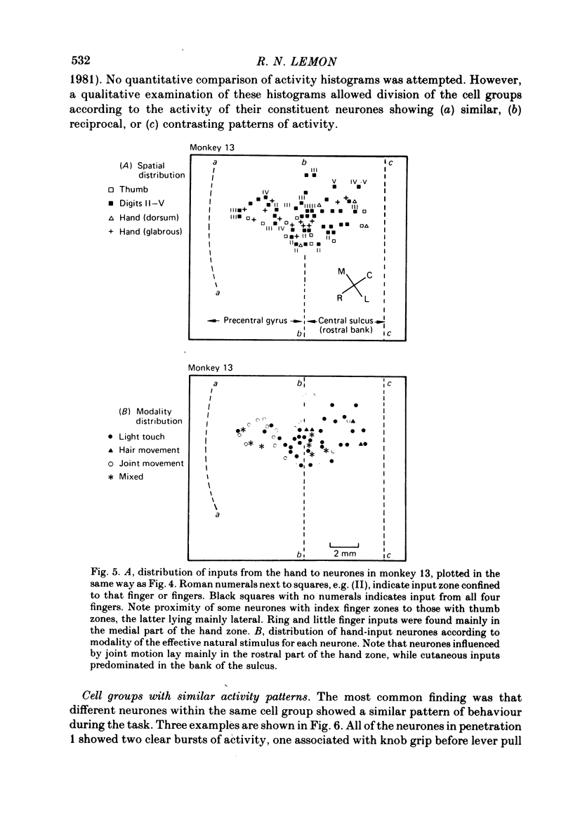
Abstract
1. Single-unit recordings have been made from 606 neurones in the arm region of area 4 in five conscious monkeys. Their activity during a stereotyped motor task and their responses to passive natural stimulation of the limb have been investigated.
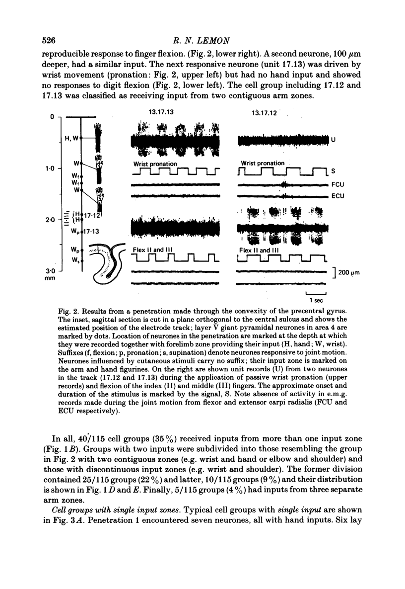
2. 88% of area 4 neurones responsive to natural stimulation received their afferent input from a restricted region of the contralateral arm.
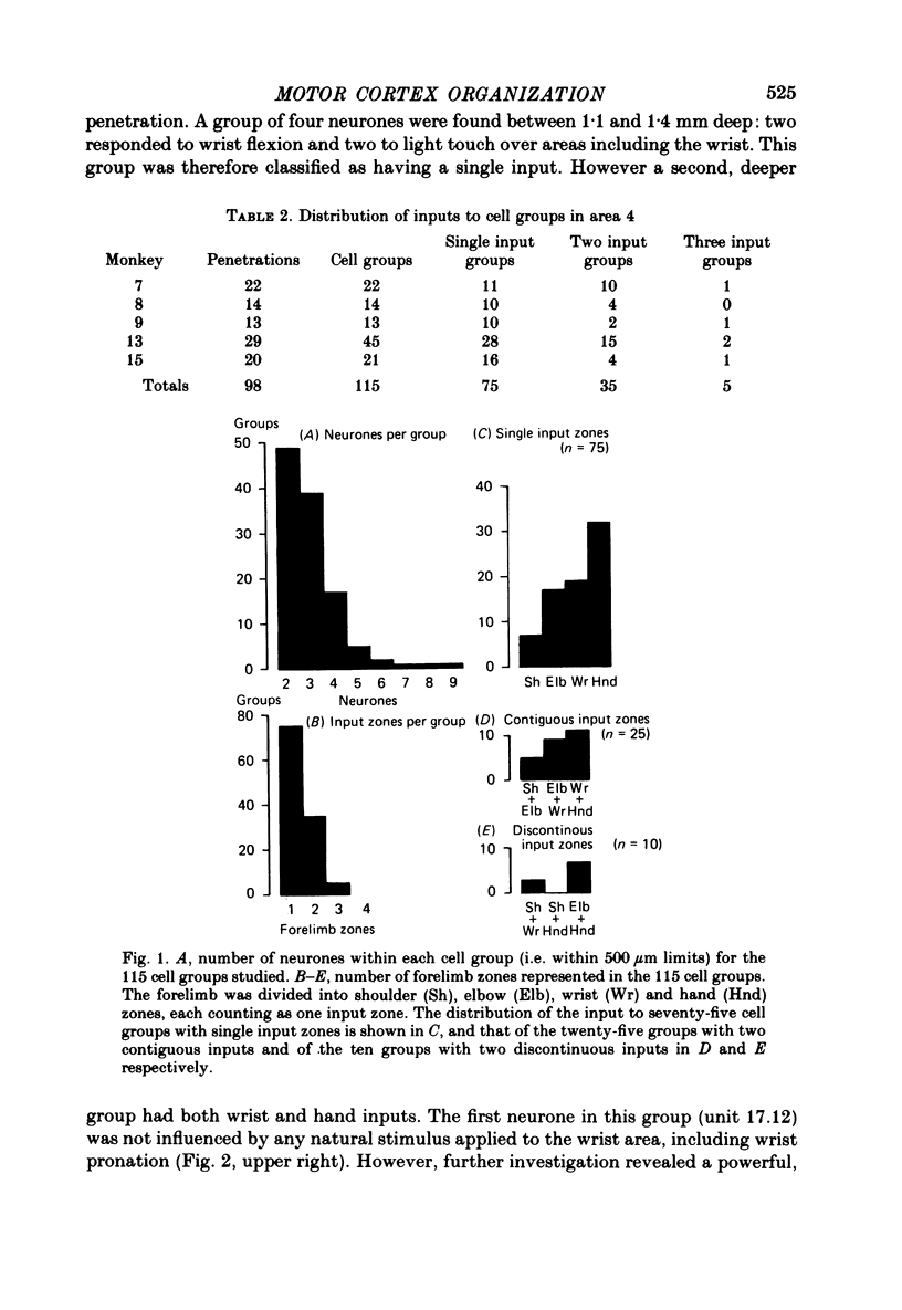
3. The activity and afferent input to cell groups have been determined by comparing the properties of neurones located within 500 μm of each other and recorded in one and the same micro-electrode penetration. 115 such cell groups containing 344 neurones were investigated.
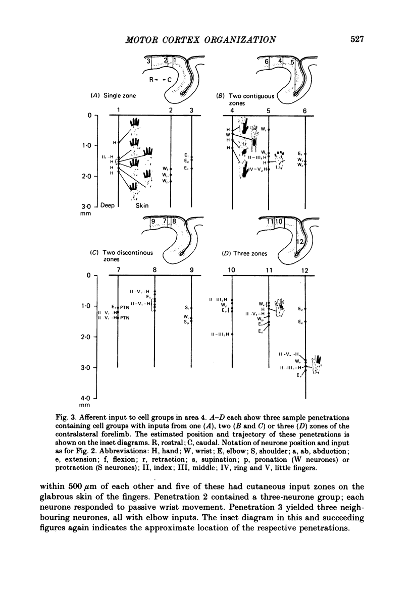
4. 75/115 cell groups (65%) contained neurones with input from the same arm zone (shoulder, elbow, wrist or hand) and with a similar pattern of task-related activity. Cell groups containing neurones with identical afferent inputs never showed contrasting behaviour during movement.
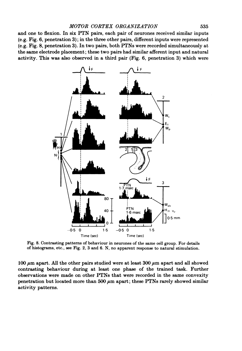
5. 40/115 cell groups (35%) contained neurones receiving inputs from more than one arm zone. Twenty-five cell groups (22%) had two contiguous zones (e.g. wrist and hand) represented and ten groups had input from two discontinuous zones (e.g. elbow and hand). These differences in input within a cell group were usually reflected in contrasting behaviour of its constituent neurones during movement.
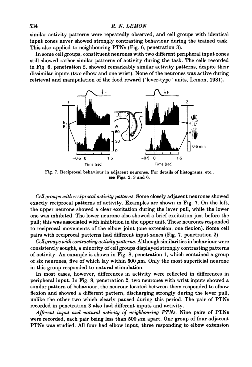
6. Pyramidal tract neurones (PTNs) lying immediately adjacent to one another usually received similar inputs and exhibited matching behaviour. PTNs lying further apart in the same penetration often showed different activity and responded to different stimuli.
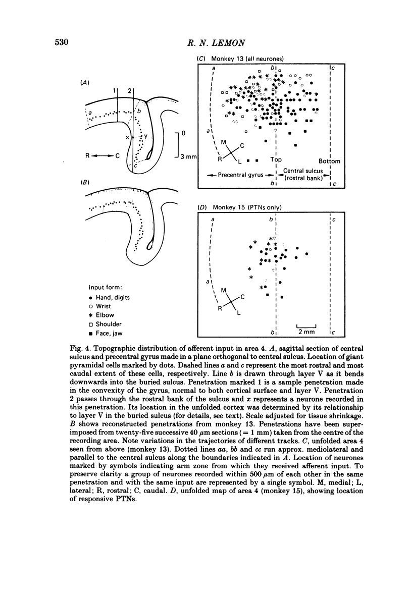
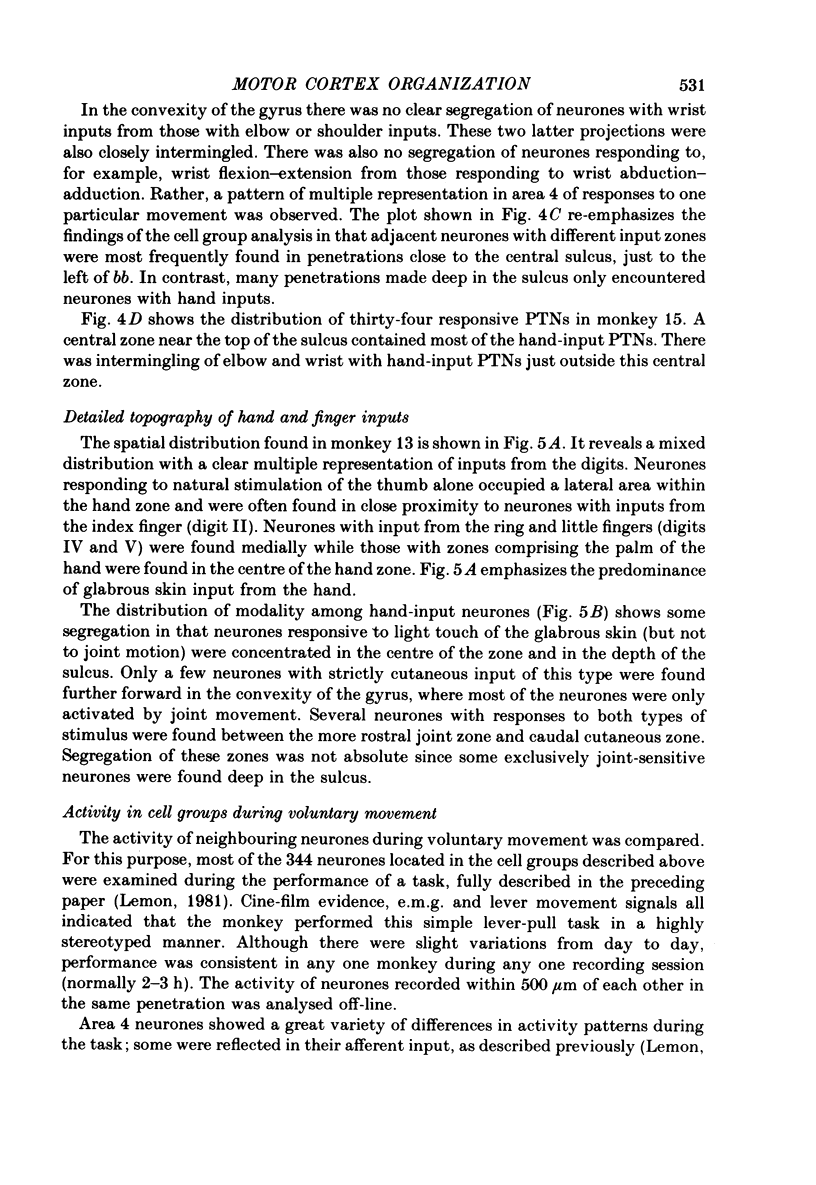
7. The topographic distribution of afferent input to area 4 revealed multiple representation of input from a single zone combined with considerable intermingling of input from all four zones. Neurones with shoulder and elbow inputs surrounded those with wrist inputs which in turn lay scattered around a central zone. This central zone only contained neurones with hand inputs, although neurones with hand inputs were found outside this central zone.
8. The significance of this complex organization is discussed in terms of motor cortex input and output.
Full text
PDF

Selected References
These references are in PubMed. This may not be the complete list of references from this article.
- Andersen P., Hagan P. J., Phillips C. G., Powell T. P. Mapping by microstimulation of overlapping projections from area 4 to motor units of the baboon's hand. Proc R Soc Lond B Biol Sci. 1975 Jan 21;188(1090):31–36. doi: 10.1098/rspb.1975.0002. [DOI] [PubMed] [Google Scholar]
- Asanuma H., Fernandez J., Scheibel M. E., Scheibel A. B. Characteristics of projections from the nucleus ventralis lateralis to the motor cortex in the cats: an anatomical and physiological study. Exp Brain Res. 1974;20(4):315–330. doi: 10.1007/BF00237378. [DOI] [PubMed] [Google Scholar]
- Asanuma H., Larsen K. D., Yumiya H. Receptive fields of thalamic neurons projecting to the motor cortex in the cat. Brain Res. 1979 Aug 24;172(2):217–228. doi: 10.1016/0006-8993(79)90534-1. [DOI] [PubMed] [Google Scholar]
- Asanuma H. Recent developments in the study of the columnar arrangement of neurons within the motor cortex. Physiol Rev. 1975 Apr;55(2):143–156. doi: 10.1152/physrev.1975.55.2.143. [DOI] [PubMed] [Google Scholar]
- Asanuma H., Zarzecki P., Jankowska E., Hongo T., Marcus S. Projection of individual pyramidal tract neurons to lumbar motor nuclei of the monkey. Exp Brain Res. 1979 Jan 2;34(1):73–89. doi: 10.1007/BF00238342. [DOI] [PubMed] [Google Scholar]
- Fetz E. E., Cheney P. D., German D. C. Corticomotoneuronal connections of precentral cells detected by postspike averages of EMG activity in behaving monkeys. Brain Res. 1976 Sep 24;114(3):505–510. doi: 10.1016/0006-8993(76)90973-2. [DOI] [PubMed] [Google Scholar]
- Fetz E. E., Finocchio D. V. Correlations between activity of motor cortex cells and arm muscles during operantly conditioned response patterns. Exp Brain Res. 1975 Sep 29;23(3):217–240. doi: 10.1007/BF00239736. [DOI] [PubMed] [Google Scholar]
- HUBEL D. H., WIESEL T. N. Receptive fields, binocular interaction and functional architecture in the cat's visual cortex. J Physiol. 1962 Jan;160:106–154. doi: 10.1113/jphysiol.1962.sp006837. [DOI] [PMC free article] [PubMed] [Google Scholar]
- Horne M. K., Tracey D. J. The afferents and projections of the ventroposterolateral thalamus in the monkey. Exp Brain Res. 1979 Jun 1;36(1):129–141. doi: 10.1007/BF00238473. [DOI] [PubMed] [Google Scholar]
- Jankowska E., Padel Y., Tanaka R. Projections of pyramidal tract cells to alpha-motoneurones innervating hind-limb muscles in the monkey. J Physiol. 1975 Aug;249(3):637–667. doi: 10.1113/jphysiol.1975.sp011035. [DOI] [PMC free article] [PubMed] [Google Scholar]
- Jones E. G., Coulter J. D., Hendry S. H. Intracortical connectivity of architectonic fields in the somatic sensory, motor and parietal cortex of monkeys. J Comp Neurol. 1978 Sep 15;181(2):291–347. doi: 10.1002/cne.901810206. [DOI] [PubMed] [Google Scholar]
- Kwan H. C., MacKay W. A., Murphy J. T., Wong Y. C. Spatial organization of precentral cortex in awake primates. II. Motor outputs. J Neurophysiol. 1978 Sep;41(5):1120–1131. doi: 10.1152/jn.1978.41.5.1120. [DOI] [PubMed] [Google Scholar]
- Lemon R. N. Functional properties of monkey motor cortex neurones receiving afferent input from the hand and fingers. J Physiol. 1981 Feb;311:497–519. doi: 10.1113/jphysiol.1981.sp013601. [DOI] [PMC free article] [PubMed] [Google Scholar]
- Lemon R. N., Hanby J. A., Porter R. Relationship between the activity of precentral neurones during active and passive movements in conscious monkeys. Proc R Soc Lond B Biol Sci. 1976 Oct 29;194(1116):341–373. doi: 10.1098/rspb.1976.0083. [DOI] [PubMed] [Google Scholar]
- Lemon R. N., Porter R. Afferent input to movement-related precentral neurones in conscious monkeys. Proc R Soc Lond B Biol Sci. 1976 Oct 29;194(1116):313–339. doi: 10.1098/rspb.1976.0082. [DOI] [PubMed] [Google Scholar]
- Lemon R. N., van der Burg J. Short-latency peripheral inputs to thalamic neurones projecting to the motor cortex in the monkey. Exp Brain Res. 1979 Aug 1;36(3):445–462. doi: 10.1007/BF00238515. [DOI] [PubMed] [Google Scholar]
- MOUNTCASTLE V. B. Modality and topographic properties of single neurons of cat's somatic sensory cortex. J Neurophysiol. 1957 Jul;20(4):408–434. doi: 10.1152/jn.1957.20.4.408. [DOI] [PubMed] [Google Scholar]
- PHILLIPS C. G., PORTER R. THE PYRAMIDAL PROJECTION TO MOTONEURONES OF SOME MUSCLE GROUPS OF THE BABOON'S FORELIMB. Prog Brain Res. 1964;12:222–245. doi: 10.1016/s0079-6123(08)60625-1. [DOI] [PubMed] [Google Scholar]
- Pandya D. N., Kuypers H. G. Cortico-cortical connections in the rhesus monkey. Brain Res. 1969 Mar;13(1):13–36. doi: 10.1016/0006-8993(69)90141-3. [DOI] [PubMed] [Google Scholar]
- Rispal-Padel L., Massion J., Grangetto A. Relations between the ventrolateral thalamic nucleus and motor cortex and their possible role in the central organization of motor control. Brain Res. 1973 Sep 28;60(1):1–20. doi: 10.1016/0006-8993(73)90847-0. [DOI] [PubMed] [Google Scholar]
- Rosén I., Asanuma H. Peripheral afferent inputs to the forelimb area of the monkey motor cortex: input-output relations. Exp Brain Res. 1972;14(3):257–273. doi: 10.1007/BF00816162. [DOI] [PubMed] [Google Scholar]
- Shinoda Y., Zarzecki P., Asanuma H. Spinal branching of pyramidal tract neurons in the monkey. Exp Brain Res. 1979 Jan 2;34(1):59–72. doi: 10.1007/BF00238341. [DOI] [PubMed] [Google Scholar]
- Strick P. L., Preston J. B. Multiple representation in the primate motor cortex. Brain Res. 1978 Oct 13;154(2):366–370. doi: 10.1016/0006-8993(78)90707-2. [DOI] [PubMed] [Google Scholar]
- Strick P. L., Preston J. B. Sorting of somatosensory afferent information in primate motor cortex. Brain Res. 1978 Nov 10;156(2):364–368. doi: 10.1016/0006-8993(78)90520-6. [DOI] [PubMed] [Google Scholar]
- Thompson W. D., Stoney S. D., Jr, Asanuma H. Characteristics of projections from primary sensory cortex to motorsensory cortex in cats. Brain Res. 1970 Aug 12;22(1):15–27. doi: 10.1016/0006-8993(70)90399-9. [DOI] [PubMed] [Google Scholar]
- Wong Y. C., Kwan H. C., MacKay W. A., Murphy J. T. Spatial organization of precentral cortex in awake primates. I. Somatosensory inputs. J Neurophysiol. 1978 Sep;41(5):1107–1119. doi: 10.1152/jn.1978.41.5.1107. [DOI] [PubMed] [Google Scholar]