

Abstract
The adherence of Porphyromonas gingivalis to host cells is likely a prerequisite step in the pathogenesis of P. gingivalis-induced periodontal disease. P. gingivalis binds to and invades epithelial cells, and fimbriae are shown to be involved in this process. Little is known regarding epithelial receptor(s) involved in binding of P. gingivalis fimbriae. Using an overlay assay with purified P. gingivalis fimbriae as a probe, two major epithelial cell proteins with masses of 50 and 40 kDa were identified by immunoblotting with fimbria-specific antibodies. Iodinated purified fimbriae also bound to the same two epithelial cell proteins. An affinity chromatography technique was utilized to isolate and purify the epithelial components to which P. gingivalis fimbriae bind. Purified fimbriae were coupled to CNBr-activated Sepharose-4B, and the solubilized epithelial cell extract proteins bound to the immobilized fimbriae were isolated from the column. A major 50-kDa component and a minor 40-kDa component were purified and could be digested with trypsin, suggesting that they were proteins. These affinity-eluted 50- and 40-kDa proteins were then subjected to amino-terminal sequencing, and no sequence could be determined, suggesting that these proteins have blocked amino-terminal residues. CNBr digestion of the 50-kDa component resulted in an internal sequence homologous to that of Keratin I molecules. Further evidence that P. gingivalis fimbriae bind to cytokeratin molecule(s) comes from studies showing that multicytokeratin rabbit polyclonal antibodies cross-react with the affinity-purified 50-kDa epithelial cell surface component. Also, binding of purified P. gingivalis fimbriae to epithelial components can be inhibited in an overlay assay by multicytokeratin rabbit polyclonal antibodies. Furthermore, we showed that biotinylated purified fimbriae bind to purified human epidermal keratin in an overlay assay. These studies suggest that the surface-accessible epithelial cytokeratins may act as receptor(s) for P. gingivalis fimbriae. We hypothesize that adherence of P. gingivalis fimbriae to cytokeratin may be important for colonization of oral mucous membranes and possibly also for activation of epithelial cells.
Porphyromonas gingivalis plays an important role in the initiation and progression of periodontal disease. P. gingivalis has been shown to colonize the periodontal pocket surface epithelium and attach to and invade oral epithelial cells in vitro. A variety of cell surface structures have been postulated to play a role in the interaction of P. gingivalis with host cells. P. gingivalis fimbriae have been reported to induce the expression of inflammatory cytokines in human gingival fibroblasts and mouse peritoneal macrophages (7, 8), strongly suggesting that fimbriae play a crucial role in bacterial interactions with host tissues. Several reports have documented the role of P. gingivalis fimbriae in mediating adherence to epithelial cells (13, 18, 31) and endothelial cells (2). A P. gingivalis strain with a reduced level of fimbriae surface protein has been shown to have a diminished capacity for adherence to oral epithelial cells in an in vitro assay. Also, antifimbrial monoclonal antibodies have been shown to block the adhesion of P. gingivalis to human buccal epithelial cells in an in vitro assay (11). A mutant of P. gingivalis 33277 (MPG1) lacking fimbriae does not adhere to gingival fibroblasts or epithelial cells (6), strongly implicating fimbriae in adherence to these mammalian cells. We found that a domain corresponding to amino acid residues 49 to 90 of the fimbrial protein is a major epithelial cell binding domain of P. gingivalis fimbriae (28). Although P. gingivalis fimbriae have received much attention, little is known about the epithelial cell receptor(s) for these adhesins.
Since fimbriae of P. gingivalis appear to be critical for binding and subsequent invasion of epithelial cells, our goal was to purify and characterize cell surface component(s) of epithelial cells involved in fimbriae binding. In the present study, we provide biochemical and immunological identification of a likely epithelial receptor for P. gingivalis fimbriae, which by its size, nature, amino acid sequence, and immunologic reactivity appears to be a cytokeratin. Our findings may have significance for understanding the role of P. gingivalis epithelial cell interaction and its virulence.
MATERIALS AND METHODS
Bacterial culture conditions.
P. gingivalis strain 381 was grown in half-strength (18 mg/ml) brain heart infusion broth (Difco) supplemented with 5 mg of yeast extract per ml and buffered at pH 7.4; cells were incubated for 2 days in an anaerobic chamber (85% N2, 10% H2, 5% CO2).
Fimbriae and major membrane antigen preparation.
Fimbriae were purified by the method of Lee et al. (15) as described earlier. Briefly, sonic extracts were treated with 40% ammonium sulfate, and the insolubilized fimbriae were purified on a Sepharose CL-6B column in the presence of guanidine-hydrochloride. Purity was confirmed by sodium dodecyl sulfate (SDS)-polyacrylamide gel electrophoresis.
A major membrane 75-kDa antigen from P. gingivalis was prepared (27) for use as control.
Iodination of fimbriae and major membrane antigen.
Purified fimbriae were iodinated using chloramine T (10). Ten micrograms of purified fimbriae and the 75-kDa major membrane antigen were labeled using 0.5 mCi of sodium iodine-125 (Amersham Corp.) in 0.5 M phosphate buffer, pH 7.2, in the presence of 10 μl of chloramine T (1 mg/ml) for 60 s. Iodination was terminated by adding 20 μl of sodium metabisulfite (2 mg/ml). After termination, 100 μl of phosphate buffer containing 10% sucrose and 10% potassium iodide was added, and the mixture was loaded on a Sephadex G75 column (1 by 30 cm) saturated with 1% bovine serum albumin (BSA) to prevent nonspecific binding. The column was extensively washed after saturation with BSA. The iodinated fimbriae peak was collected without carrier protein.
Biotinylation of fimbriae and the major membrane antigen.
Proteins were biotinylated using Sulfo-NHS-Biotin (Pierce Chemicals, Rockford, Ill.) as per the manufacturer’s directions.
Epithelial cell extract preparation.
The epithelial KB cell line ATCC CCL17 (American Type Culture Collection, Rockville, Md.) was cultured in Dulbecco’s modified Eagle medium (DMEM) supplemented with 10% fetal calf serum. Epithelial cells were grown to confluent monolayers, the medium was removed, and cells were washed with the same medium without serum. Cells were solubilized in 1% Triton X-100 and 20% glycerol in 50 mM Tris-Cl [pH 7.5] buffer at 4°C for 1 h. After solubilization, the extract was centrifuged at 10,000 rpm for 30 min at 4°C, and the clear supernatant was used for further studies.
SDS-PAGE and overlay assay.
SDS-polyacrylamide gel electrophoresis (PAGE) was performed according to the method of Laemmli (12) using a 12.5% gel (Mini-tall; Hoffer Scientific). For immunoblot analysis, the epithelial cell extract was separated by SDS-PAGE and transferred to nitrocellulose membranes, using the polyBlot Transfer System. After the transfer, membranes were blocked with 1% fatty-acid-free BSA and incubated with purified and iodinated P. gingivalis fimbriae. The membrane was incubated overnight at 4°C and washed extensively with phosphate-buffered saline (PBS). After the washing, the membrane was dried and exposed for autoradiography. Identical results were obtained when earlier prepared (26) specific antifimbriae antibodies or biotinylated fimbriae were used to detect P. gingivalis fimbriae bound to epithelial cell components on the membranes. For the inhibition assay, the blot was preincubated at room temperature for 1 h with the respective inhibitor, such as anticytokeratin antibodies, prior to incubation with biotinylated fimbriae.
Silver staining of SDS-polyacrylamide gel.
SDS-PAGE was performed according to the method of Laemmli (12) using a 15% gel (Mini-tall; Hoffer Scientific). Gels were stained with silver by the method of Merril et al. (16).
Western immunoblot analysis.
For Western immunoblot analysis, proteins separated by SDS-PAGE were transferred to nitrocellulose membranes by the method of Towbin et al. (30). The unoccupied sites on the nitrocellulose membranes were blocked with a 4.5% aqueous suspension of dry fat-free milk powder. The nitrocellulose membrane was then treated with an appropriate dilution of the respective rabbit polyclonal antibodies in 50 mM Tris-Cl [pH 7.5] buffer at 4°C overnight. After five to six washes with 50 mM Tris-Cl buffer containing 0.5 M NaCl and 0.1% Tween 20 (wash buffer), the nitrocellulose membranes were incubated with horseradish peroxidase (HRP)-conjugated goat anti-rabbit immunoglobulin G for 1 h at room temperature. The membranes were extensively washed with the wash buffer five to six times, and the bound HRP-conjugated antibodies were visualized using 4-chloro-1-naphthol as the color-developing reagent.
Preparation of immobilized fimbriae.
Cyanogen bromide-activated Sepharose 4B was swollen in distilled water and washed with 1 mM HCl. The gel was then washed with ice-cold distilled water and twice with coupling buffers containing 0.1 M NaHCO3, at pH 8.3. Purified fimbriae were dissolved in the same buffer and left for 2 h with gentle shaking in the cyanogen bromide-activated Sepharose. The gel was filtered and then washed successively with the same coupling buffer containing 0.5 M NaCl. After the gel was washed with distilled water, a solution of 0.2 M glycine, pH 8.0, was added to the gel to block the free amino groups. The gel was washed extensively with coupling buffer followed by 0.1 M acetate buffer (pH 4.0) containing 1 M NaCl. The gel was washed in binding buffer and stored at 4°C.
Affinity purification of epithelial receptor(s) using immobilized fimbriae on Sepharose 4B.
The gel containing the immobilized fimbriae was suspended and washed with the solubilizing buffer (1% Triton X-100 and 20% glycerol in 50 mM Tris-Cl, pH 7.5). After equilibration of the gel, the solubilized epithelial cell preparation was added and the gel was incubated at 4°C for an hour. Unbound sample was collected, and the gel was washed with the same buffer containing 1 M NaCl. The elution buffer of low pH (pH 2.5), 50 mM sodium acetate, was added to the gel to elute the bound protein. The eluted fraction was immediately neutralized with 1 M ammonium bicarbonate, pH 8.0 (25). As a control, a major cell surface antigen, the 75-kDa protein, was coupled to a Sepharose 4B gel and the affinity chromatography was carried out under identical conditions.
Analysis of affinity-eluted components.
Affinity-eluted fractions were analyzed by 15% gel analysis. The gel was stained with 0.2% Coomassie blue and by silver staining. Eluted fractions were checked with P.gingivalis antifimbriae antibodies using Western immunoblot methods described above.
Tryptic digestion of fimbriae binding protein.
Aliquots of the affinity-eluted components were incubated with 5% Trypsin at 37°C for 1 h. After 1 h of incubation, samples were analyzed on an SDS-polyacrylamide gel. Trypsin only and samples without trypsin were used as controls.
The amino-terminal sequence of fimbriae binding protein.
The affinity-eluted fraction was run on an SDS-polyacrylamide gel according to the method of Laemmli (12) using a 15% gel (Mini-tall; Hoffer Scientific). For amino-terminal sequence determination, the protein separated by SDS-PAGE was transferred to Immobilon membranes using the polyBlot Transfer System. After transfer, the membranes were stained quickly and washed extensively with high-performance liquid chromatography-grade water. The 50- and 40-kDa epithelial cell protein bands were removed from the membranes and subjected to amino-terminal sequencing (ProSeq, Inc., Salem, Mass.).
Immunoblot with multicytokeratin rabbit polyclonal antibodies.
Rabbit, anti-human cytokeratin polyclonal antibodies were purchased from Novocastra Laboratories Limited, Newcastle upon Tyne, United Kingdom. Immunoblot analysis was done as described above. HRP-conjugated goat antirabbit antibodies were used as a secondary antibody.
Dot blot analysis with epidermal keratin and biotinylated purified fimbriae.
Human epidermal keratin was purchased from Sigma Chemical Company (St. Louis, Mo.). Zero to 40 ng, in increments of 10 ng, of human epidermal keratin was spotted on nitrocellulose membranes, and the membranes were blocked with 1% BSA. The membranes were then incubated with biotinylated purified fimbriae at 37°C for 1 h, and as a control another identical membrane was incubated with biotinylated major cell surface P. gingivalis 75-kDa antigen. The membranes were washed extensively five to six times with PBS and incubated with avidin-HRP (Bio-Rad Laboratories, Hercules, Calif.) at room temperature for 1 h. The membranes were then washed five to six times with PBS and developed with HRP color developing reagent (Bio-Rad Laboratories). The biotinylated major cell surface 75-kDa component was used as a negative control.
RESULTS
Fimbrial and major cell surface antigen.
Fimbriae were purified by the above-mentioned methods, and purity was confirmed by silver staining, which showed a single component with no detectable contaminants (Fig. 1, lane 4). A major cell surface antigen, the 75-kDa component, was purified from P. gingivalis for use as a control (Fig. 1, lane 3). Purified fimbriae and the 75-kDa major cell surface components were iodinated using the chloramine T method, and the integrity of labeled proteins was confirmed by autoradiography (data not shown).
FIG. 1.

SDS-PAGE analysis of purified fimbriae and major cell surface antigen from P. gingivalis on 15% polyacrylamide gels stained with silver. Lane 1, molecular mass standard proteins; lane 2, 40% ammonium sulfate cell extract of P. gingivalis; lane 3, purified major cell surface component from cell surface of P. gingivalis; lane 4, purified fimbriae from P. gingivalis.
Overlay assay with iodinated fimbriae.
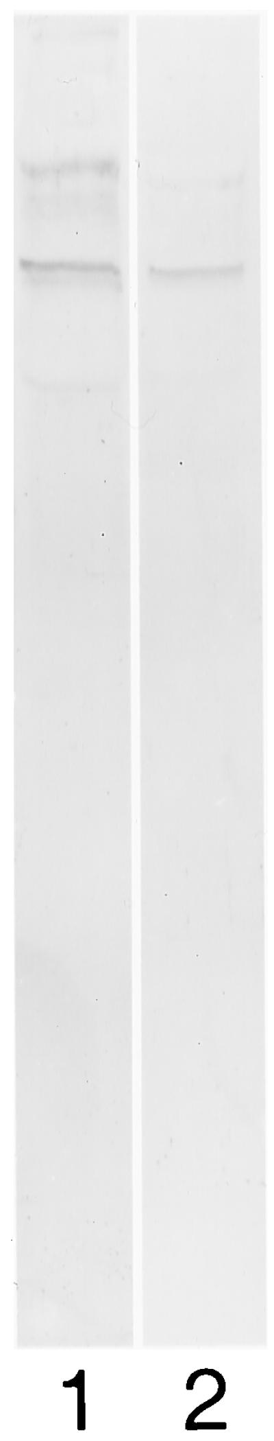
Soluble extracts of KB cells without heating were run on an SDS-polyacrylamide gel and transferred to nitrocellulose membranes. The membranes were blocked with 1% fatty-acid-free BSA. When the membrane was incubated with iodinated fimbriae, two distinct bands of 50 and 40 kDa were observed by autoradiography (Fig. 2). However, the major cell surface 75-kDa antigen of P. gingivalis failed to detectably bind to KB cell components under the same conditions (data not shown).
FIG. 2.

Autoradiogram of overlay assay. KB cell extract was treated with and without β-mercaptoethanol and electrophoresed on a 12.5% gel. Lane 1, epithelial cell extract under nonreducing conditions without β-mercaptoethanol; lane 2, epithelial cell extract under reducing conditions with β-mercaptoethanol.
Affinity column using immobilized purified P. gingivalis fimbriae.
The gel containing the immobilized fimbriae was used for affinity purification of fimbriae binding component(s) from KB cell solubilized extracts. After the bound protein was eluted at low pH, all fractions were run on an SDS-15% polyacrylamide gel. Silver staining of the eluted fraction revealed two protein bands: a major band of molecular size 50 kDa and a minor component of 40 kDa (Fig. 3, lane 5). When the 75-kDa-cell-surface-antigen-coupled Sepharose was used as a control affinity column, no bands were observed in eluted fractions after treatment of the gel with solubilized epithelial extracts (Fig. 3, lane 9).
FIG. 3.

SDS-PAGE analysis of samples at various steps of affinity purification. Samples were electrophoresed on a 15% gel with β-mercaptoethanol and stained with silver. Lane 1, standard molecular mass markers; lane 2, total KB cell extract; lane 3, unbound sample; lane 4, wash; lane 5, bound components; lane 6, incubation of affinity-purified component without trypsin; lane 7, incubation of trypsin only; lane 8, incubation of affinity-purified component with trypsin; lane 9, bound component with 75-kDa-protein-coupled Sepharose 4B.
Analysis of affinity-eluted components.
To exclude the possibility that eluted material contained fimbriae that had leached from the affinity column, affinity-eluted components were checked with P. gingivalis antifimbriae antibodies by immunoblot analysis (Fig. 4, lane 2).
FIG. 4.

Immunoblot of affinity-purified epithelial components with P. gingivalis antifimbria antibodies. A 15% polyacrylamide gel was electrophoretically transferred to a nitrocellulose membrane. Lane 1, isolated P. gingivalis fimbriae; lane 2, affinity-purified components.
Tryptic digestion of affinity-purified components.
To confirm the proteinaceous nature of the epithelial cell components, the fractions eluted from the affinity column were incubated with trypsin. After incubation the digested samples were run on an SDS-15% polyacrylamide gel. Both epithelial cell components eluted from the affinity column (50 and 40 kDa) were degraded as shown in Fig. 3. Tryptic digestion of both components suggested the proteinaceous nature of fimbriae binding KB-cell component(s).
Amino-terminal sequence of fimbriae binding protein.
The affinity-eluted epithelial cell fractions were transferred to polyvinylidene difluoride membranes and stained quickly. The membranes were washed extensively with high-performance liquid chromatography-grade water. The 50- and 40-kDa protein bands were cut from the membrane and subjected to amino-terminal sequencing (ProSeq, Inc.). No sequence could be determined, suggesting that these proteins have blocked amino-terminal residues. However, CNBr digestion of the 50-kDa component resulted in an internal sequence, suggesting that it has at least a cytokeratin-like domain. The 50-kDa CNBr digest had a 15-amino-acid internal sequence that was found to be DQXEQXAEKNRNKDV. The FASTA database for homology to known sequences was searched, and it was found that about 80% of the 50-kDa internal 15 amino acid was homologous to the keratin 14 domain and about 73% was homologous to the Keratin 16 peptide domain (Table 1).
TABLE 1.
Homologies of amino acid sequence from the 50-kDa protein to keratin 14 and keratin 16 molecules
| Protein (Swall Database accession no.) | Sequencea | No. with identity/total no. (%) in sequence |
|---|---|---|
| 50 kDa | DQXEQXAEKNRNKDV | 12/15 (80) |
| ** ** ***** *** | ||
| Keratin 14 (035813) | DQYEQMAEKNR--KDV | |
| 50 kDa | DQXEQXAEKNRNKDV | 11/15 (73) |
| ** ** ***** *** | ||
| Keratin 16 (09Z2K1) | DQYEQMAEKNR--RDV |
X denotes an amino acid that could not be determined. Identical amino acids are bold with * symbol in between.
The 40-kDa epithelial cell component was a minor eluted component and could not be reliably processed for internal amino acid sequence. The unidentified 40-kDa component appeared to be less abundant in the eluted fractions from the affinity column (Fig. 3, lane 5), and it appeared to have a greater affinity for fimbriae in the overlay assay (Fig. 2) than did the 50-kDa putative cytokeratin, suggesting that the unidentified 40-kDa component may be very significant in the host cell-bacterium interaction.
Immunoblot with multicytokeratin rabbit polyclonal antibodies.
A rabbit, anti-human cytokeratin polyclonal antibody which has specificity to multiple cytokeratins was used for immunoblot analysis of the fimbriae-affinity-purified KB-cell component. Extensive immunohistochemical evaluation of this antibody by Novocastra Laboratories has indicated specific staining in various cells of epithelial origin. Tissues tested with positive staining that were observed include bile duct, intralobular liver ducts, cuboidal epithelia of the pancreas, epithelial cells of the breast and uterine glands, basal cells of the skin, hair follicles, mucosa of the gallbladder, and the cornea of the eye. Staining has also been observed in bronchial mucosa of the lung, squamous cell carcinoma of the skin, and oral mucous membranes.
In immunoblot analysis, this anti-human cytokeratin polyclonal antibody recognized the affinity-purified 50-kDa component of KB cells isolated from the P. gingivalis fimbriae affinity column (Fig. 5B). However, anticytokeratin antibodies recognized many components at about and above 50 kDa when total epithelial cell extract was used in immunoblot analysis (Fig. 5A) In addition, preincubation of the blot with multicytokeratin polyclonal antibodies reduced the binding of fimbriae to the 50-kDa component of KB cells (Fig. 6).
FIG. 5.

Immunoblot of affinity-purified epithelial components with multicytokeratin antibodies. A 15% polyacrylamide gel was electrophoretically transferred to a nitrocellulose membrane. (A) Lane 1, prestained standard molecular mass markers; lane 2, total KB cell extract. (B) Lane 1, prestained standard molecular weight markers; lane 2, affinity-purified components.
FIG. 6.
Overlay assay of affinity-purified epithelial components using biotinylated fimbriae with (lane 2) and without (lane 1) prior incubation with antikeratin antibodies.
Dot blot analysis with epidermal keratin and biotinylated purified fimbriae.
Human epidermal keratin was used to confirm that it can bind to biotinylated purified P. gingivalis fimbriae. In dot blot analysis, the biotinylated purified fimbriae bound to human epidermal keratin very strongly at nanogram levels of fimbriae (Fig. 7A). However, the biotinylated 75-kDa major cell surface component of P. gingivalis did not bind to keratin (Fig. 7B).
FIG. 7.

Dot blot analysis of epidermal keratin. Zero to forty nanograms of human epidermal cytokeratin was spotted on 1- by 1-cm squares of nitrocellulose membrane in duplicate. Unoccupied sites on membranes were blocked with 1% BSA. Lanes 1 to 5, 0, 10, 20, 30, and 40 ng of keratin. (A) Membrane incubated with biotinylated P. gingivalis fimbriae. (B) Membrane incubated with biotinylated P. gingivalis major cell surface component.
DISCUSSION
Adherence to and invasion of epithelial cells by P. gingivalis may play an important role in the pathogenesis of periodontal disease, especially in the initial stages of the infection. P. gingivalis major fimbriae have been shown to be involved in adherence and invasion of P. gingivalis in epithelial cells. In order to characterize adhesin receptor(s) on epithelial cells, we used Triton X-100-solubilized KB cells to which purified fimbriae bind in overlay assays. Purified P. gingivalis fimbriae bound to two specific proteins present in KB cell extract. The present studies indicate that two major epithelial cell components (with masses of 50 and 40 kDa) bind to P. gingivalis fimbriae. Both of these components appear proteinaceous in nature, as they can be digested with trypsin. The 50-kDa component has a size and internal peptide sequence comparable to those of cytokeratins and cytokeratin 16. Multicytokeratin antibody recognizes the 50-kDa component, and the binding of purified fimbriae to this epithelial component can be inhibited by multicytokeratin rabbit polyclonal antibodies. Moreover, purified fimbriae bind to purified human epidermal keratin, suggesting interaction between P. gingivalis fimbriae and cytokeratin. The unidentified 40-kDa component appeared to be less abundant in the eluted fractions from the affinity column but appeared to have greater affinity for fimbriae in the overlay assay than did the 50-kDa putative cytokeratin, suggesting that the unidentified 40-kDa component may also be very significant in the host cell-bacterium interaction.
Studies by Godfroid et al. (5), Diaz et al. (3), and Hembrough et al. (9) showed that cytokeratin 8 and cytokeratin 18 are present on the surfaces of cultured human and mouse keratinocytes. Some cytokeratins were also identified on injured corneal epithelial cells to which pili of P. aeruginosa bind (33). Among the identified major P. aeruginosa pilus-binding proteins, three proteins with masses of 45, 55, and 66 kDa were identified as cytokeratins (33). Recently, Sajjan et al. (21) showed that cable-piliated Burkholderia cepacia binds to cytokeratin 13 of epithelial cells. Group B streptococci and other gram-positive cocci also were found to bind cytokeratin 8 on epithelial cells (29). It is possible, therefore, that cytokeratins are more common at cell surfaces than is usually assumed, and they may play a role in initial interaction with microorganisms.
The role of adherence of P. gingivalis fimbriae to cytokeratin in the epithelial binding is unclear. In general, an interacting adhesin or receptor must be present on the surfaces of epithelial cells to interact with bacterial fimbriae, and this interaction likely leads to colonization of the surface. There are several mechanisms by which adherence of P. gingivalis fimbriae to cytokeratin may play a role in pathogenesis (20), including binding to cytokeratin present on epithelial cell surfaces. Godfroid et al. (5) showed that cytokeratins are exposed on the outer surfaces of established human mammary carcinoma cells. Hembrough et al. (9) also showed that a cytokeratin 8-like protein with plasminogen-binding activity is present on the external surfaces of hepatocytes, HepG2 cells, and breast carcinoma cells. Another possibility is that P. gingivalis may adhere to cytokeratin present on the surfaces of damaged or altered epithelial cells. P. gingivalis bacteria secrete many proteases and hemolysin, which can damage the surfaces of epithelial cells (14). Group B streptococcus beta-hemolysin expression is associated with injury to lung epithelial cells (17). P. gingivalis may thus adhere to altered keratinized epithelium through keratin molecules exposed by prior injury or enzymatic activity (1). Yet another possibility is that binding of P. gingivalis to cytoplasmic keratin occurs after invasion of epithelial cells. P. gingivalis is able to invade epithelial cells (19, 22, 23, 24), and it appears that cytoskeletal components are an important part of the cellular systems for cytoplasmic transport of vesicles and other organelles. It is possible, therefore, that P. gingivalis fimbriae may take advantage of these systems by adhering to cytoskeletal components, such as keratin, as part of the invasion process.
The cellular consequences of cytokeratin-bacterium interactions are unknown, but further studies will certainly help us in understanding how binding to the cytokeratin network of epithelial cells leads to important intracellular changes. Doorbar et al. (4) have shown specific interactions between human papillomavirus and cytokeratins that result in the collapse of the epithelial cell intermediate filament network. White and Cipriani (32) showed that adenovirus E1B protein binding to cytokeratin causes rearrangement and disruption of intermediate filaments. Similar changes may occur when P. gingivalis fimbriae bind to epithelial cells. Further studies to explore the possible mechanisms are in progress.
In summary, we demonstrate here that P. gingivalis fimbriae bind to cytokeratin molecules of KB cells. These data may provide new insight in elucidating the steps in the pathogenesis of P. gingivalis which involve interaction with oral mucosal epithelial cells.
Acknowledgments
This study was supported in part by U.S. Public Health Service grant no. DE04898.
We thank Darlene Badgett for her excellent technical assistance.
Editor: V. J. DiRita
REFERENCES
- 1.Bosch, F. X., R. E. Leube, T. Achstatter, T. Moll, and W. W. Franke. 1988. Expression of simple epithelial type cytokeratins in stratified epithelia as detected by immunolocalization and hybridization in situ. J. Cell Biol. 106: 1635–1648. [DOI] [PMC free article] [PubMed] [Google Scholar]
- 2.Deshpande, R., M. Khan, and C. A. Genco. 1998. Invasion of heart and aortic endothelial cells by Porphyromonas gingivalis. Infect. Immun. 66: 5337–5343. [DOI] [PMC free article] [PubMed] [Google Scholar]
- 3.Diaz, L. A., S. A. P. Sampaio, C. R. Martins, E. A. Rivitti, M. L. Macca, J. T. Roscoe, Y. Takahashi, R. S. Labib, H. P. Patel, D. F. Nutasim, E. M. Kugan, and G. J. Anhalt. 1987. An autoantibody in pemphigus serum, specific for the 59 kDa keratin, selectively binds the surface of keratinocytes: evidence for an extracellular keratin domain. J. Investig. Dermatol. 89: 287–295. [DOI] [PubMed] [Google Scholar]
- 4.Doorbar, J., S. Ely, J. Sterling, C. McLean, and J. Crawford. 1991. Specific interaction between HPV-16 E1–E4 and cytokeratins results in collapse of epithelial cell intermediate filament network. Nature 352: 824–827. [DOI] [PubMed] [Google Scholar]
- 5.Godfroid, E., M. Geuskens, T. Dupressoir, I. Parent, and C. Szpirer. 1991. Cytokeratins are exposed on the outer surface of established human mammary carcinoma cells. J. Cell Sci. 99: 565–607. [DOI] [PubMed] [Google Scholar]
- 6.Hamada, N., K. Watanabe, C. Sasakawa, M. Yoshikawa, F. Yoshimura, and T. Umemoto. 1994. Construction and characterization of a fimA mutant of Porphyromonas gingivalis. Infect. Immun. 62: 1696–1704. [DOI] [PMC free article] [PubMed] [Google Scholar]
- 7.Hanazawa, S., K. Hirose, Y. Ohmori, S. Amano, and S. Kitano. 1988. Bacteroides gingivalis fimbriae stimulate production of thymocyte-activating factor by human gingival fibroblasts. Infect. Immun. 56: 272–274. [DOI] [PMC free article] [PubMed] [Google Scholar]
- 8.Hanazawa, S., Y. Murakami, K. Kirose, S. Amano, Y. Ohmori, S. Higuchi, and S. Kitano. 1991. Bacteroides (Porphyromonas) gingivalis fimbriae activate mouse peritoneal macrophages and induce gene expression and production of interleukin-1. Infect. Immun. 59: 1972–1977. [DOI] [PMC free article] [PubMed] [Google Scholar]
- 9.Hembrough, T. A., J. Vasudevan, M. M. Allietta, W. F. Glass II, and S. L. Gonias. 1995. A cytokeratin 8-like protein with plasminogen-binding activity is present on the external surfaces of hepatocytes, HepG2 cells and breast carcinoma cell lines. J. Cell Sci. 108: 1071–1082. [DOI] [PubMed] [Google Scholar]
- 10.Hunter, W. M., and F. C. Greenwood. 1962. Preparation of 131iodine labeled growth hormone of high specific activity. Nature (London) 194: 495–496. [DOI] [PubMed] [Google Scholar]
- 11.Isogai, H., E. Isogai, F. Yoshimura, T. Suzuki, W. Kagota, and K. Takano. 1988. Specific inhibition of adherence of an oral strain of Bacteroides gingivalis 381 to epithelial cells by monoclonal antibodies against the bacterial fimbriae. Arch. Oral Biol. 33: 479–485. [DOI] [PubMed] [Google Scholar]
- 12.U. K. Laemmli. 1970. Cleavage of structural proteins during the assembly of the head of bacteriophage T4. Nature (London) 194: 680–685. [DOI] [PubMed] [Google Scholar]
- 13.Lamont, R. J., A. Chan, C. M. Belton, K. T. Izutsu, D. Vasel, and A. Weinberg. 1995. Porphyromonas gingivalis invasion of gingival epithelial cells. Infect. Immun. 63: 3878–3885. [DOI] [PMC free article] [PubMed] [Google Scholar]
- 14.Lantz, M. S., R. D. Allen, T. A. Vail, L. M. Switalski, and M. Hook. 1991. Specific cell components of Bacteroides gingivalis mediate binding and degradation of human fibrinogen. J. Bacteriol. 173: 495–504. [DOI] [PMC free article] [PubMed] [Google Scholar]
- 15.Lee, J. Y., H. T. Sojar, A. Amano, and R. J. Genco. 1995. Purification of major fimbrial proteins of Porphyromonas gingivalis. Protein Expr. Purif. 6: 496–500. [DOI] [PubMed] [Google Scholar]
- 16.Merril, C. R., D. Goldman, and M. L. VanKeuren. 1984. Gel protein stains: silver stain. Methods Enzymol. 104: 441–447. [DOI] [PubMed] [Google Scholar]
- 17.Nizet, V. R., L. Gibson, E. Y. Chi, P. E. Framson, M. Hulse, and C. E. Rubens. 1996. Group B streptococcal beta-hemolysin expression is associated with injury of lung epithelial cells. Infect. Immun. 64: 3818–3826. [DOI] [PMC free article] [PubMed] [Google Scholar]
- 18.Njogore, T., R. J. Genco, H. T. Sojar, N. Hamada, and C. A. Genco. 1997. A role for fimbriae in Porphyromonas gingivalis invasion of oral epithelial cells. Infect. Immun. 65: 1980–1984. [DOI] [PMC free article] [PubMed] [Google Scholar]
- 19.Papapanou, P. N., J. Sandros, K. Lindberg, M. J. Duncan, R. Niederman, and U. Nannmark. 1994. Porphyromonas gingivalis may multiply and advance within stratified human functional epithelium in vitro. J. Periodontal Res. 29: 374–375. [DOI] [PubMed] [Google Scholar]
- 20.Rosenshine, I., and B. B. Finlay. 1993. Exploitation of host signal transduction pathways and cytoskeletal functions by invasive bacteria. Bio-Essays 15: 17–24. [DOI] [PubMed] [Google Scholar]
- 21.Sajjan, U. S., F. A. Sylvester, and J. F. Forstner. 2000. Cable-piliated Burkholderia cepacia binds to cytokeratin 13 of epithelial cells. Infect. Immun. 68: 1787–1795. [DOI] [PMC free article] [PubMed] [Google Scholar]
- 22.Sandros, J., P. N. Papapanou, and G. Dahlen. 1993. Porphyromonas gingivalis invades oral epithelial cells in vitro. J. Periodontal Res. 28: 219–226. [DOI] [PubMed] [Google Scholar]
- 23.Sandros, J., P. N. Papapanou, U. Nannmark, and G. Dahlen. 1994. Porphyromonas gingivalis invades pocket epithelium in vitro. J. Periodontal Res. 29: 62–69. [DOI] [PubMed] [Google Scholar]
- 24.Sansonetti, P. J. 1993. Bacterial pathogens, from adherence to invasion: comparative strategies. Med. Microbiol. Immunol. 182: 223–232. [DOI] [PubMed] [Google Scholar]
- 25.Sojar, H. T., and O. P. Bahl. 1989. Characterization of rat ovarian lutropin receptor: role of thiol groups in receptor association. J. Biol. Chem. 264: 2552–2559. [PubMed] [Google Scholar]
- 26.Sojar, H. T., J. Y. Lee, G. S. Bedi, M. I. Cho, and R. J. Genco. 1991. Purification, characterization and immunolocalization of fimbrial protein from Porphyromonas (Bacteroides) gingivalis. Biochem. Biophys. Res. Commun. 175: 713–719. [DOI] [PubMed] [Google Scholar]
- 27.Sojar, H. T., J. Y. Lee, G. S. Bedi, M. I. Cho, and R. J. Genco. 1991. Purification, characterization, and localization of a major membrane protein antigen from Porphyromonas (Bacteroides) gingivalis. Biochem. Int. 25: 437–446. [PubMed] [Google Scholar]
- 28.Sojar, H. T., Y. Han, N. Hamada, A. Sharma, and R. J. Genco. 1999. Role of amino-terminal region of Porphyromonas gingivalis fimbriae in adherence to epithelial cells. Infect. Immun. 67: 6173–6176. [DOI] [PMC free article] [PubMed] [Google Scholar]
- 29.Tamura, G. S., and A. Nittayajaran. 2000. Group B streptococci and other gram-positive cocci bind to cytokeratin 8. Infect. Immun. 68: 2129–2134. [DOI] [PMC free article] [PubMed] [Google Scholar]
- 30.Towbin, H. T., T. Staehelin. and J. Gordon. 1979. Electrophoretic transfer of proteins. Proc. Natl. Acad. Sci. USA 76: 4350–4354. [DOI] [PMC free article] [PubMed] [Google Scholar]
- 31.Weinberg, A., C. M. Belton, Y. Park, and R. J. Lamont. 1997. Role of Porphyromonas gingivalis fimbriae in invasion of gingival epithelial cells. Infect. Immun. 65: 313–316. [DOI] [PMC free article] [PubMed] [Google Scholar]
- 32.White, E., and R. Cipriani. 1990. Role of adenovirus E1B proteins in transformation: altered organization of intermediate filaments in transformed cells that express the 19-kilodalton protein. Mol. Cell. Biol. 10: 120–130. [DOI] [PMC free article] [PubMed] [Google Scholar]
- 33.Wu, X., M. Kurpakus, and L. D. Hazlett. 1996. Some P. aeruginosa pilus binding proteins of human corneal epithelium are cytokeratins. Curr. Eye Res. 15: 1194–1198. [DOI] [PubMed] [Google Scholar]