Abstract
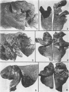
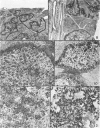
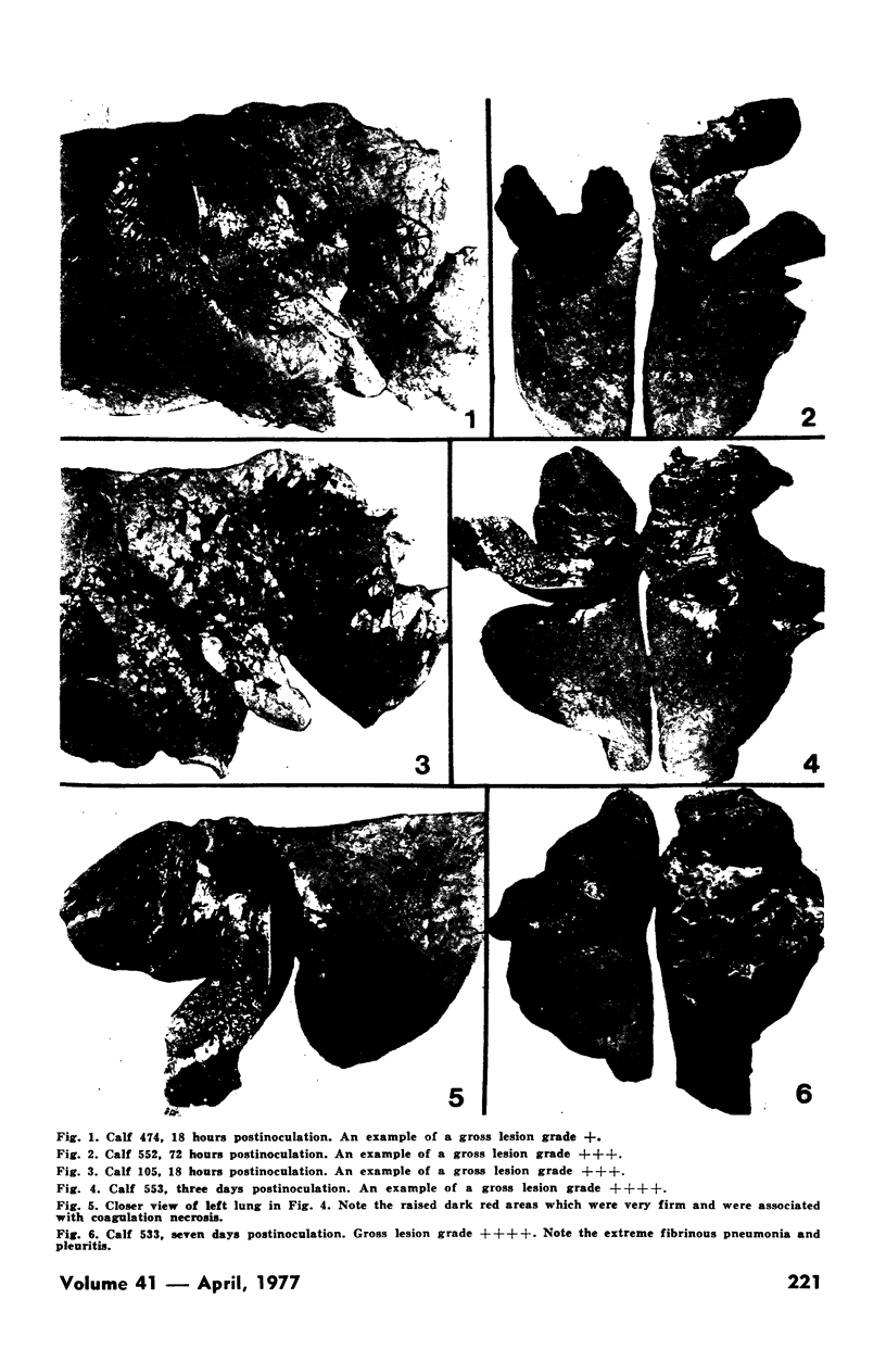
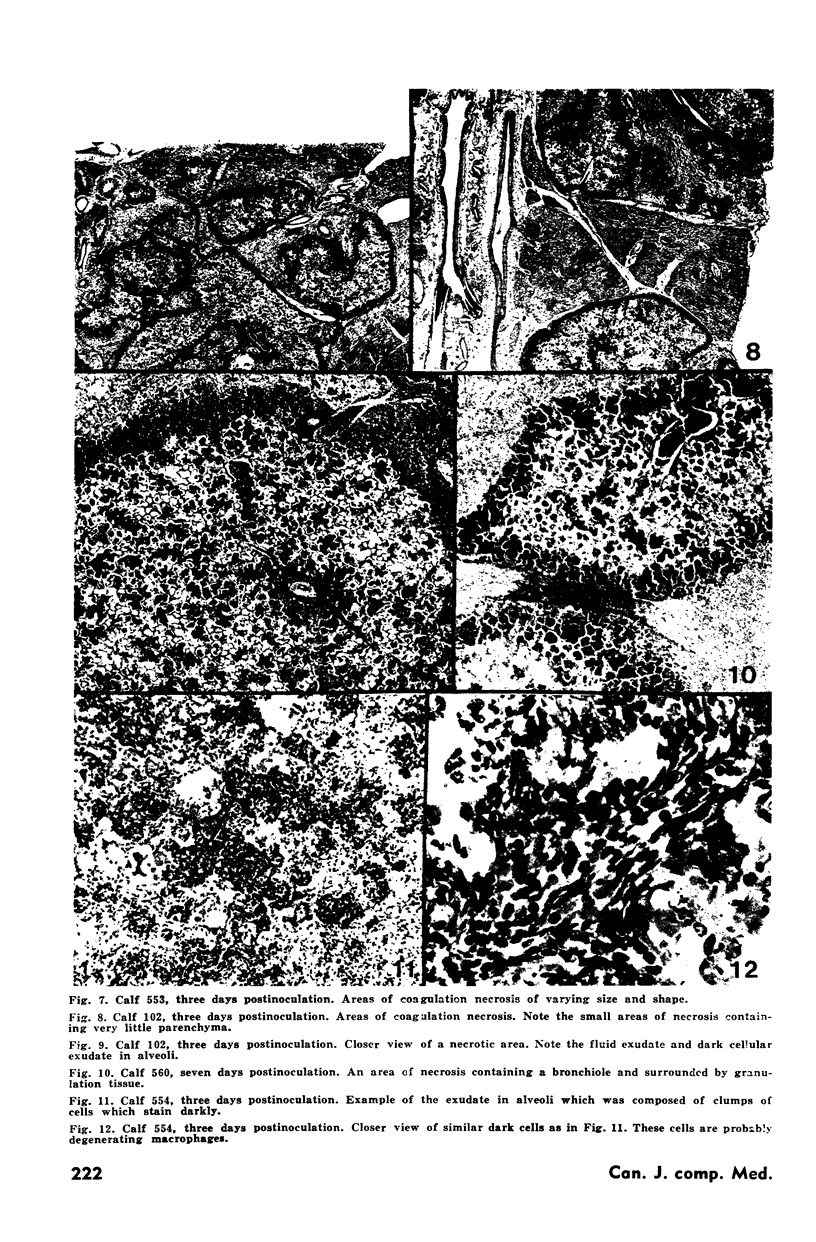
Twelve four month old calves were inoculated intratracheally with a large dose of Pasteurella hemolytica and the lungs examined 18 hours, three days and seven days later. The gross and microscopic lesions were graded in each calf. The most extensive gross lesions were present at three days and were characteristic of a fibrinous pneumonia. Discrete areas of coagulation necrosis were present at three days and granulation tissue had formed around these by seven days. Characteristic swirly dark cells accumulated in alveoli and alveolar ducts and were observed from three days onwards. The gross and microscopic lesions were similar to those seen in field cases of pneumonic pasteurellosis.
Full text
PDF
Images in this article
Selected References
These references are in PubMed. This may not be the complete list of references from this article.
- Baskerville A., Dow C. Pathology of experimental pneumonia in pigs produced by Salmonella cholerae-suis. J Comp Pathol. 1973 Apr;83(2):207–215. doi: 10.1016/0021-9975(73)90044-3. [DOI] [PubMed] [Google Scholar]
- Bygrave A. C., Moulton J. E., Shifrine M. Clinical, serological and pathological findings in an outbreak of contagious bovine pleuropneumonia. Bull Epizoot Dis Afr. 1968 Mar;16(1):21–46. [PubMed] [Google Scholar]
- Friend S. C., Wilkie B. N., Thomson R. G., Barnum D. A. Bovine pneumonic pasteurellosis: experimental induction in vaccinated and nonvaccinated calves. Can J Comp Med. 1977 Jan;41(1):77–83. [PMC free article] [PubMed] [Google Scholar]
- SHOPE R. E. PORCINE CONTAGIOUS PLEUROPNEUMONIA. I. EXPERIMENTAL TRANSMISSION, ETIOLOGY, AND PATHOLOGY. J Exp Med. 1964 Mar 1;119:357–368. doi: 10.1084/jem.119.3.357. [DOI] [PMC free article] [PubMed] [Google Scholar]


