Abstract
The heat shock protein DnaK is essential for intramacrophagic replication of Brucella suis. The replacement of the stress-inducible, native dnaK promoter of B. suis by the promoter of the constitutively expressed bla gene resulted in temperature-independent synthesis of DnaK. In contrast to a dnaK null mutant, this strain grew at 37°C, with a thermal cutoff at 39°C. However, the constitutive dnaK mutant, which showed high sensitivity to H2O2-mediated stress, failed to multiply in murine macrophage-like cells and was rapidly eliminated in a mouse model of infection, adding strong arguments to our hypothesis that stress-mediated and heat shock promoter-dependent induction of dnaK is a crucial event in the intracellular replication of B. suis.
We have described the important role of the heat shock protein and molecular chaperone DnaK in intramacrophagic growth of Brucella suis and its acid-induced expression (12). Brucella spp., facultative intracellular, gram-negative bacteria which are the causative agents of brucellosis in humans and animals (6), resist intracellular killing and replicate within the phagosome of macrophages (3, 11, 16). It has been observed recently that this phagosome is acidic (pH 4.0 to 4.5), suggesting a stressful environment, and that early acidification is essential for intracellular multiplication of B. suis (19). Work on the identification of proteins induced under stress conditions in Brucella abortus and Brucella melitensis confirmed our results (17, 20, 23). From previous data (12), we concluded that DnaK from B. suis may play an essential role as part of protein repair systems in protecting the bacteria from the environment encountered in the phagosome. Another hypothesis is that DnaK may be directly involved in folding and proper localization of virulence factors, as intracellular multiplication is abolished in the null mutant.
The earlier work was done with a dnaK null mutant, allowing us to conclude only that the chaperone participated in intracellular multiplication of B. suis (12). On the other hand, dnaJ, which is located downstream of dnaK and forms an operon with the latter, is not involved in resistance to acid stress and intracellular multiplication of the pathogen (12). As a dnaJ knockout mutant of B. suis behaves like the wild-type strain with respect to these properties, we concluded that dnaJ was of no relevance in the context of the work presented here. Western blot analysis showed induction of dnaK during heat and acid shock and under intramacrophagic growth conditions (12). We therefore hypothesized that low-level, constitutive expression of dnaK was not sufficient for B. suis to resist macrophage attack. To verify this hypothesis, we replaced the original dnaK promoter on the B. suis chromosome by the constitutive promoter of the β-lactamase gene blaTEM (Pbla) from pUC18, and we studied the phenotype of the mutant obtained. Pbla was chosen, as we and others (13, 14) have observed that bla is expressed in brucellae, conferring ampicillin resistance to strains bearing this gene on a plasmid. Furthermore, bla and its promoter region are well characterized, in contrast to promoters of constitutively expressed Brucella genes.
Replacement of the native heat shock promoter of dnaK by the β-lactamase promoter
To perform promoter exchange, three distinct sets of cloning steps were performed in Escherichia coli DH5α (Life Technologies, Cergy Pontoise, France). (i) A 580-bp DNA fragment containing the promoter region and the sequence coding for the N-terminal 36 amino acids of the bla protein was isolated from pUC18 (24) following digestion by Sau3AI, and cloned into the promoter selection vector pKK232-8 (Amersham Pharmacia Biotech, Orsay, France). Activation of the reporter gene cat, leading to chloramphenicol resistance, determined the orientation of the bla promoter. We then inserted a blunted kanamycin resistance gene isolated from pUC4K (Amersham Pharmacia Biotech, Orsay, France) in the SmaI site upstream of the bla promoter and in the opposite direction of transcription. (ii) Based on the sequence from B. ovis described earlier (4), a 1-kb DNA fragment containing 330 bp upstream of the start codon of dnaK from B. suis and the first 670 bp of the same gene was obtained by PCR with the primers 5′-GCGGGGTGAAAATGTGCCGC-3′ (positions 108 to 127), and 5′-ACCGCCAAGGAACGTGTCGC-3′ (1113 to 1094) and cloned blunt-ended into pUC18 (Amersham Pharmacia Biotech). This DNA was then digested by the restriction enzymes AgeI and NdeI, resulting in the excision of a 60-bp fragment from positions 330 to 390 of the published sequence carrying the promoter region of the dnaK/dnaJ operon (4, 21). (iii) In a final step, the AgeI-NdeI gap was blunted and filled with a 1.8-kb DNA fragment containing the kanamycin resistance gene and the bla promoter from step 1 in the proper orientation, i.e., the bla promoter supporting transcription of dnaK. For allelic exchange between this modified 5′ region of dnaK and the wild-type gene of B. suis, we recloned the final construct from pUC18 into the brucella suicide vector pCVD442 and performed electroporation of B. suis 1330 and selection for homologous recombination on sucrose-containing tryptic soy (TS) agar plates as described previously (7). Southern blot analysis (22) of the resulting dnaKmut mutant with a dnaK probe revealed the correct insertion of the Kanr-Pbla sequence into the expected site of the chromosomal dnaK gene (data not shown).
Constitutive, temperature-independent expression of dnaK and growth of B. suis at various temperatures
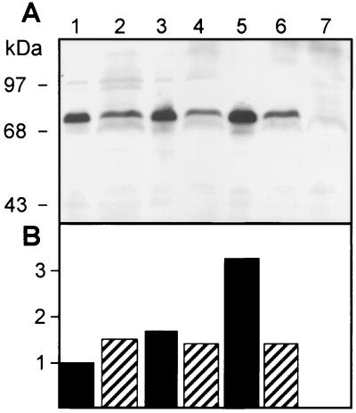
The expression of dnaK in the mutant strain at 30, 37, and 46°C compared to that in wild-type B. suis 1330 was analyzed by Western blotting with monoclonal anti-DnaK antibody as described previously (5, 12). Briefly, strains were grown to an optical density at 600 nm (OD600) of 0.5 at 30°C, concentrated fourfold in preheated TS broth, and incubated for 30 min at the appropriate temperatures. Bacteria were immediately heat killed at 65°C for 45 min, washed once in phosphate-buffered saline (PBS), and resuspended in sodium dodecyl sulfate-polyacrylamide gel electrophoresis sample buffer. Total cell lysates equivalent to 5 × 10⁸ bacteria per lane were loaded onto a sodium dodecyl sulfate-12.5% polyacrylamide gel. Total proteins were visualized by Coomassie staining after transfer onto a PolyScreen membrane (NEN) prior to antibody incubation and detection of DnaK by enhanced chemiluminescence (Amersham). As determined with SigmaGel software (SPSS Science, Chicago, Ill.), B. suis DnaK was induced approximately 1.7-fold between 30 and 37°C and another 2-fold between 37 and 46°C in the wild type, whereas the amounts of DnaK produced by the mutant containing the bla promoter remained constant (Fig. 1). The dnaK null mutant, obtained by gene knockout (12), did not produce any DnaK (Fig. 1A, lane 7). This result provided evidence that promoter replacement led to the noninducible expression of dnaK in this B. suis mutant.
FIG. 1.
(A) Western blot analysis of dnaK expression in total cell lysates of wild-type B. suis (lanes 1, 3, and 5) and the dnaKmut strain (lanes 2, 4, and 6) with monoclonal anti-DnaK antibody. Growth temperatures were 30°C (lanes 1, 2, and 7), 37°C (lanes 3 and 4), and 46°C (lanes 5 and 6). The dnaK null mutant was used as a control (lane 7). (B) Quantification of DnaK in panel A, using SigmaGel software, relative to the amount of DnaK present in the wild-type lysate at 30°C (lane 1).
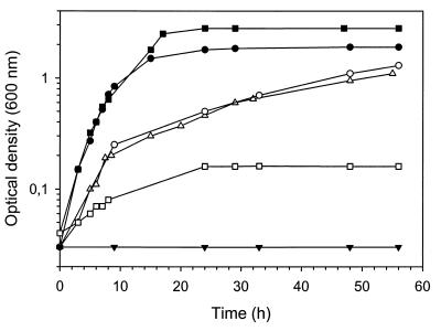
The absence of DnaK in the null mutant of B. suis described earlier resulted in temperature sensitivity for growth at 37°C and above (12). We compared this observation to the behavior of the dnaKmut strain at different temperatures: As we observed that the strain grew well on agar plates at 37 but not at 42°C (data not shown), we performed growth curve experiments in TS broth at 37, 38, and 39°C. B. suis 1330 (wild type) and the dnaKmut strain were grown to stationary phase, diluted to an OD600 of 0.03, and grown at the appropriate temperatures for 56 h. We observed lower growth rates for the mutant than for the wild-type at 37 and 38°C. At both temperatures, wild-type B. suis entered stationary phase at about 15 h postdilution, whereas the dnaKmut strain reached a similar OD600 only at 56 h (Fig. 2). The dnaK null mutant (12), in contrast, did not show any growth at 37°C. The temperature limit for normal growth of B. suis dnaKmut was 39°C. Constitutive, low-level production of DnaK therefore appeared to be sufficient for growth at temperatures of up to 38°C. In contrast, heat stress conditions such as growth at 39°C and above necessitate higher-level synthesis of DnaK, which is not possible in the dnaKmut strain (Fig. 1, lane 6).
FIG. 2.
Growth rates in TS broth of the B. suis wild-type strain at 37°C (•) and 39°C (▪) and of the dnaKmut strain at 37°C (○), 38°C (▵), and 39°C (□). The absence of growth of the dnaK null mutant at 37°C is indicated (▾). Growth curves are from one representative experiment out of three performed under identical conditions.
Sensitivity of the dnaKmut strain and of the dnaK null mutant to acid pH and oxidative stress
Additional phenotypic characteristics that might be linked to a modification of dnaK expression, such as sensitivity to acid pH and to oxidative stress, were investigated according to previously published protocols (8, 12). Briefly, for pH sensitivity experiments B. suis 1330 and the dnaKmut strain were grown to stationary phase (OD600 = 1.5) in TS broth, and the number of CFU per milliliter was determined prior to dilution 1:50 in TS broth adjusted to pH 4.5. At 0, 6, 24, 48, and 54 h postdilution, culture samples were diluted appropriately and plated on TS agar for colony enumeration. In contrast to the very high sensitivity of the dnaK null mutant to acid pH, as expressed by a 5,000-fold reduction in viable bacteria after 24 h of incubation (12), viability of the dnaKmut strain decreased only 1.8-fold over the same period compared to the wild-type and 5-fold following incubation for 54 h. The mutant constitutively expressing low levels of DnaK therefore resisted acid pH significantly better than the null mutant but did not reach the wild-type level of resistance. We concluded from these data that an acid pH stress could not by itself account for the rapid elimination of the dnaKmut strain in the macrophage.
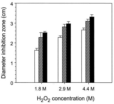
To investigate the sensitivity of the different strains to oxidative killing, strains were grown to stationary phase and rediluted to an OD600 of 0.25 each. Each strain (75 μl) was plated on TS agar, and sterile paper disks saturated with 10 μl of H2O2 at concentrations of 1.8, 2.9, and 4.4 M were layered on top prior to incubation at 30°C for 2 days and measurement of inhibition zone diameters (8). Interestingly, both the dnaK null mutant and the dnaKmut strain were significantly more sensitive to the three concentrations of H2O2 than the parental strain (Fig. 3) (P < 0.001). The sensitivities of both mutants were comparable, with inhibition zone diameters that were not significantly different from one another. The results therefore showed (i) that DnaK participated in the resistance of B. suis to oxidative stress and (ii) that noninducible dnaK expression was not sufficient for protection from oxidative stress.
FIG. 3.
Sensitivities of wild-type B. suis 1330 (open bars), the dnaKmut strain (hatched bars), and the dnaK null mutant (solid bars) to various concentrations of H2O2. Six disk assays were performed per strain per concentration. Error bars indicate standard deviations.
The dnaKmut strain is rapidly eliminated in J774 murine macrophages and in BALB/c mice
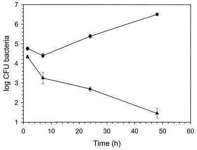
In order to assess the capacity of the dnaKmut strain of B. suis to survive within the host cell, we infected J774 murine macrophage-like cells with the mutant and the wild-type strain. Bacteria were added to adherent cells at a multiplicity of infection of 20 and incubated for 30 min at 37°C in the presence of 5% CO2, followed by three washes with PBS and incubation of the cells in RPMI medium supplemented with 10% fetal calf serum and gentamicin at a concentration of 30 μg/ml for at least 1 h. At 1.5, 7, 24, and 48 h postinfection, cells were washed once with PBS and lysed in 0.2% Triton X-100. CFU were enumerated by plating of serial dilutions on TS agar plates and incubation at 37°C. Low-level, noninducible expression of dnaK resulted in rapid elimination of the mutant strain in J774 cells, whereas the wild-type strain showed a profile typical of intracellular multiplication (Fig. 4). This is in contrast to the mutant's extracellular growth capacities at 37°C. These results therefore made it clear that production of DnaK necessary and sufficient for B. suis growth at 37°C in culture medium is not sufficient for intracellular replication of the pathogen, and they added support for our previous hypothesis that induction of dnaK is crucial for intramacrophagic replication of B. suis (12). The rapid elimination of B. suis dnaKmut in macrophages could not easily be explained by an eventual sensitivity of the strain to low pH, encountered in the phagosome at least during the first hours of infection, as there was no obvious correlation between its 40-fold reduction during the first 24 h of infection and the only 2-fold loss in viability at acid pH in vitro during the same period. The high sensitivity of the dnaKmut strain to oxidative stress, in contrast, suggested that this type of stress may participate in the antimicrobial defense mechanisms of murine macrophages against brucellae.
FIG. 4.
Intracellular growth of B. suis in J774 murine macrophage-like cells. Adherent cells were infected with the wild-type strain (•) or the dnaKmut strain (▴). Experiments were performed in triplicate. Error bars indicate standard deviations.
With the well-established murine model of infection (9, 10), the fate of the bacteria was monitored in vivo by the enumeration of B. suis 1330 and the dnaKmut strain in the spleens of BALB/c mice at various times postinfection. An infectious dose of 5 × 10⁴ bacteria was injected intraperitoneally, and residual virulence of the strains was evaluated following killing of four mice per point and strain at 1 day and 1, 4, 5, and 8 weeks postinoculation, homogenization of the spleens, and plating of serial dilutions on TS agar plates. Virulent B. suis multiplied rapidly in the spleen, and numbers reached a maximum at 1 week postinoculation before slightly declining until the end of the experiment (Fig. 5). In contrast, the dnaKmut strain was very quickly eliminated from the spleen, and only a few bacteria were recovered at 1 week in only one mouse out of four (Fig. 5). The 2-log difference observed at 1 day postinfection in intrasplenic survival of the wild-type versus the dnaKmut strain was in agreement with the 100-fold reduction of the intramacrophagic survival of the mutant 24 h after infection of J774 cells (Fig. 4). We therefore concluded that the effect observed in the complex in vivo model could be attributed mainly to the murine macrophage. In analogy to what we observed in J774 macrophage-like cells, these results confirmed the necessity of the presence of the native, inducible promoter of dnaK for survival of B. suis in mice.
FIG. 5.
Course of infection in BALB/c mice of wild-type B. suis (•) and the dnaKmut strain (▴). Growth of the bacteria in spleens was determined at various time points for 8 weeks. Data are means and standard deviations for four mice.
Conclusions.
It was observed previously that intracellular pathogens such as Salmonella enterica serovar Typhimurium and Legionella pneumophila have adapted to the hostile environment of the macrophage by selective induction of a multitude of proteins, among which are many stress proteins (1, 2, 15). Virulence gene transcription in salmonellae is activated after phagosome acidification, illustrating the importance of a coordinated gene activation in response to the microenvironment encountered by the bacterium (18). Based on the previous finding that a dnaK null mutant lost its capacity for intramacrophagic multiplication (12), our aim in this work on the pathogen B. suis was to determine whether basic levels of DnaK were sufficient for intracellular resistance. We concluded that a constitutive but noninducible expression of dnaK in B. suis was not sufficient to allow survival in a macrophage model of infection and in a host organism, despite extracellular growth of the mutant at 37 and 38°C. The increased sensitivity to oxidative stress of the strain producing constitutively low levels of DnaK therefore suggested that the induction of the gene via its native heat shock promoter was an essential event in the resistance of the pathogen not only to high temperature but also to the stressful environment of the macrophage in the host animal.
Acknowledgments
We thank Patrick Michel for excellent assistance in the laboratory work and A. Cloeckaert for his kind gift of monoclonal anti-DnaK antibody.
This work was partly supported by grant QLK2-CT-1999-00014 from the European Union.
REFERENCES
- 1.Abshire, K. Z., and F. C. Neidhardt. 1993. Analysis of proteins synthesized by Salmonella typhimurium during growth within a host macrophage. J. Bacteriol. 175:3734-3743. [DOI] [PMC free article] [PubMed] [Google Scholar]
- 2.Buchmeier, N. A., and F. Heffron. 1990. Induction of Salmonella stress proteins upon infection of macrophages. Science 248:730-732. [DOI] [PubMed] [Google Scholar]
- 3.Caron, E., J. P. Liautard, and S. Köhler. 1994. Differentiated U937 cells exhibit increased bactericidal activity upon LPS activation and discriminate between virulent and avirulent Listeria and Brucella species. J. Leukoc. Biol. 56:174-181. [DOI] [PubMed] [Google Scholar]
- 4.Cellier, M. F., J. Teyssier, M. Nicolas, J. P. Liautard, J. Marti, and J. Sri Widada. 1992. Cloning and characterization of the Brucella ovis heat shock protein DnaK functionally expressed in Escherichia coli. J. Bacteriol. 174:8036-8042. [DOI] [PMC free article] [PubMed] [Google Scholar]
- 5.Cloeckaert, A., O. Grépinet, H. Salih-Alj Debbarh, and M. S. Zygmunt. 1996. Overproduction of the Brucella melitensis heat shock protein DnaK in Escherichia coli and its localization by use of specific monoclonal antibodies in B. melitensis cells and fractions. Res. Microbiol. 147:145-157. [DOI] [PubMed] [Google Scholar]
- 6.Corbel, M. J. 1990. Brucella, p. 339-353. In M. T. Parker and L. H. Collier (ed.), Principles of bacteriology, virology, and immunity, vol. 2. E. Arnold, London, United Kingdom. [Google Scholar]
- 7.Ekaza, E., L. Guilloteau, J. Teyssier, J. P. Liautard, and S. Köhler. 2000. Functional analysis of the ClpATPase ClpA of Brucella suis, and persistence of a knockout mutant in BALB/c mice. Microbiology 146:1605-1616. [DOI] [PubMed] [Google Scholar]
- 8.Ekaza, E., J. Teyssier, S. Ouahrani-Bettache, J. P. Liautard, and S. Köhler. 2001. Characterization of Brucella suis clpB and clpAB mutants and participation of the genes in stress responses. J. Bacteriol. 183:2677-2681. [DOI] [PMC free article] [PubMed] [Google Scholar]
- 9.Endley, S., D. McMurray, and T. A. Ficht. 2001. Interruption of the cydB locus in Brucella abortus attenuates intracellular survival and virulence in the mouse model of infection. J. Bacteriol. 183:2454-2462. [DOI] [PMC free article] [PubMed] [Google Scholar]
- 10.Godfroid, F., B. Taminiau, I. Danese, P. Denoel, A. Tibor, V. Weynants, A. Cloeckaert, J. Godfroid, and J. J. Letesson. 1998. Identification of the perosamine synthetase gene of Brucella melitensis 16M and involvement of lipopolysaccharide O side chain in Brucella survival in mice and in macrophages. Infect. Immun. 66:5485-5493. [DOI] [PMC free article] [PubMed] [Google Scholar]
- 11.Harmon, B. G., L. G. Adams, and M. Frey. 1988. Survival of rough and smooth strains of Brucella abortus in bovine mammary gland macrophages. Am. J. Vet. Res. 49:1092-1097. [PubMed] [Google Scholar]
- 12.Köhler, S., J. Teyssier, A. Cloeckaert, B. Rouot, and J. P. Liautard. 1996. Participation of the molecular chaperone DnaK in intracellular growth of Brucella suis within U937-derived phagocytes. Mol. Microbiol. 20:701-712. [DOI] [PubMed] [Google Scholar]
- 13.Kovach, M. E., P. H. Elzer, D. S. Hill, G. T. Robertson, M. A. Farris, R. M. Roop II, and K. M. Peterson. 1995. Four new derivatives of the broad-host-range cloning vector pBBR1MCS, carrying different antibiotic-resistance cassettes. Gene 166:175-176. [DOI] [PubMed] [Google Scholar]
- 14.Kovach, M. E., R. W. Phillips, P. H. Elzer, R. M. Roop II, and K. M. Peterson. 1994. pBBR1MCS: a broad-host-range cloning vector. BioTechniques 16:800-802. [PubMed] [Google Scholar]
- 15.Kwaik, Y. A., B. I. Eisenstein, and N. C. Engleberg. 1993. Phenotypic modulation by Legionella pneumophila upon infection of macrophages. Infect. Immun. 61:1320-1329. [DOI] [PMC free article] [PubMed] [Google Scholar]
- 16.Liautard, J. P., A. Gross, J. Dornand, and S. Köhler. 1996. Interactions between professional phagocytes and Brucella spp. Microbiol. SEM 12:197-206. [PubMed] [Google Scholar]
- 17.Lin, J., and T. A. Ficht. 1995. Protein synthesis in Brucella abortus induced during macrophage infection. Infect. Immun. 63:1409-1414. [DOI] [PMC free article] [PubMed] [Google Scholar]
- 18.Miller, J. F., J. J. Mekalanos, and S. Falkow. 1989. Coordinate regulation and sensory transduction in the control of bacterial virulence. Science 243:916-922. [DOI] [PubMed] [Google Scholar]
- 19.Porte, F., J. P. Liautard, and S. Köhler. 1999. Early acidification of phagosomes containing Brucella suis is essential for intracellular survival in murine macrophages. Infect. Immun. 67:4041-4047. [DOI] [PMC free article] [PubMed] [Google Scholar]
- 20.Rafie-Kolpin, M., R. C. Essenberg, and J. H. Wyckoff III. 1996. Identification and comparison of macrophage-induced proteins and proteins induced under various stress conditions in Brucella abortus. Infect. Immun. 64:5274-5283. [DOI] [PMC free article] [PubMed] [Google Scholar]
- 21.Robertson, G. T., M. E. Kovach, C. A. Allen, T. A. Ficht, and R. M. Roop II. 2000. The Brucella abortus Lon functions as a generalized stress response protease and is required for wild-type virulence in BALB/c mice. Mol. Microbiol. 35:577-588. [DOI] [PubMed] [Google Scholar]
- 22.Sambrook, J., E. F. Fritsch, and T. Maniatis. 1989. Molecular cloning: a laboratory manual, 2nd ed. Cold Spring Harbor Laboratory, Cold Spring Harbor, N.Y.
- 23.Teixeira-Gomes, A. P., A. Cloeckaert, and M. S. Zygmunt. 2000. Characterization of heat, oxidative, and acid stress responses in Brucella melitensis. Infect. Immun. 68:2954-2961. [DOI] [PMC free article] [PubMed] [Google Scholar]
- 24.Yanisch-Perron, C., J. Vieira, and J. Messing. 1985. Improved M13 phage cloning vectors and host strains: nucleotide sequences of the M13mp18 and pUC19 vectors. Gene 33:103-119. [DOI] [PubMed] [Google Scholar]


