Subcutaneous emphysema is often seen after thoracic surgical procedures. In most cases it is due to a leak from the lung parenchyma and is self-limiting, requiring no specific treatment. Massive subcutaneous emphysema, however, should be treated both to reduce discomfort and to prevent respiratory embarrassment.
CASE HISTORY
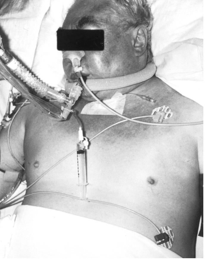
A man aged 71 with severe chronic obstructive pulmonary disease who had suffered recurrent right-sided pneumothoraces was admitted to a district general hospital for elective pleurectomy and apical bullectomy. The procedure was performed thoracoscopically and was uneventful; an area of bullous emphysema was identified in the right upper lobe and was stapled and excised. Postoperatively, the patient was noted to have a continuous air-leak and was maintained on 5 kPa of thoracic suction via apical and basal intercostal drains. Chest radiography showed a 10% pneumothorax. Subcutaneous emphysema of the thorax and neck was noted, but it was causing no symptoms. However, over the next two days, the subcutaneous emphysema worsened, involving the face, arms and abdomen. Despite an increase in the suction to 10 kPa, the patient was uncomfortable and had an obvious rise in the pitch of his voice. The pneumothorax was unaltered. On the third postoperative night he became acutely distressed and had first a respiratory arrest then a cardiac arrest. Initial attempts at intubation failed because of vocal cord and soft-tissue swelling (seen at laryngoscopy), but cricothyroidotomy allowed ventilation to be established. After resuscitation he was successfully intubated by the orotracheal route. At this point he had massive subcutaneous emphysema extending from his face to his lower extremities and scrotum (Figure 1). The pneumothorax remained small. Two further intercostal drains were inserted and connected to suction. A substantial air-leak continued for three days and the subcutaneous emphysema persisted. The patient was transferred to the regional cardiothoracic unit. At right thoracotomy, an air-leak was identified from the staple-line on the lung, which was considerably diseased. A right upper lobectomy was performed. Postoperatively there was no further air-leak and the lung expanded well. The subcutaneous emphysema resolved over several days and the patient recovered well.
Figure 1.

Massive subcutaneous emphysema involving the face and trunk. The lower limbs were also affected
COMMENT
The simplest explanation for the respiratory compromise would be restriction of thoracic expansion by a large volume of air in the subcutaneous tissues1. An alternative explanation would be direct compression of the airway itself. Air in the subcutaneous tissues, originating from the lung, may get there by two routes. First, if the parietal pleura is torn, air which has entered the pleural space may pass directly into the chest wall and subcutaneous tissues. Alternatively, alveolar air may track proximally within the bronchovascular sheath towards the hilum of the lungs where it may pass superficial to the endothoracic fascia, producing subcutaneous emphysema2. It may also pass into the mediastinum and then into the cervical visceral space which invests the trachea and oesophagus3. Whether air in this fascial compartment can actually compress the airway is debatable. The exact nature of the airway compression in this case is largely speculation based on laryngoscopic findings at the time of respiratory arrest—bulging vocal cords occluding the airway. Air in the cervical visceral space may have entered the submucosa of the trachea. The laryngeal sinus is the site of loosest attachment to the surrounding skeletal tissues and may therefore bulge into the airway. Certainly, a rise in the pitch of the voice is frequently seen in patients with subcutaneous emphysema originating in the lungs, pointing to laryngeal disturbance. The presence of subcutaneous emphysema in the cervical region is usually taken as evidence of decompression of the mediastinal and cervical visceral spaces. However, in this case the pressure was clearly sufficient to cause dissection throughout the body, right to the toes. The presence of a one-way valve in the pathway of tracking air could allow a substantial volume to accumulate with each breath, leading to progressive build-up of pressure beyond the valve. Air in the mediastinum, accumulating by a similar mechanism, has been associated with a picture of cardiac tamponade4, and several case reports describe respiratory distress with pneumomediastinum although the mechanism is difficult to ascertain.
Whatever the exact mechanism of airway compression in this case, the patient would clearly have benefited from earlier decompression of the subcutaneous tissues. Various approaches have been described, including the use of subcutaneous incisions, needles or drains5,6,7. Cervical mediastinotomy is an option when these interventions do not relieve increasing respiratory distress8.
References
- 1.Tonnesen AS, Wagner W, Mackey-Hargadine J. Tension subcutaneous emphysema. Anaesthesiology 1985;62: 90-2 [DOI] [PubMed] [Google Scholar]
- 2.Macklin CC. Transport of air along sheaths of pulmonic blood vessels from alveoli to mediastinum. Clinical implications. Arch Intern Med 1939;64: 913-26 [Google Scholar]
- 3.Maunder RJ, Pierson DJ, Hudson LD. Subcutaneous and mediastinal emphysema. Pathophysiology, diagnosis, and management. Arch Intern Med 1984;144: 1447-53 [PubMed] [Google Scholar]
- 4.Beg MH, Reyazuddin, Ansari MM. Traumatic tension pneumomediastinum mimicking cardiac tamponade. Thorax 1988;43: 576-7 [DOI] [PMC free article] [PubMed] [Google Scholar]
- 5.Herlan DB, Landreneau RJ, Ferson PF. Massive spontaneous subcutaneous emphysema. Acute management with infraclavicular ‘blow holes’. Chest 1992;102: 503-5 [DOI] [PubMed] [Google Scholar]
- 6.Nair KK, Neville E, Rajesh P, Papaliya H. A simple method of palliation for gross subcutaneous surgical emphysema. J R Coll Surg Edinb 1989;34: 163-4 [PubMed] [Google Scholar]
- 7.Terada Y, Matsunobe S, Nemoto T, Tsuda T, Shimizu Y. Palliation of severe subcutaneous emphysema with use of trocar-type chest tube as a subcutaneous drain [Letter]. Chest 1993;103: 323. [DOI] [PubMed] [Google Scholar]
- 8.Rydell JR, Jennings WK. Emergency cervical mediastinotomy for massive mediastinal emphysema. Arch Surg 1955;70: 647-53 [DOI] [PubMed] [Google Scholar]


