Abstract
Treatment of slipped upper femoral epiphysis (SUFE) is directed at preventing progressive slippage, minimizing deformity and avoiding avascular necrosis and chondrolysis. Delay in treatment adversely affects long-term outcomes. In a retrospective study we assessed delays between symptom onset and evaluation of the patient in an orthopaedic department. 27 patients aged 10-16 years were grouped by source of referral (general practitioner or accident and emergency department), and hips were classified as stable or unstable according to ability to bear weight.
The 27 children had 37 affected hips, 31 stable and 6 unstable. In the 20 patients referred by general practitioners, mean delay from symptom onset to orthopaedic evaluation was 119 days (range 2-504); in the 7 referred from accident and emergency departments it was 95 days (1-482). In the latter group the slips were more likely to be acute and unstable. 9 (45%) of the patients in the general-practitioner group had hip radiography before referral, all correctly diagnosed though not all the examinations included the recommended frog-lateral views.
Long delays between onset and diagnosis of SUFE are most likely in patients with mild symptoms, able to bear weight on the hip. Any adolescent with undiagnosed hip or knee pain that has lasted more than a week should undergo radiological investigation of the hip, with frog-lateral as well as anteroposterior views.
INTRODUCTION
Slipped upper femoral epiphysis (SUFE) is one of the common causes of hip pain in adolescence1. The presentation is often subtle and clinicians need to bear the condition constantly in mind. Untreated SUFE tends to progress, with increasing risk of hip deformity and osteoarthritis. Hence early diagnosis and treatment is essential for good long-term results. Yet long delays still arise between onset of symptoms and diagnosis. We have examined the records of a teaching-hospital orthopaedic department to elucidate these delays.
METHODS
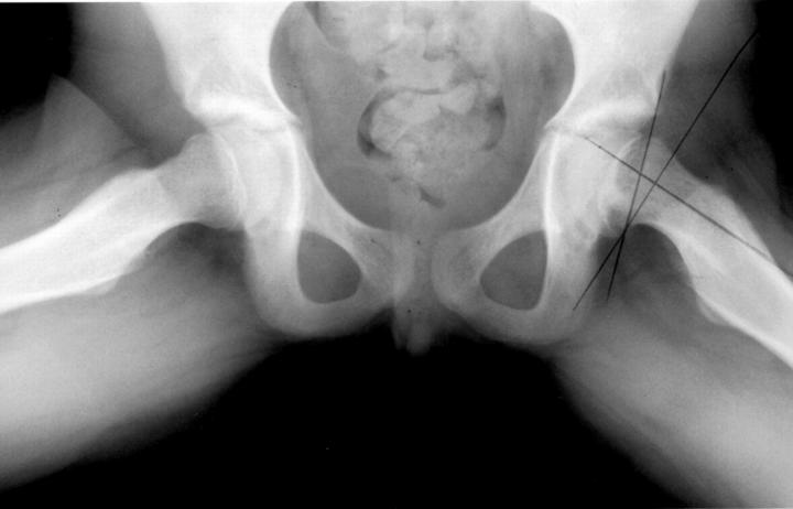
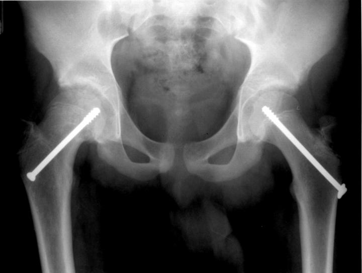
We examined the medical records and radiographs of all 30 children who had been treated surgically for SUFE in St James's University Hospital, Leeds, over the past 7 years. 3 children who were referred from other hospitals were excluded because we could not determine the interval before presentation to the initial hospital. The slips were divided into stable and unstable according to the patient's ability to bear weight on the hip2. Patients were also grouped into acute (symptoms <3 weeks), chronic (symptoms ≥3 weeks) and acute-on-chronic1. The degree of slip was measured by calculating the head—shaft angle on a lateral radiograph and was graded as mild (<30°), moderate (30-50°) or severe (>50°)3 (Figure 1). All measurements were made by the senior author to limit interobserver variation. Delay in diagnosis was recorded as the time from initial symptom to evaluation by an orthopaedic surgeon. All patients with SUFE were treated in a standard manner with a single percutaneous in-situ pin (Figure 2). They were followed up in paediatric outpatient clinics with clinical and radiological assessments until the proximal femoral epiphysis fused. Any patient with complications directly related to the condition or its operative treatment was followed up for longer as indicated.
Figure 1.
Measurement of the extent of epiphyseal displacement on the lateral radiograph. The degree of slip is reflected by the angle between the perpendicular of a line drawn along the long axis of the femoral neck and a line produced from the anterior to the posterior border of the epiphysis
Figure 2.
Anteroposterior radiograph showing mild slip of the right hip fixed with a single percutaneous screw, and prophylactic fixation of the left hip
The patients were put in two groups according to the source of primary referral. Group 1 were direct orthopaedic referrals from a general practitioner, either with a confirmed radiological diagnosis of SUFE or with a history of hip or leg pain which proved on further evaluation in hospital to be due to SUFE. Group 2 were patients who had their diagnosis confirmed after presenting directly in the emergency department.
RESULTS
Of the 27 patients in groups 1 and 2, 17 were boys; mean age of the children was 12.8 years (range 10-16). 37 hips were affected. The most common presenting symptom was pain localized to the hip region (14 children); in 10 children the first complaint was of thigh discomfort and 3 others had knee pain.
20 patients were originally seen by a general practitioner (group 1), 7 in an emergency department. Table 1 shows the distribution of presentations and severity in these two groups. In group 1 there was a higher proportion of stable or chronic SUFE—perhaps because those with severe pain went directly to the hospital emergency department. For 9 (45%) of the patients in group 1 the diagnosis had already been confirmed radiographically at the time of referral; in 4 the diagnosis had been made with anteroposterior views alone and in 5 with additional frog-lateral views. All 9 of these patients had stable slips. The single patient in group 1 with an unstable hip (acute painful limp) was referred urgently to hospital without radiography. In group 2 all patients had radiographs when first seen.
Table 1.
Patients' details
| Group 1 | Group 2 | |
|---|---|---|
| No. of hips | 24 (20 patients) | 13 (7 patients) |
| Stable slips | 23 | 8 |
| Unstable slips | 1 | 5 |
| Mild slip | 16 (66.7%) | 10 (76.9%) |
| Moderate slip | 3 (12.5%) | 2 (15.4%) |
| Severe slip | 5 (20.8%) | 1 (7.7%) |
| Acute slip | 0 | 4 (3 patients) |
| Acute-on-chronic slip | 4 (3 patients) | 4 (3 patients) |
| Chronic slip | 20 (15 patients) | 5 (3 patients) |
| No. of patients who had radiographs on initial consultation | 9 (45%) | 7 (100%) |
| Mean delay in diagnosis (days) | 119 (2-504) | 95 (1-482) |
Group 1=referrals from general practitioners; group 2=referrals from emergency departments
In the general-practitioner group the mean time from onset of symptoms to definitive diagnosis was 119 days (range 2-504, median 102). In group 2 it was 95 days (range 1-482, median 73). The mean delay between referral and orthopaedic assessment was 5 days (range 3-8).
DISCUSSION
The initial symptoms of SUFE are often vague—an ache in the thigh or pain around the ipsilateral knee4. The fact that, in this study, delays in diagnosis were somewhat greater in the general-practitioner group probably reflects a tendency for those with acute pain to go direct to hospital. The final outcome in SUFE is closely related to delay in diagnosis and the degree of the slip5,6; and the severity of the slip is related to the duration of symptoms7. Cowell5 described poorer short-term clinical results in those with delayed diagnosis, especially a delay of more than six months. For the general-practitioner group in this series, the mean delay of four months before evaluation in an orthopaedic department exceeds that reported by Green et al.8 in the USA, but clearly it will be influenced by the proportion of acute and unstable cases. It is satisfying that in nearly half these cases the general practitioner sent the patient for radiography and thus established the correct diagnosis. Other general practitioners may have hesitated because of concern about radiation hazards in this age group.
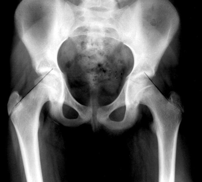
This study clearly shows that there is still a delay in establishing the diagnosis of SUFE in the community. We believe that obese older children and young adolescents with hip, thigh or knee pain should have their hips examined clinically and if there is any likelihood of SUFE, should undergo radiographic assessment of both hips. Sometimes either the patient or the parents will relate the onset of symptoms to some trivial trauma, and where soft-tissue injury is suspected to be the cause of symptoms, there is an argument for delaying radiological assessment for about two weeks provided there are no clinical signs of SUFE. If the pain does not improve, a radiological evaluation with anteroposterior and frog-lateral views of the hip is then indicated. It is important to look for subtle signs of SUFE in a radiograph. In a normal antero-posterior radiograph of the hip, a line drawn along the upper border of the femoral neck should cut off a segment of the superior epiphysis. In a minor slip, this line will skirt the superior margin of the femoral epiphysis (Figure 3), indicating postero-inferior displacement of the femoral epiphysis. Alertness to the possibility of SUFE, in any adolescent with hip, thigh or knee pain, will lead to earlier diagnosis and improve long-term outcomes.
Figure 3.
Minor slip. A line drawn along the upper femoral neck in an antero-posterior radiograph does not cut off a segment of epiphysis on the left side
References
- 1.Fahey JJ, O'Brien ET. Acute slipped femoral epiphysis: review of literature and report of ten cases. J Bone Joint Surg 1965;47A: 1105-27 [PubMed] [Google Scholar]
- 2.Lorder RT, Richards BS, Shapiro PS, et al. Acute slipped capital femoral epiphysis: the importance of physeal stability. J Bone Joint Surg 1993;75A: 113A. [DOI] [PubMed] [Google Scholar]
- 3.Weiner DS, Weiner S, Melby A, Hoyt WA. A 30-year experience with bone graft epiphysiodesis in the treatment of slipped capital femoral epiphysis. J Pediatr Orthop 1984;4: 145-52 [DOI] [PubMed] [Google Scholar]
- 4.Matava MJ, Patton CM, Luhman S, et al. Knee pain as the initial symptoms of slipped capital femoral epiphysis: an analysis of initial presentation and treatment. J Pediatr Orthop 1999;19: 455-60 [DOI] [PubMed] [Google Scholar]
- 5.Cowell HR. The significance of early diagnosis and treatment of slipping of the capital femoral epiphysis. Clin Orthop 1966;48: 89-94 [PubMed] [Google Scholar]
- 6.Wilson PD, Jacobs B, Schecter L. Slipped capital femoral epiphysis—on end-result study. J Bone Joint Surg Am 1965;47: 1128-45 [PubMed] [Google Scholar]
- 7.Jacobs B. Diagnosis and natural history of slipped femoral capital epiphysis. Instr Course Lect 1972;21: 167-73 [Google Scholar]
- 8.Green DW, Reynolds RAK, Khan S, Tolo VT. Delay in diagnosis for slipped capital femoral epiphysis. Pediatrics 1999;104: 713-14 [Google Scholar]


