

Abstract
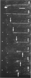
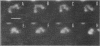
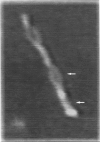
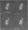
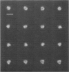
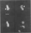
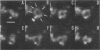
A fluorescence microscopy technique was used to image the dynamics of individual DNA molecules. Lambda, calf thymus, cosmid (circular), and T4 DNA were studied with the fluorescent dye acridine orange. Experiments with DNAase I were conducted, and the results indicate that these observations correspond to DNA molecules. The results of experiments with circular DNA provide strong evidence that these were single DNA molecules. Molecules were observed free in solution or attached to a glass or copper surface at one or several points. The Brownian motion of these molecules was observed, indicating that DNA in solution exists in a partially supercoiled state. Some molecules appeared stretched and were attached to the surface by their termini; the lengths of these molecules were measured. Such molecules also exhibited elastic behavior upon breaking. The power of this technique is demonstrated in images of cosmid DNA molecules, catenanes, and DNA extending from T4 phage particles. These results suggest immediate applications to molecular biology, such as examining the dynamics of protein-DNA interactions. Areas of ongoing research are discussed.
Full text
PDF


Images in this article
Selected References
These references are in PubMed. This may not be the complete list of references from this article.
- Barton J. K. Metals and DNA: molecular left-handed complements. Science. 1986 Aug 15;233(4765):727–734. doi: 10.1126/science.3016894. [DOI] [PubMed] [Google Scholar]
- CAIRNS J. The application of autoradiography to the study of DNA viruses. Cold Spring Harb Symp Quant Biol. 1962;27:311–318. doi: 10.1101/sqb.1962.027.001.029. [DOI] [PubMed] [Google Scholar]
- Dervan P. B. Design of sequence-specific DNA-binding molecules. Science. 1986 Apr 25;232(4749):464–471. doi: 10.1126/science.2421408. [DOI] [PubMed] [Google Scholar]
- Dickerson R. E., Drew H. R., Conner B. N., Wing R. M., Fratini A. V., Kopka M. L. The anatomy of A-, B-, and Z-DNA. Science. 1982 Apr 30;216(4545):475–485. doi: 10.1126/science.7071593. [DOI] [PubMed] [Google Scholar]
- FREIFELDER D., DAVISON P. F., GEIDUSCHEK E. P. Damage by visible light to the acridine orange--DNA complex. Biophys J. 1961 May;1:389–400. doi: 10.1016/s0006-3495(61)86897-5. [DOI] [PMC free article] [PubMed] [Google Scholar]
- Matsumoto S., Morikawa K., Yanagida M. Light microscopic structure of DNA in solution studied by the 4',6-diamidino-2-phenylindole staining method. J Mol Biol. 1981 Oct 25;152(2):501–516. doi: 10.1016/0022-2836(81)90255-2. [DOI] [PubMed] [Google Scholar]
- Morikawa K., Yanagida M. Visualization of individual DNA molecules in solution by light microscopy: DAPI staining method. J Biochem. 1981 Feb;89(2):693–696. doi: 10.1093/oxfordjournals.jbchem.a133247. [DOI] [PubMed] [Google Scholar]
- Slater G. W., Rousseau J., Noolandi J. On the stretching of DNA in the reptation theories of gel electrophoresis. Biopolymers. 1987 Jun;26(6):863–872. doi: 10.1002/bip.360260607. [DOI] [PubMed] [Google Scholar]
- Vollmer S. J., Yanofsky C. Efficient cloning of genes of Neurospora crassa. Proc Natl Acad Sci U S A. 1986 Jul;83(13):4869–4873. doi: 10.1073/pnas.83.13.4869. [DOI] [PMC free article] [PubMed] [Google Scholar]
- Yanagida M., Hiraoka Y., Katsura I. Dynamic behaviors of DNA molecules in solution studied by fluorescence microscopy. Cold Spring Harb Symp Quant Biol. 1983;47(Pt 1):177–187. doi: 10.1101/sqb.1983.047.01.023. [DOI] [PubMed] [Google Scholar]