

Abstract
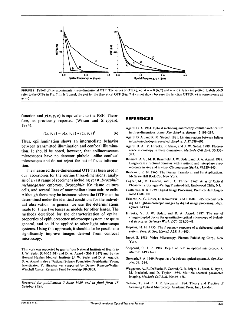
We have determined the three-dimensional image-forming properties of an epifluorescence microscope for use in obtaining very high resolution three-dimensional images of biological structures by image processing methods. Three-dimensional microscopic data is collected as a series of two-dimensional images recorded at different focal planes. Each of these images contains not only in-focus information from the region around the focal plane, but also out-of-focus contributions from the remainder of the specimen. Once the imaging properties of the microscope system are characterized, powerful image processing methods can be utilized to remove the out-of-focus information and to correct for image distortions. Although theoretical calculations for the behavior of an aberration-free microscope system are available, the properties of real lenses under the conditions used for biological observation are often far from an ideal. For this reason, we have directly determined the image-forming properties of an epifluorescence microscope under conditions relevant to biological observations. Through-focus series of a point object (fluorescently-coated microspheres) were recorded on a charge-coupled device image detector. From these images, the three-dimensional point spread function and its Fourier transform, the optical transfer function, were derived. There were significant differences between the experimental results and the theoretical models which have important implications for image processing. The discrepancies can be explained by imperfections of the microscope system, nonideal observation conditions, and partial confocal effects found to occur with epifluorescence illumination. Understanding the optical behavior of the microscope system has indicated how to optimize specimen preparation, data collection, and processing protocols to obtain significantly improved images.
Full text
PDF
Images in this article
Selected References
These references are in PubMed. This may not be the complete list of references from this article.
- Agard D. A., Hiraoka Y., Shaw P., Sedat J. W. Fluorescence microscopy in three dimensions. Methods Cell Biol. 1989;30:353–377. doi: 10.1016/s0091-679x(08)60986-3. [DOI] [PubMed] [Google Scholar]
- Agard D. A. Optical sectioning microscopy: cellular architecture in three dimensions. Annu Rev Biophys Bioeng. 1984;13:191–219. doi: 10.1146/annurev.bb.13.060184.001203. [DOI] [PubMed] [Google Scholar]
- Agard D. A., Stroud R. M. Linking regions between helices in bacteriorhodopsin revealed. Biophys J. 1982 Mar;37(3):589–602. [PMC free article] [PubMed] [Google Scholar]
- Belmont A. S., Braunfeld M. B., Sedat J. W., Agard D. A. Large-scale chromatin structural domains within mitotic and interphase chromosomes in vivo and in vitro. Chromosoma. 1989 Aug;98(2):129–143. doi: 10.1007/BF00291049. [DOI] [PubMed] [Google Scholar]
- Hiraoka Y., Sedat J. W., Agard D. A. The use of a charge-coupled device for quantitative optical microscopy of biological structures. Science. 1987 Oct 2;238(4823):36–41. doi: 10.1126/science.3116667. [DOI] [PubMed] [Google Scholar]
- Waggoner A., DeBiasio R., Conrad P., Bright G. R., Ernst L., Ryan K., Nederlof M., Taylor D. Multiple spectral parameter imaging. Methods Cell Biol. 1989;30:449–478. doi: 10.1016/s0091-679x(08)60990-5. [DOI] [PubMed] [Google Scholar]