Abstract
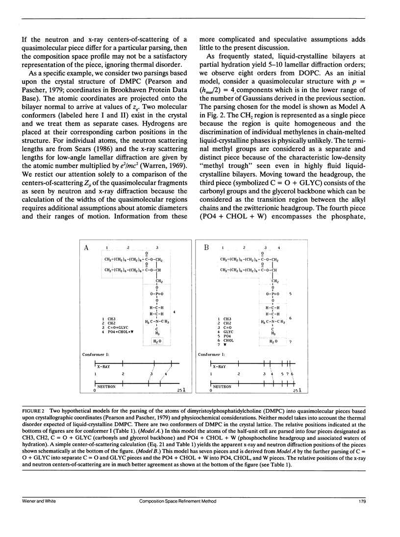
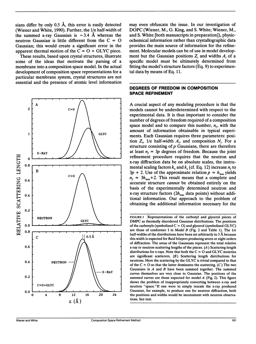
This is the second of two papers describing a method for the joint refinement of the structure of fluid bilayers using x-ray and neutron diffraction data. We showed in the first paper (Wiener, M. C., and S. H. White. 1990. Biophys. J. 59:162-173) that fluid bilayers generally consist of a nearly perfect lattice of thermally disordered unit cells and that the canonical resolution d/hmax is a measure of the widths of quasimolecular components represented by simple Gaussian functions. The thermal disorder makes possible a "composition space" representation in which the quasimolecular Gaussian distributions describe the number or probability of occupancy per unit length across the width of the bilayer of each component. This representation permits the joint refinement of neutron and x-ray lamellar diffraction data by means of a single quasimolecular structure that is fit simultaneously to both diffraction data sets. Scaling of each component by the appropriate neutron or x-ray scattering length maps the composition space profile to the appropriate scattering length space for comparison to experimental data. Other extensive properties, such as mass, can also be obtained by an appropriate scaling of the refined composition space structure. Based upon simple bilayer models involving crystal and liquid crystal structural information, we estimate that a fluid bilayer with hmax observed diffraction orders will be accurately represented by a structure with approximately hmax quasimolecular components. Strategies for assignment of quasimolecular components are demonstrated through detailed parsing of a phospholipid molecule based upon the one-dimensional projection of the crystal structure of dimyristoylphosphatidylcholine. Finally, we discuss in detail the number of experimental variables required for the composition space joint refinement. We find fluid bilayer structures to be marginally determined by the experimental data. The analysis of errors, which takes on particular importance under these circumstances, is also discussed.
Full text
PDF
Selected References
These references are in PubMed. This may not be the complete list of references from this article.
- Büldt G., Gally H. U., Seelig A., Seelig J., Zaccai G. Neutron diffraction studies on selectively deuterated phospholipid bilayers. Nature. 1978 Jan 12;271(5641):182–184. doi: 10.1038/271182a0. [DOI] [PubMed] [Google Scholar]
- Büldt G., Gally H. U., Seelig J., Zaccai G. Neutron diffraction studies on phosphatidylcholine model membranes. I. Head group conformation. J Mol Biol. 1979 Nov 15;134(4):673–691. doi: 10.1016/0022-2836(79)90479-0. [DOI] [PubMed] [Google Scholar]
- Büldt G., Seelig J. Conformation of phosphatidylethanolamine in the gel phase as seen by neutron diffraction. Biochemistry. 1980 Dec 23;19(26):6170–6175. doi: 10.1021/bi00567a034. [DOI] [PubMed] [Google Scholar]
- Coppens P. Comparative X-Ray and Neutron Diffraction Study of Bonding Effects in s-Triazine. Science. 1967 Dec 22;158(3808):1577–1579. doi: 10.1126/science.158.3808.1577. [DOI] [PubMed] [Google Scholar]
- Dorset D. L. Electron diffraction structure analysis of phospholipids. J Electron Microsc Tech. 1987 Sep;7(1):35–46. doi: 10.1002/jemt.1060070105. [DOI] [PubMed] [Google Scholar]
- Franks N. P., Arunachalam T., Caspi E. A direct method for determination of membrane electron density profiles on an absolute scale. Nature. 1978 Nov 30;276(5687):530–532. doi: 10.1038/276530a0. [DOI] [PubMed] [Google Scholar]
- Franks N. P. Structural analysis of hydrated egg lecithin and cholesterol bilayers. I. X-ray diffraction. J Mol Biol. 1976 Jan 25;100(3):345–358. doi: 10.1016/s0022-2836(76)80067-8. [DOI] [PubMed] [Google Scholar]
- Gruen D. W. A statistical mechanical model of the lipid bilayer above its phase transition. Biochim Biophys Acta. 1980 Jan 25;595(2):161–183. doi: 10.1016/0005-2736(80)90081-4. [DOI] [PubMed] [Google Scholar]
- Hauptman H. The Direct Methods of X-ray Crystallography. Science. 1986 Jul 11;233(4760):178–183. doi: 10.1126/science.233.4760.178. [DOI] [PubMed] [Google Scholar]
- Hendrickson W. A. Stereochemically restrained refinement of macromolecular structures. Methods Enzymol. 1985;115:252–270. doi: 10.1016/0076-6879(85)15021-4. [DOI] [PubMed] [Google Scholar]
- Herbette L., DeFoor P., Fleischer S., Pascolini D., Scarpa A., Blasie J. K. The separate profile structures of the functional calcium pump protein and the phospholipid bilayer within isolated sarcoplasmic reticulum membranes determined by X-ray and neutron diffraction. Biochim Biophys Acta. 1985 Jul 11;817(1):103–122. doi: 10.1016/0005-2736(85)90073-2. [DOI] [PubMed] [Google Scholar]
- Hitchcock P. B., Mason R., Shipley G. G. Phospholipid arrangements in multilayers and artificial membranes: quantitative analysis of the X-ray diffraction data from a multilayer of 1,2-dimyristoyl-DL-phosphatidylethanolamine. J Mol Biol. 1975 May 15;94(2):297–299. doi: 10.1016/0022-2836(75)90084-4. [DOI] [PubMed] [Google Scholar]
- Hitchcock P. B., Mason R., Thomas K. M., Shipley G. G. Structural chemistry of 1,2 dilauroyl-DL-phosphatidylethanolamine: molecular conformation and intermolecular packing of phospholipids. Proc Natl Acad Sci U S A. 1974 Aug;71(8):3036–3040. doi: 10.1073/pnas.71.8.3036. [DOI] [PMC free article] [PubMed] [Google Scholar]
- Jacobs R. E., White S. H. The nature of the hydrophobic binding of small peptides at the bilayer interface: implications for the insertion of transbilayer helices. Biochemistry. 1989 Apr 18;28(8):3421–3437. doi: 10.1021/bi00434a042. [DOI] [PubMed] [Google Scholar]
- Johnson M. L. Evaluation and propagation of confidence intervals in nonlinear, asymmetrical variance spaces. Analysis of ligand-binding data. Biophys J. 1983 Oct;44(1):101–106. doi: 10.1016/S0006-3495(83)84281-7. [DOI] [PMC free article] [PubMed] [Google Scholar]
- King G. I., Jacobs R. E., White S. H. Hexane dissolved in dioleoyllecithin bilayers has a partial molar volume of approximately zero. Biochemistry. 1985 Aug 13;24(17):4637–4645. doi: 10.1021/bi00338a024. [DOI] [PubMed] [Google Scholar]
- McCaughan L., Krimm S. Biochemical profiles of membranes from x-ray and neutron diffraction. Biophys J. 1982 Feb;37(2):417–426. doi: 10.1016/S0006-3495(82)84687-0. [DOI] [PMC free article] [PubMed] [Google Scholar]
- Mischel M., Seelig J., Braganza L. F., Büldt G. A neutron diffraction study of the headgroup conformation of phosphatidylglycerol from Escherichia coli membranes. Chem Phys Lipids. 1987 May;43(4):237–246. doi: 10.1016/0009-3084(87)90020-x. [DOI] [PubMed] [Google Scholar]
- Nagle J. F., Wiener M. C. Relations for lipid bilayers. Connection of electron density profiles to other structural quantities. Biophys J. 1989 Feb;55(2):309–313. doi: 10.1016/S0006-3495(89)82806-1. [DOI] [PMC free article] [PubMed] [Google Scholar]
- Nagle J. F., Wiener M. C. Structure of fully hydrated bilayer dispersions. Biochim Biophys Acta. 1988 Jul 7;942(1):1–10. doi: 10.1016/0005-2736(88)90268-4. [DOI] [PubMed] [Google Scholar]
- Norvell J. C., Nunes A. C., Schoenborn B. P. Neutron diffraction analysis of myoglobin: structure of the carbon monoxide derivative. Science. 1975 Nov 7;190(4214):568–570. doi: 10.1126/science.1188354. [DOI] [PubMed] [Google Scholar]
- Pearson R. H., Pascher I. The molecular structure of lecithin dihydrate. Nature. 1979 Oct 11;281(5731):499–501. doi: 10.1038/281499a0. [DOI] [PubMed] [Google Scholar]
- Rand R. P., Luzzati V. X-ray diffraction study in water of lipids extracted from human erythrocytes: the position of cholesterol in the lipid lamellae. Biophys J. 1968 Jan;8(1):125–137. doi: 10.1016/S0006-3495(68)86479-3. [DOI] [PMC free article] [PubMed] [Google Scholar]
- Strenk L. M., Westerman P. W., Doane J. W. A model of orientational ordering in phosphatidylcholine bilayers based on conformational analysis of the glycerol backbone region. Biophys J. 1985 Nov;48(5):765–773. doi: 10.1016/S0006-3495(85)83834-0. [DOI] [PMC free article] [PubMed] [Google Scholar]
- White S. H., Jacobs R. E., King G. I. Partial specific volumes of lipid and water in mixtures of egg lecithin and water. Biophys J. 1987 Oct;52(4):663–665. doi: 10.1016/S0006-3495(87)83259-9. [DOI] [PMC free article] [PubMed] [Google Scholar]
- Wiener M. C., Suter R. M., Nagle J. F. Structure of the fully hydrated gel phase of dipalmitoylphosphatidylcholine. Biophys J. 1989 Feb;55(2):315–325. doi: 10.1016/S0006-3495(89)82807-3. [DOI] [PMC free article] [PubMed] [Google Scholar]
- Wiener M. C., White S. H. Fluid bilayer structure determination by the combined use of x-ray and neutron diffraction. I. Fluid bilayer models and the limits of resolution. Biophys J. 1991 Jan;59(1):162–173. doi: 10.1016/S0006-3495(91)82208-1. [DOI] [PMC free article] [PubMed] [Google Scholar]
- Worcester D. L., Franks N. P. Structural analysis of hydrated egg lecithin and cholesterol bilayers. II. Neutrol diffraction. J Mol Biol. 1976 Jan 25;100(3):359–378. doi: 10.1016/s0022-2836(76)80068-x. [DOI] [PubMed] [Google Scholar]
- Zaccai G., Büldt G., Seelig A., Seelig J. Neutron diffraction studies on phosphatidylcholine model membranes. II. Chain conformation and segmental disorder. J Mol Biol. 1979 Nov 15;134(4):693–706. doi: 10.1016/0022-2836(79)90480-7. [DOI] [PubMed] [Google Scholar]


