

Abstract
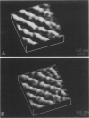
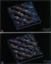
We present images of the polar or headgroup regions of bilayers of dimyristoyl-phosphatidylethanolamine (DMPE), deposited by Langmuir-Blodgett deposition onto mica substrates at high surface pressures and imaged under water at room temperature with the optical lever atomic force microscope. The lattice structure of DMPE is visualized with sufficient resolution that the location of individual headgroups can be determined. The forces are sufficiently small that the same area can be repeatedly imaged with a minimum of damage. The DMPE molecules in the bilayer appear to have relatively good long-range orientational order, but rather short-range and poor positional order. These results are in good agreement with x-ray measurements of unsupported lipid monolayers on the water surface, and with electron diffraction of adsorbed monolayers.
Full text
PDF


Images in this article
Selected References
These references are in PubMed. This may not be the complete list of references from this article.
- Binnig G, Quate CF, Gerber C. Atomic force microscope. Phys Rev Lett. 1986 Mar 3;56(9):930–933. doi: 10.1103/PhysRevLett.56.930. [DOI] [PubMed] [Google Scholar]
- Drake B., Prater C. B., Weisenhorn A. L., Gould S. A., Albrecht T. R., Quate C. F., Cannell D. S., Hansma H. G., Hansma P. K. Imaging crystals, polymers, and processes in water with the atomic force microscope. Science. 1989 Mar 24;243(4898):1586–1589. doi: 10.1126/science.2928794. [DOI] [PubMed] [Google Scholar]
- Helm C. A., Möhwald H., Kjaer K., Als-Nielsen J. Phospholipid monolayers between fluid and solid states. Biophys J. 1987 Sep;52(3):381–390. doi: 10.1016/S0006-3495(87)83226-5. [DOI] [PMC free article] [PubMed] [Google Scholar]
- Kjaer K, Als-Nielsen J, Helm CA, Laxhuber LA, Möhwald H. Ordering in lipid monolayers studied by synchrotron x-ray diffraction and fluorescence microscopy. Phys Rev Lett. 1987 May 25;58(21):2224–2227. doi: 10.1103/PhysRevLett.58.2224. [DOI] [PubMed] [Google Scholar]
- Mamin HJ, Ganz E, Abraham DW, Thomson RE, Clarke J. Contamination-mediated deformation of graphite by the scanning tunneling microscope. Phys Rev B Condens Matter. 1986 Dec 15;34(12):9015–9018. doi: 10.1103/physrevb.34.9015. [DOI] [PubMed] [Google Scholar]
- Ribi H. O., Ludwig D. S., Mercer K. L., Schoolnik G. K., Kornberg R. D. Three-dimensional structure of cholera toxin penetrating a lipid membrane. Science. 1988 Mar 11;239(4845):1272–1276. doi: 10.1126/science.3344432. [DOI] [PubMed] [Google Scholar]
- Tamm L. K., McConnell H. M. Supported phospholipid bilayers. Biophys J. 1985 Jan;47(1):105–113. doi: 10.1016/S0006-3495(85)83882-0. [DOI] [PMC free article] [PubMed] [Google Scholar]
- Zasadzinski J. A. Scanning tunneling microscopy with applications to biological surfaces. Biotechniques. 1989 Feb;7(2):174–187. [PubMed] [Google Scholar]