Hypoperfusion retinopathy, also known as venous stasis or slow flow retinopathy, is a severe and blinding manifestation of carotid artery disease. Treatment by carotid endarterectomy, combined with panretinal photocoagulation, can stabilize vision.
CASE HISTORY
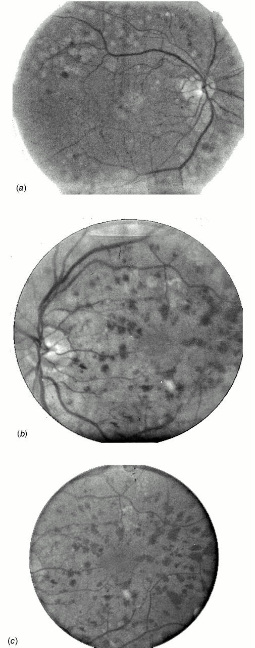
A man aged 83 described bilateral progressive visual loss over three weeks, worse in the left than the right eye and with a notable deterioration during the past three days. Medical history included non-insulin-dependent diabetes mellitus, hypertension, ischaemic heart disease, and peripheral vascular disease. On examination corrected visual acuity (VA) was 6/9 in the right eye and 6/12 in the left. The intraocular pressure was slightly raised in the left eye. Fundoscopy revealed small intraretinal haemorrhages in all quadrants in the right eye. In the left eye, superficial and deep intraretinal haemorrhages were densely scattered throughout the fundus and one cotton-wool spot was seen (Figure 1). The retinal veins were dilated and of irregular calibre. The initial diagnosis was of right background diabetic retinopathy and left non-ischaemic central retinal vein occlusion. Blood tests were normal except for moderately raised cholesterol, glucose and HbA1c.
Figure 1.

Fundus photographs obtained on second visit. (a) Right fundus: some midperipheral retinal haemorrhages superotemporally to the vascular arcade. (b) Left fundus: numerous posterior pole and midperipheral retinal haemorrhages. (c) Higher magnification of left posterior pole. One cotton-wool spot can be seen below the macula
Within three days the vision had deteriorated to 6/18 in the right eye and 6/60 in the left. The pupillary margin of both irides showed small dilated vessels, consistent with early neovascularization of the iris, leading to a diagnosis of bilateral hypoperfusion retinopathy. Bilateral retinal argon laser photocoagulation was begun immediately. A carotid duplex ultrasound scan revealed calcified atheromatous plaques in both common carotid arteries and a 90-99% stenosis of the right internal carotid artery (ICA). No flow could be seen in the proximal left ICA; the distal ICA could not be displayed. A consultation with a vascular surgeon, with a view to possible carotid endarterectomy, was arranged, but this would have been a high-risk procedure for a person in such poor general health and the patient opted for conservative management. Within two months of presentation he underwent three sessions of panretinal photocoagulation to both eyes. The progression of ischaemia was stopped in the right eye, but the left developed iris neovascularization and rubeotic glaucoma, necessitating cryoablation of the anterior retina and cyclocryotherapy to the ciliary body to bring the intraocular pressure under control. Visual acuity is currently stable at 6/18 in the right eye and hand movements in the left eye.
COMMENT
Chronic hypoperfusion retinopathy is seen in severe stenosis or complete occlusion of an internal carotid artery. Patients often have a history of hypertension, peripheral vascular, cerebrovascular or ischaemic heart disease, or diabetes1. The condition is usually ipsilateral to the more severely affected carotid artery. The pathophysiology is that chronic low arterial perfusion pressure leads to retinal hypoxia. Slowing of the retinal circulation time causes dilatation and tortuosity of the retinal veins, breakdown of capillary walls, superficial (flame-shaped) and deep (dot-blot) retinal haemorrhages, macular oedema and eventual neovascular proliferation in retina and iris which occurs as a response to the release of angiogenic factors from the ischaemic retina2,3. The fundoscopic picture resembles diabetic retinopathy but there are two distinguishing signs: in the early stages, hypoperfusion retinopathy affects the retinal midperiphery rather than the posterior pole; and it is usually unilateral2,3. Contrasting features to a central retinal vein occlusion are the absence of optic disc swelling and the midperipheral location of the haemorrhages. In more profound ocular hypoperfusion, known as ocular ischaemic syndrome, the retinopathy is associated with anterior segment ischaemia as reflected in corneal oedema and ischaemic uveitis, neovascularization of the iris and raised intraocular pressure secondary to neovascular glaucoma. A poorly reactive pupil is often seen and patients complain of severe orbital pain2. Visual prognosis is poor1.
The differential diagnosis includes diabetic retinopathy, non-ischaemic central retinal vein occlusion, hyperviscosity syndromes such as polycythaemia, Waldenström's macroglobulinaemia, haemoglobinopathies, myelomatosis, and lymphoma3, all of which need to be excluded by investigations.
Chronic ocular ischaemia is treated by panretinal photocoagulation, which reduces the production of angiogenic factors by the hypoxic retina2. Reduction of intraocular pressure to improve ocular perfusion can be achieved by topical β-blockers and/or topical or systemic carbonic anhydrase inhibitors1. If possible, this is followed by management of the carotid occlusion by endarterectomy or a bypass from the superficial temporal artery to the middle cerebral artery. Reports on the outcome of carotid endarterectomy in patients with ocular ischaemic syndrome show conflicting results1,4,5. The decision about cerebrovascular surgery needs to be made on an individual basis, and the risk of perioperative complications, especially strokes, needs to be balanced against the expected benefit—namely, stabilization of vision2.
References
- 1.Mizener JB, Podhajsky P, Hayreh SS. Ocular ischemic syndrome. Ophthalmology 1997;104: 859-64 [DOI] [PubMed] [Google Scholar]
- 2.Dugan JD, Green WR. Ophthalmic manifestations of carotid occlusive disease. Eye 1991;5: 226-38 [DOI] [PubMed] [Google Scholar]
- 3.McCrary JA. Venous stasis retinopathy of stenotic or occlusive carotid origin. J Clin Neuro-Ophthalmol 1989;9: 195-9 [PubMed] [Google Scholar]
- 4.Johnston ME, Gonder JR, Canny CL. Successful treatment of the ocular ischemic syndrome with panretinal photocoagulation and cerebrovascular surgery. Can J Ophthalmol 1988;23: 114-19 [PubMed] [Google Scholar]
- 5.Neupert JR, Brubaker RF, Kearns TP, Sundt TM. Rapid resolution of venous stasis retinopathy after carotid endarterectomy. Am J Ophthalmol 1976;81: 600-2 [DOI] [PubMed] [Google Scholar]


