Abstract
Yersinia pestis strain Yreka was grown at 27 or 37°C, and the lipid A structures (lipid A-27°C and lipid A-37°C) of the respective lipopolysaccharides (LPS) were investigated by matrix-assisted laser desorption ionization-time-of-flight (MALDI-TOF) mass spectrometry. Lipid A-27°C consisted of a mixture of tri-acyl, tetra-acyl, penta-acyl, and hexa-acyl lipid A's, of which tetra-acyl lipid A was most abundant. Lipid A-37°C consisted predominantly of tri- and tetra-acylated molecules, with only small amounts of penta-acyl lipid A; no hexa-acyl lipid A was detected. Furthermore, the amount of 4-amino-arabinose was substantially higher in lipid A-27°C than in lipid A-37°C. By use of mouse and human macrophage cell lines, the biological activities of the LPS and lipid A preparations were measured via their abilities to induce production of tumor necrosis factor alpha (TNF-α). In both cell lines the LPS and the lipid A from bacteria grown at 27°C were stronger inducers of TNF-α than those from bacteria grown at 37°C. However, the difference in activity was more prominent in human macrophage cells. These results suggest that in order to reduce the activation of human macrophages, it may be more advantageous for Y. pestis to produce less-acylated lipid A at 37°C.
Yersinia pestis was isolated as a causative agent of plague in Hong-Kong in 1894 independently by A. Yersin and S. Kitasato (3). Since then, this highly pathogenic bacterium has been investigated, and many virulence factors have been identified, including fraction 1 antigen, murine toxin, Yop proteins, pH 6 antigen, and iron acquisition systems (5, 28). Lipopolysaccharide (LPS) has also been studied for many decades as one of the virulence factors of Y. pestis, and its composition and endotoxic activity have been examined in earlier studies (1, 40, 41). By use of modern analytical methods, the LPS of Y. pestis was proven to be a rough type LPS without O-antigenic polysaccharide (7, 8, 25, 29, 30, 35) that contains 3-hydroxy-myristic acid (3-OH-C14:0) as a main fatty acid in the lipid A portion. However, there was a discrepancy in the amounts of the nonhydroxy-fatty acids found. Hartley et al. reported that nearly 90% of fatty acids in the lipid A of Y. pestis strain 195/R (a virulent strain) consisted of 3-OH-C14:0 (14). On the other hand, considerable amounts of lauric acid (C12:0), palmitic acid (C16:0), and palmitoleic acid (C16:1) were detected in the lipid A of Y. pestis strain EV40 (an avirulent strain) (4, 39). Using the same strain, Aussel et al. (2) proposed a hexa-acylated lipid A structure (four molecules of 3-OH-C14:0, one of C12:0, and one of C16:1). In our preliminary experiment we isolated the lipid A from Y. pestis strain Yreka (virulent strain) grown at 37°C and found that it contained 3-OH-C14:0 and only trace amounts of other fatty acids. A possible explanation of the controversy on fatty acids may be differences in strains or culture conditions.
Darveau et al. (8) reported that the molecular size of the LPS from Y. pestis strain EV76 changed slightly when the growth temperature was shifted from 26 to 37°C. Modification of the antigenicity by growth temperature was also reported by another group (13). It was previously reported that the amount of C16:1 in the lipid A of Escherichia coli increased when it was grown at lower temperatures (38). The palmitoleoyl transferase that functions at low temperatures was identified, and its character was studied (6). Since C16:1 was also present in the lipid A of Y. pestis (2, 4, 39), we assumed that a similar phenomenon could occur in this bacterium. Furthermore, it was proved by several groups (15, 32, 36) using synthetic lipid A compounds that the amounts, kinds, and linkages of fatty acids in lipid A influenced its biological activity. Therefore, if the composition and the structure of fatty acids in lipid A are modified, the biological activity may also be changed. This possibility attracted our interest in relation to pathogenicity.
In this study modification of the structure and activity of Y. pestis lipid A by growth temperature was investigated, and the possible association of such modification with the pathogenesis of Y. pestis is discussed.
MATERIALS AND METHODS
Bacterial strain and culture condition.
Y. pestis strain Yreka (a virulent strain; National Institute of Infectious Diseases, Tokyo, Japan) was cultured on brain heart infusion agar (Difco Laboratories, Detroit, Mich.) at 27 or 37°C for 48 h. Bacterial cells were suspended in saline (30 mg [wet weight]/ml) and heated at 90°C for 30 min for killing.
Extraction of LPS and preparation of lipid A.
Killed bacterial cells were washed with distilled water, ethanol, acetone, and diethyl ether to prepare dry cells. LPS was extracted from the dry cells by a conventional phenol-water method (42), and the aqueous phase was dialyzed extensively to remove phenol and then lyophilized. The crude LPS preparation was treated with nucleases and proteases as described previously (34) and was purified by repeated ultracentrifugation. The LPS in the precipitate from centrifugation was further purified by extraction with 45% phenol plus triethylamine and sodium deoxycholate (23) and was used for the macrophage stimulation assay. In order to release lipid A, the purified LPS was hydrolyzed in 0.1 M sodium acetate buffer (pH 4.4) at 100°C for 2 h. The hydrolysate was dialyzed, and the retentate was ultracentrifuged to obtain lipid A in the precipitate.
SDS-PAGE.
Sodium dodecyl sulfate-polyacrylamide gel electrophoresis (SDS-PAGE) was performed by using a 4 to 20% gradient gel (Tefco, Tokyo, Japan) with the buffer system of Laemmli (20). The gel was oxidized with periodate and silver stained as described by Tsai and Frasch (37). LPS samples from Salmonella enterica were used as references. Smooth type LPS of Salmonella enterica serovar Abortus-equi, kindly donated by C. Galanos (Max-Planck-Institut für Immunbiologie, Freiburg, Germany), was purified as described elsewhere (11). Ra type LPS of Salmonella enterica serovar Minnesota R60 was prepared by phenol-chloroform-petroleum ether extraction (10). Re type LPS of Salmonella serovar Minnesota R595 was purchased from Sigma Chemical Co. (St. Louis, Mo.).
Chemical analysis.
Neutral sugars were determined by gas-liquid chromatography (GLC) in the form of alditol acetate after they were released from LPS by hydrolysis in 0.1 M HCl (at 100°C for 48 h). d-Glucosamine (GlcN) was determined by the same method used for neutral sugars after it was released by hydrolysis in 4 M HCl (at 100°C for 16 h). Fatty acids were determined by GLC in the methyl ester derivatives after hydrolysis in 4 M HCl (at 100°C for 5 h). For GLC analysis, a GC-14A chromatograph equipped with a 25-m CBP1 capillary column (both from Shimadzu, Kyoto, Japan) was used. Sugar derivatives were analyzed by a temperature program of 170°C for 2 min raised to 270°C by 5°C/min, and fatty acid methyl esters were analyzed by a program of 150°C for 2 min raised to 230°C by 5°C/min. 3-Deoxy-d-manno-oct-2-ulosonic acid (Kdo) and phosphorus were determined colorimetrically as described previously (17).
Mass spectrometry.
Lipid A preparations were analyzed by matrix-assisted laser desorption ionization-time-of-flight (MALDI-TOF) mass spectrometry with a Bruker-Reflex III (Bruker-Franzen Analytik, Bremen, Germany) in a linear TOF configuration at an acceleration voltage of 20 kV. Details of the applied methods have been described elsewhere (21). In general, the compounds were dissolved in distilled water at a concentration of 10 μg/μl and treated with an ion exchanger (Amberlite IR-120; Merck, Darmstadt, Germany) to remove disturbing cations. A 1-μl aliquot of the sample was then mixed with 0.5 M 2,5-dihydroxybenzoic acid (Aldrich, Milwaukee, Wis.) in methanol, and 0.5-μl aliquots were deposited on a metallic sample holder. Mass scale calibration was performed externally with similar compounds of known chemical structure.
Cell culture.
The murine macrophage cell line RAW 264.7 (American Type Culture Collection, Manassas, Va.) and the human macrophage cell line U937 (Japanese Cancer Research Resources Bank, Tokyo, Japan) were used for induction of tumor necrosis factor alpha (TNF-α) upon stimulation with LPS or lipid A as reported previously (9, 24). For culture of cells, RPMI 1640 medium (Flow Laboratories, Inc., Rockville, Md.) supplemented with 10 mM HEPES, 2 mM l-glutamine, 100 U of penicillin per ml, 100 μg of streptomycin per ml, and 0.2% NaHCO3 was used as the basic medium, and heat-inactivated fetal calf serum (FCS; Flow Laboratories) was added at a concentration of 5 or 10% (5 or 10% FCS-RPMI medium). RAW 264.7 cells were suspended in 5% FCS-RPMI medium at 106 cells per ml. These cell suspensions were dispensed (0.5 ml) to each well of a 48-well culture plate and cultured for 2 h. The cells in each well were washed three times with 0.5 ml of Hanks' balanced salt solution (Flow Laboratories), and adherent cells were cultured with 5% FCS-RPMI medium in the presence of test samples (0.5 ml/well). The culture supernatant obtained at 24 h after stimulation was assayed for TNF-α activity. U937 cells were prepared for experiments by adding phorbol myristate acetate at a final concentration of 30 ng per ml in 10% FCS-RPMI medium (2 × 10⁵ cells/ml) and culturing cells for 3 days on a 48-well culture plate (0.5 ml/well) to induce differentiation into macrophage-like cells. Cells were washed once with 10% FCS-RPMI medium (0.5 ml/well), and adherent cells were cultured with 10% FCS-RPMI medium in the presence of test samples (0.5 ml/well). The culture supernatant obtained at 4 h after stimulation was assayed for TNF-α activity.
TNF-α assay.
TNF-α was determined by a cytotoxic assay with L-929 cells (33). Briefly, L929 cells were cultured with 5% FCS-RPMI medium in a 96-well flat-bottom culture plate for 3 h, and actinomycin D (Sigma Chemical Co.) was added to a final concentration of 1 μg/ml with serial dilution of test samples. Viable cells in the overnight culture were stained with crystal violet, and absorbance of the blue color extracted with 30% acetic acid was measured at 540 nm. TNF-α activity was calculated from the dilution factor of test samples necessary for 50% cell lysis, with correction by an internal standard of recombinant human TNF-α in each assay, and was expressed in units per milliliter.
RESULTS
Analysis of LPS and lipid A preparations from cells grown at 27 and 37°C.
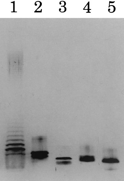
LPSs from cells grown at 27°C (LPS-27°C) and from cells grown at 37°C (LPS-37°C) were first analyzed by SDS-PAGE. As shown in Fig. 1, both LPSs showed condensed single bands, and they migrated as fast as the major band of Salmonella serovar Minnesota R60 (Ra) LPS and slightly more slowly than that of Salmonella serovar Minnesota R595 (Re) LPS. This result clearly indicates that these LPSs are rough type LPSs without O-antigenic polysaccharides, a fact which agrees with previous reports of other groups (7, 8, 25, 29, 30). Between LPS-27°C and LPS-37°C a small but reproducible difference in migration was observed (Fig. 1, lanes 4 and 5). LPS-37°C migrated slightly faster than LPS-27°C, suggesting that LPS-37°C contained smaller molecules than LPS-27°C.
FIG. 1.
SDS-PAGE of LPS from Y. pestis strain Yreka grown at 27 and 37°C. Lane 1, Salmonella serovar Abortus-equi (1 μg); lane 2, Salmonella serovar Minnesota R60 (0.2 μg); lane 3, Salmonella serovar Minnesota R595 (0.2 μg); lane 4, Y. pestis grown at 27°C (0.2 μg); lane 5, Y. pestis grown at 37°C (0.2 μg).
The chemical compositions of LPS and lipid A preparations from cells grown at both temperatures are shown in Table 1. Although no clear difference was found in the amounts of carbohydrate components, the composition of fatty acids was different between LPS-27°C and LPS-37°C, and between lipid A prepared from cells grown at 27°C (lipid A-27°C) and that from cells grown at 37°C (lipid A-37°C). In LPS-27°C and lipid A-27°C, C16:1 was present and the amount of C 12:0 was somewhat higher than in LPS-37°C and lipid A-37°C. These results indicate that at the low growth temperature (27°C), Y. pestis produces lipid A with higher amounts of nonhydroxy-fatty acids, i.e., C12:0 and C16:1.
TABLE 1.
Chemical compositions of LPS and lipid A of Y. pestis strain Yreka
| Componenta | Concnb (μmol/mg) in:
|
|||
|---|---|---|---|---|
| Crude LPS
|
Lipid A
|
|||
| 27°C | 37°C | 27°C | 37°C | |
| Glc | 0.36 | 0.21 | 0.15 | 0.06 |
| Gal | 0.11 | 0.02 | 0.04 | — |
| d,d-Hep | 0.12 | 0.13 | 0.04 | 0.03 |
| l,d-Hep | 0.57 | 0.48 | 0.20 | 0.09 |
| Kdo | 0.12 | 0.33 | — | — |
| GlcN | 0.72 | 0.65 | 0.87 | 0.70 |
| Phosphorus | 0.64 | 0.80 | 0.75 | 0.99 |
| C12:0 | 0.06 | 0.02 | 0.04 | 0.03 |
| C16:1 | 0.02 | — | 0.02 | — |
| C16:0 | 0.01 | 0.01 | — | 0.01 |
| 3-OH-C14:0 | 0.44 | 0.56 | 1.35 | 1.46 |
Ara4N was not determined in this experiment, Glc, d-glucose; Gal, d-galactose; d,d-Hep, d-glycero-d-manno-heptose; l,d-Hep, l-glycero-d-manno-heptose.
—, less than 0.01 μmol/mg.
Analysis of lipid A by MALDI-TOF mass spectrometry.
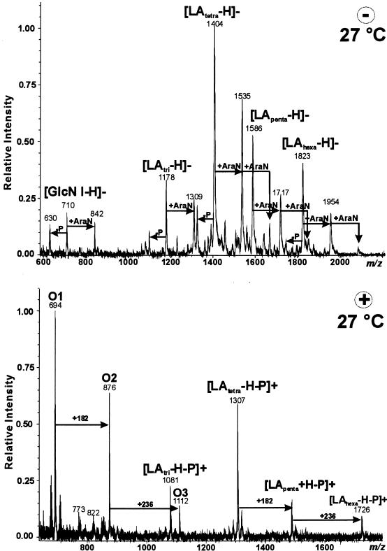
Lipid A-27°C and lipid A-37°C were analyzed by MALDI-TOF mass spectrometry to confirm the change in fatty acid content found in compositional analysis. The mass spectrum of the negative-ion mode for lipid A-27°C (Fig. 2) showed prominent mass peaks of tri-acyl, tetra-acyl, penta-acyl. and hexa-acyl lipid A, each of which was accompanied by peaks with one and, to a far lower extent, with two additional 4-amino-arabinose (Ara4N) molecules (mass difference, +131) or without one phosphate molecule (mass difference, −80). Among these, the peaks corresponding to tetra-acylated lipid A were the most abundant. Additionally, fragment ions were detected at m/z 630, 710, and 842. These ions originated from the reducing GlcN of the lipid A carrying two 3-OH-C14:0 molecules with or without Ara4N and phosphate. In the positive-ion mass spectrum (Fig. 2), oxonium ion peaks of m/z 694 (O1), 876 (O2), and 1112 (O3) were observed; these were formed from the nonreducing GlcN of the lipid A carrying one phosphate and two 3-OH-C14:0 molecules (O1), one phosphate, two 3-OH-C14:0, and one C12:0 molecule (O2), and one phosphate, two 3-OH-C14:0, one C12:0, and one C16:1 molecule (O3). These data indicate that the structure of hexa-acylated lipid A, composed of a reducing GlcN carrying two 3-OH-C14:0 molecules and a nonreducing GlcN carrying the four acyl groups mentioned above, is identical to that proposed by Aussel et al. (2), but this fully acylated molecule was only a minor component in our lipid A preparation.
FIG. 2.
MALDI-TOF mass spectra of lipid A from Y. pestis strain Yreka grown at 27°C. (Top) Negative-ion mode. (Bottom) Positive-ion mode.
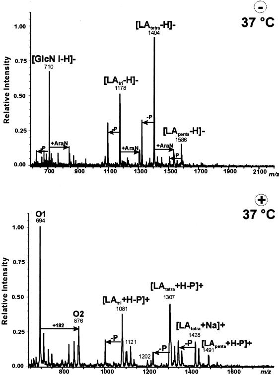
Lipid A-37°C gave the mass spectra shown in Fig. 3. The negative-ion mass spectrum showed prominent mass peaks of tri-acyl and tetra-acyl lipid A, but the peak of penta-acyl lipid A was very small, and no peak was detected for hexa-acyl lipid A. Each mass peak was accompanied by peaks of molecules with one Ara4N or without phosphate as in lipid A-27°C, but the peaks of molecules with one Ara4N of less intensity, indicating that the Ara4N content in lipid A-37°C was lower than that in lipid A-27°C. In the positive-ion mass spectrum, a large peak of oxonium ion O1 (m/z 694), carrying one phosphate and two 3-OH-C14:0 molecules, and a relatively small peak of ion O2 (m/z 876), carrying one phosphate, two 3-OH-C14:0, and one C12:0 molecule, were observed. These results agree with those obtained from the negative-ion spectrum, indicating that hexa-acylated lipid A is absent in lipid A-37°C.
FIG. 3.
MALDI-TOF mass spectra of lipid A from Y. pestis strain Yreka grown at 37°C. (Top) Negative-ion mode. (Bottom) Positive-ion mode.
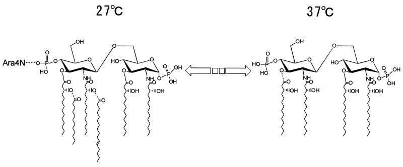
Summarizing the data obtained by MALDI-TOF mass spectrometric analysis, structural modification of the lipid A by growth temperature is shown schematically in Fig. 4. When the growth temperature of Y. pestis was shifted from 27 to 37°C, the amounts of nonhydroxy-fatty acids (C12:0 and C16:1) and Ara4N in lipid A were reduced. The amount of 3-OH-C14:0 was also reduced, increasing the amount of tri-acylated lipid A.
FIG. 4.
Structural modification of Y. pestis lipid A by growth temperature. In this figure the structure of the lipid A backbone is shown in analogy to other published structures (2, 39), although it was not precisely determined in this study. The positions of C16:1 and C12:0 may be interchanged. The second Ara4N detected by MALDI-TOF mass spectrometry as a minor component is not shown here.
TNF-inducing activities of LPS and lipid A preparations.
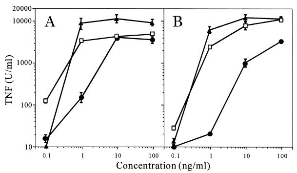
The macrophage-stimulating activities of the LPS and lipid A preparations were measured as TNF-inducing activity in murine and human macrophages. When RAW 267.4 murine macrophages were used, lipid A-27°C and LPS-27°C exhibited activities comparable to those of the synthetic lipid A 506 (Daiichi Kagaku Co., Tokyo, Japan) and S. enterica LPS, respectively (Fig. 5), while lipid A-37°C and LPS-37°C showed weaker activities. The activity of LPS-37°C was approximately 1/10 that of LPS-27°C, judging from the dose responses.
FIG. 5.
TNF-inducing activities of lipid A and LPS from Y. pestis strain Yreka in RAW 264.7 murine macrophages. (A) RAW 264.7 cells were cultured in the presence of the indicated concentrations of synthetic lipid A 506 (▴), Y. pestis lipid A grown at 27°C (□), or Y. pestis lipid A grown at 37°C (•). (B) RAW 264.7 cells were cultured in the presence of the indicated concentrations of Salmonella serovar Abortus-equi LPS (▴), LPS of Y. pestis grown at 27°C (□), and LPS of Y. pestis grown at 37°C (•). Culture supernatants obtained at 24 h were assayed for determination of TNF-α activity. Data are means ± standard errors of the means from triplicate samples. Results of one experiment representative of three independent experiments are shown.
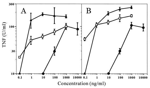
In a subsequent experiment, TNF-inducing activity in U937 human macrophages was measured. In U937 cells the activities of lipid A-27°C and LPS-27°C were somewhat weaker than those of synthetic lipid A and S. enterica LPS, respectively, as shown in Fig. 6, and the activities of lipid A-37°C and LPS-37°C were much weaker. The amount of LPS-37°C required to induce 100 U of TNF-α/ml was about 100 times greater than that of LPS-27°C (Fig. 6).
FIG. 6.
TNF-inducing activities of lipid A and LPS of Y. pestis strain Yreka in U937 human macrophages. U937 cells were cultured in the presence of the indicated concentrations of lipid A's (A) or LPSs (B). Symbols in panels A and B are as explained for the corresponding panels of Fig. 5. Culture supernatants were obtained at 4 h and assayed for TNF-α activity. Data are means ± standard errors of the means from triplicate samples. Results of one experiment representative of three independent experiments are shown.
These results indicate that LPS-37°C is a weaker stimulant of macrophages than LPS-27°C and that this difference in activity is more marked in human macrophages than in murine macrophages.
DISCUSSION
Although the basic structure of lipid A is common among bacteria belonging to the family Enterobacteriaceae, variations of fatty acids are present in each genus or species. The most common lipid A structure, that of E. coli was investigated and finally established by chemical synthesis, and it is considered to be highly active toward the mammalian immune system (15, 32, 36). It contains four 3-OH-C 14:0, one myristic acid (C14:0), and one C12:0 molecule, and two nonhydroxy-fatty acids bind to the hydroxyl group of 3-OH-C14:0 in the nonreducing GlcN molecule to form an acyloxyacyl structure. While some genera in Enterobacteriaceae that are taxonomically close to E. coli, e.g., Salmonella and Shigella, have lipid A structures similar to that of E. coli, other genera in this family have lipid A structures that differ from that of E. coli in number and linkage of fatty acids. For Yersinia, Aussel et al. (2) recently studied the lipid A structures of various species including Y. pestis. They presented the fully acylated structure of lipid A as a major molecule, but in other studies Yersinia was reported to have less-acylated lipid A (19, 26). In our preliminary experiments we found that Y. pestis strain Yreka (a virulent strain) produced a lipid A containing 3-OH-C14:0 as the only fatty acid component when grown at 37°C. Controversial results were reported by other groups using the avirulent strain EV40 grown at 28°C (4, 39). In this study, to clarify this discrepancy, we analyzed the lipid A from cells grown at 27°C or 37°C by MALDI-TOF mass spectrometry, and we found that at the low growth temperature (27°C) Y. pestis produced a lipid A containing larger amounts of C16:1 and C12:0 than the lipid A produced at the high growth temperature (37°C), as shown in Fig. 4. Additionally, the amount of Ara4N was also greater in LPS-27°C. These compositional differences were also observed by SDS-PAGE analysis (Fig. 1), as a slight difference in the mobility and width of LPS bands.
For E. coli, it was found that acyltransferase encoded on the lpxP gene transfers C16:1 to the intermediate of lipid A biosynthesis at a low temperature (6). A similar thermoregulation mechanism may also be present in acyltransferases in the biosynthesis of Y. pestis lipid A. This should be investigated in the future in comparison with genes encoding E. coli acyltransferases by using the genomic sequence data of Y. pestis (27).
The proposed structure of lipid A-37°C shown in Fig. 4 is similar to that of the lipid A biosynthesis intermediate lipid IVA (precursor Ia) (31) or synthetic compound 406 (16). Lipid IVA has been reported to act as an LPS agonist to murine cells but to behave as an antagonist to human cells (12, 18, 22). From this structure and the reported data, we expected, and proved in this study, that lipid A-37°C or LPS-37°C, in comparison with lipid A-27°C or LPS-27°C, would exhibit weaker TNF-inducing activity in murine macrophages, and much weaker activity in human macrophages.
It is extremely interesting that Y. pestis produces lipid A with reduced biological activity at 37°C, i.e., the body temperature of humans and other host animals. This phenomenon may be related to the infectious ability of Y. pestis. When this pathogen is injected into mammalian tissue through a flea bite, the environmental temperature for the pathogen shifts up to 37°C, giving a signal to modify the lipid A structure. The modified structure of lipid A will then stimulate the host immune system less actively. This reduction in activity may be more prominent in the human immune system than in the mouse system and may be of great advantage to Y. pestis, enabling the organism to succeed in infection by escaping from the host defense systems. In order to elucidate the complete mechanism of lipid A modification and its effects on the host immune system during infection by and proliferation of Y. pestis, factors other than growth temperature, such as culture medium, pH, or oxygen supply, should also be considered in future studies.
Editor: R. N. Moore
REFERENCES
- 1.Albizo, J. M., and M. J. Surgalla. 1970. Isolation and biological characterization of Pasteurella pestis endotoxin. Infect. Immun. 2:229-236. [DOI] [PMC free article] [PubMed] [Google Scholar]
- 2.Aussel, L., H. Thérisod, D. Karibian, M. B. Perry, M. Bruneteau, and M. Caroff. 2000. Novel variation of lipid A structures in strains of different Yersinia species. FEBS Lett. 465:87-92. [DOI] [PubMed] [Google Scholar]
- 3.Bibel, D. J., and T. H. Chen. 1976. Diagnosis of plague: an analysis of the Yersin-Kitasato controversy. Bacteriol. Rev. 40:633-651. [DOI] [PMC free article] [PubMed] [Google Scholar]
- 4.Bordet, C., M. Bruneteau, and G. Michel. 1977. Lipopolysaccharides d'une souche à virulence atténuée de Yersinia pestis. Eur. J. Biochem. 79:443-449. [DOI] [PubMed] [Google Scholar]
- 5.Brubaker, R. R. 1991. Factors promoting acute and chronic diseases caused by yersiniae. Clin. Microbiol. Rev. 4:309-324. [DOI] [PMC free article] [PubMed] [Google Scholar]
- 6.Carty, S. M., K. R. Sreekumar, and C. R. H. Raetz. 1999. Effect of cold shock on lipid A biosynthesis in Escherichia coli. Induction at 12°C of an acyltransferase specific for palmitoleoyl-acyl carrier protein. J. Biol. Chem. 274:9677-9685. [DOI] [PubMed] [Google Scholar]
- 7.Chart, H., T. Cheasty, and B. Rowe. 1995. Differentiation of Yersinia pestis and Y. pseudotuberculosis by SDS-PAGE analysis of lipopolysaccharide. Lett. Appl. Microbiol. 20:369-370. [DOI] [PubMed] [Google Scholar]
- 8.Darveau, R. P., W. T. Charnetzky, R. E. Hurlbert, and R. E. W. Hancock. 1983. Effects of growth temperature, 47-megadalton plasmid, and calcium deficiency on the outer membrane protein porin and lipopolysaccharide composition of Yersinia pestis EV76. Infect. Immun. 42:1092-1101. [DOI] [PMC free article] [PubMed] [Google Scholar]
- 9.Funatogawa, K., M. Matsuura, M. Nakano, M. Kiso, and A. Hasegawa. 1998. Relationship of structure and biological activity of monosaccharide lipid A analogues to induction of nitric oxide production by murine macrophage RAW 264.7 cells. Infect. Immun. 66:5792-5798. [DOI] [PMC free article] [PubMed] [Google Scholar]
- 10.Galanos, C., O. Lüderitz, and O. Westphal. 1969. A new method for the extraction of R lipopolysaccharides. Eur. J. Biochem. 9:245-249. [DOI] [PubMed] [Google Scholar]
- 11.Galanos, C., O. Lüderitz, and O. Westphal. 1979. Preparation and properties of a standardized lipopolysaccharide from Salmonella abortus equi. Zentbl. Bakteriol. Microbiol. Hyg. Abt. 1 Orig. A 243:226-244. [PubMed] [Google Scholar]
- 12.Golenbock, D. T., R. Y. Hampton, N. Qureshi, K. Takayama, and C. R. H. Raetz. 1991. Lipid A-like molecules that antagonize the effect of endotoxins on human monocytes. J. Biol. Chem. 266:19490-19498. [PubMed] [Google Scholar]
- 13.Gremiakova, T. A., K. I. Volkovoi, R. Z. Shaikhutdinova, and A. V. Stepanov. 1999. Temperature-dependent changes in immunochemical properties of lipopolysaccharides of Yersinia pestis. Vestn. Ross. Acad. Med. Nauk. 12:32-34. [PubMed] [Google Scholar]
- 14.Hartley, J. L., G. A. Adams, and T. G. Tornabene. 1974. Chemical and physical properties of lipopolysaccharide of Yersinia pestis. J. Bacteriol. 118:848-854. [DOI] [PMC free article] [PubMed] [Google Scholar]
- 15.Homma, J. Y., M. Matsuura, and Y. Kumazawa. 1989. Studies on lipid A, the active center of endotoxin-structure-activity relationship. Drugs Future 14:645-665. [PubMed] [Google Scholar]
- 16.Inage, M., H. Chaki, S. Kusumoto, and T. Shiba. 1981. Chemical synthesis of phosphorylated fundamental structure of lipid A. Tetrahedron Lett. 22:2281-2284. [Google Scholar]
- 17.Kawahara, K., H. Brade, E. T. Rietschel, and U. Zähringer. 1987. Studies on the chemical structure of the core-lipid A region of the lipopolysaccharide of Acinetobacter calcoaceticus NCTC 10305. Detection of a new 2-octulosonic acid interlinking the core oligosaccharide and lipid A component. Eur. J. Biochem. 163:489-495. [DOI] [PubMed] [Google Scholar]
- 18.Kovach, N., E. Yee, R. S. Munford, C. R. H. Raetz, and J. M. Harland. 1990. Lipid IVa inhibits synthesis and release of tumor necrosis factor induced by lipopolysaccharide in human blood ex vivo. J. Exp. Med. 172:77-84. [DOI] [PMC free article] [PubMed] [Google Scholar]
- 19.Krasikova, I. N., S. I. Bakholdina, S. V. Khotimchenko, and T. F. Solov'eva. 1999. Effect of growth temperature and pVM82 plasmid on fatty acids of lipid A from Yersinia pseudotuberculosis. Biochemistry (Moscow) 64:338-344. [PubMed] [Google Scholar]
- 20.Laemmli, U. K. 1970. Cleavage of structural proteins during the assembly of the head of bacteriophage T4. Nature 227:680-685. [DOI] [PubMed] [Google Scholar]
- 21.Lindner, B. 2000. Matrix-assisted laser desorption/ionization time-of-flight mass spectrometry of lipopolysaccharides, p. 311-325. In O. Holst (ed.), Bacterial toxins: methods and protocols. Humana Press, Totowa, N.J.
- 22.Loppnow, H., H. Brade, I. Durrbaum, C. A. Dinarello, S. Kusumoto, E. T. Rietschel, and H.-D. Flad. 1989. IL-1 induction-capacity of defined lipopolysaccharide partial structures. J. Immunol. 142:3229-3238. [PubMed] [Google Scholar]
- 23.Manthey, C. L., and S. N. Vogel. 1994. Elimination of trace endotoxin protein from rough chemotype LPS. J. Endotoxin Res. 1:84-91. [Google Scholar]
- 24.Matsuura, M., M. Kiso, and A. Hasegawa. 1999. Activity of monosaccharide lipid A analogues in human monocytic cells as agonists or antagonists of bacterial lipopolysaccharide. Infect. Immun. 67:6286-6292. [DOI] [PMC free article] [PubMed] [Google Scholar]
- 25.Minka, S., and M. Bruneteau. 1998. Isolement et caractérisation chemique des lipopolysaccharides de type R dans une souche hypovirulente de Yersinia pestis. Can. J. Microbiol. 44:477-481. [PubMed] [Google Scholar]
- 26.Oertelt, C., B. Lindner, M. Skurnik, and O. Holst. 2001. Isolation and structural characterization of an R-form lipopolysaccharide from Yersinia enterocolitica serotype O:8. Eur. J. Biochem. 268:554-564. [DOI] [PubMed] [Google Scholar]
- 27.Parkhill, J., B. W. Wren, N. R. Thomson, R. W. Titball, M. T. Holden, M. B. Prentice, M. Sebaihia, K. D. James, C. Churcher, K. L. Mungall, S. Baker, D. Basham, S. D. Bentley, K. Brooks, A. M. Cerdeno-Tarraga, T. Chillingworth, A. Cronin, R. M. Davies, P. Davis, G. Dougan, T. Feltwell, N. Hamlin, S. Holroyd, K. Jagels, A. V. Karlyshev, S. Leather, S. Moule, P. C. Oyston, M. Quail, K. Rutherfold, M. Simmonds, J. Skelton, K. Stevens, S. Whitehead, and B. G. Barrell. 2001. Genome sequence of Yersinia pestis, the causative agent of plague. Nature 413:523-527. [DOI] [PubMed] [Google Scholar]
- 28.Perry, R., and J. D. Fetherston. 1997. Yersinia pestis—etiologic agent of plague. Clin. Microbiol. Rev. 10:35-66. [DOI] [PMC free article] [PubMed] [Google Scholar]
- 29.Prior, J. L., P. G. Hitchen, E. D. Williamson, A. J. Reason, H. R. Morris, A. Dell, B. W. Wren, and R. W. Titball. 2001. Characterization of the lipopolysaccharide of Yersinia pestis. Microb. Pathog. 30:49-57. [DOI] [PubMed] [Google Scholar]
- 30.Prior, J. L., J. Parkhill, P. G. Hitchen, K. L. Mungall, K. Stevens, H. R. Morris, A. J. Reason, P. C. F. Oyston, A. Dell, B. W. Wren, and R. W. Titball. 2001. The failure of different strains of Yersinia pestis to produce lipopolysaccharide O-antigen under different growth conditions is due to mutations in the O-antigen gene cluster. FEMS Microbiol. Lett. 197:229-233. [DOI] [PubMed] [Google Scholar]
- 31.Rick, P. D., L. W.-M. Fung, C. Ho, and M. J. Osborn. 1977. Lipid A mutants of Salmonella typhimurium. Purification and characterization of a lipid A precursor produced by a mutant in 3-deoxy-d-mannooctulosonate-8-phosphate synthetase. J. Biol. Chem. 252:4904-4912. [PubMed] [Google Scholar]
- 32.Rietschel, E. T., L. Brade, K. Brandenburg, H.-D. Flad, J. de Jong-Leuveninck, K. Kawahara, B. Lindner, H. Loppnow, T. Lüderitz, U. Schade, U. Seydel, Z. Sidorczyk, A. Tacken, U. Zähringer, and H. Brade. 1987. Chemical structure and biological activity of bacterial and synthetic lipid A. Rev. Infect. Dis. 9(Suppl. 5):527-536. [DOI] [PubMed] [Google Scholar]
- 33.Ruff, M. R., and G. E. Gifford. 1980. Purification and physico-chemical characterization of rabbit tumor necrosis factor. J. Immunol. 125:1671-1677. [PubMed] [Google Scholar]
- 34.Shimomura, H., M. Matsuura, S. Saito, Y. Hirai, Y. Isshiki, and K. Kawahara. 2001. Lipopolysaccharide of Burkholderia cepacia and its unique character to stimulate murine macrophages with relative lack of interleukin-1β-inducing ability. Infect. Immun. 69:3663-3669. [DOI] [PMC free article] [PubMed] [Google Scholar]
- 35.Skurnik, M., A. Peippo, and E. Ervelä. 2000. Characterization of the O-antigen gene clusters of Yersinia pseudotuberculosis and the cryptic O-antigen gene cluster of Yersinia pestis shows that the plague bacillus is most closely related to and has evolved from Y. pseudotuberculosis serotype O:1b. Mol. Microbiol. 37:316-330. [DOI] [PubMed] [Google Scholar]
- 36.Takada, H., and S. Kotani. 1992. Structure-function relationships of lipid A, p. 107-134. In D. C. Morrison and J. L. Ryan (ed.), Bacterial endotoxic lipopolysaccharides, vol. 1. Molecular biochemistry and cellular biology. CRC Press, Boca Raton, Fla.
- 37.Tsai, C.-M., and C. E. Frasch. 1982. A sensitive silver stain for detecting lipopolysaccharides in polyacrylamide gels. Anal. Biochem. 119:115-119. [DOI] [PubMed] [Google Scholar]
- 38.Van Alphen, L., B. Lugtenberg, E. T. Rietschel, and C. Mombers. 1979. Architecture of the outer membrane of Escherichia coli K12. Phase transitions of the bacteriophage K3 receptor complex. Eur. J. Biochem. 101:571-579. [DOI] [PubMed] [Google Scholar]
- 39.Venezia, N. D., S. Minka, M. Bruneteau, H. Mayer, and G. Michel. 1985. Lipopolysaccharide from Yersinia pestis. Studies on lipid A of lipopolysaccharides I and II. Eur. J. Biochem. 151:399-404. [DOI] [PubMed] [Google Scholar]
- 40.Walker, R. V. 1968. Comparative physiopathology of plague endotoxin in mice, guinea pigs and monkeys. J. Infect. Dis. 118:188-196. [DOI] [PubMed] [Google Scholar]
- 41.Walker, R. V., M. G. Barnes, and E. D. Higgins. 1966. Composition of and physiopathology produced by plague endotoxins. Nature 209:1246. [DOI] [PubMed] [Google Scholar]
- 42.Westphal, O., and K. Jann. 1965. Bacterial lipopolysaccharides. Extraction with phenol-water and further applications of the procedure. Methods Carbohydr. Chem. 5:83-91. [Google Scholar]


